Search
- Page Path
- HOME > Search
- Garre’s osteomyelitis of the mandible managed by nonsurgical re-endodontic treatment
- Heegyun Kim, Jiyoung Kwon, Hyun-Jung Kim, Soram Oh, Duck-Su Kim, Ji-Hyun Jang
- Restor Dent Endod 2024;49(2):e13. Published online March 18, 2024
- DOI: https://doi.org/10.5395/rde.2024.49.e13

-
 Abstract
 PDF
PDF PubReader
 ePub
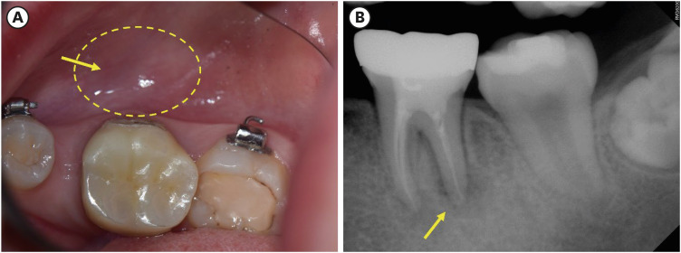
ePub Chronic osteomyelitis with proliferative periostitis, known as Garre’s osteomyelitis, is a type of osteomyelitis characterized by a distinctive gross thickening of the periosteum of bones. Peripheral reactive bone formation can be caused by mild irritation or infection. Garre’s osteomyelitis is usually diagnosed in children and young adults, and the mandible is more affected than the maxilla. The following is a case report of a 12-year-old female patient with Garre’s osteomyelitis of the mandible due to an infection of a root canal-treated tooth. Without surgical intervention, the patient’s symptoms were relieved through nonsurgical root canal re-treatment with long-term calcium hydroxide placement. A cone-beam computed tomography image obtained 6 months after treatment completion displayed complete healing of the periapical lesion and resolution of the peripheral reactive buccal bone. Due to the clinical features of Garre's osteomyelitis, which is characterized by thickening of the periosteum, it can be mistaken for other diseases such as fibrous dysplasia. It is important to correctly diagnose Garre's osteomyelitis based on its distinctive clinical features to avoid unnecessary surgical intervention, and it can lead to minimally invasive treatment options.
-
Citations
Citations to this article as recorded by- Focal osteomyelitis with proliferative periostitis
Zarah Yakoob
South African Dental Journal.2025; 79(09): 508. CrossRef - Garré’s osteomyelitis of the mandible in an adolescent: a case report
Wiem Feki, Imen Haddar, Marwa Bahloul, Zeineb Mnif, Thouraya Kammoun, Ines Maaloul
Journal of Medical Case Reports.2025;[Epub] CrossRef - Garré’s Chronic Sclerosing Osteomyelitis: An Overview of Clinical and Radiologic Features
Mohamed Fadil, Ayman Farouki, Rachida Saouab, Hassan En-nouali, Jamal El Fenni, Zakariya Toufga
Oxford Medical Case Reports.2025;[Epub] CrossRef
- Focal osteomyelitis with proliferative periostitis
- 7,092 View
- 181 Download
- 2 Web of Science
- 3 Crossref

- Use of temporary filling material for index fabrication in Class IV resin composite restoration
- Kun-Young Kim, Sun-Young Kim, Duck-Su Kim, Kyoung-Kyu Choi
- Restor Dent Endod 2013;38(2):85-89. Published online May 28, 2013
- DOI: https://doi.org/10.5395/rde.2013.38.2.85

-
 Abstract
 PDF
PDF PubReader
 ePub
ePub When a patient with a fractured anterior tooth visits the clinic, clinician has to restore the tooth esthetically and quickly. For esthetic resin restoration, clinician can use 'Natural Layering technique' and an index for palatal wall may be needed. In this case report, we introduce pre-restoration index technique on a Class IV defect, in which a temporary filling material is used for easy restoration. Chair-side index fabrication for Class IV restoration is convenient and makes a single-visit treatment possible.
-
Citations
Citations to this article as recorded by- Combining a CAD-CAM composite resin palatal wall with a direct composite resin layering technique for the restoration of a large Class IV fracture: A clinical report
Jingjin Liu, Junling Zhang, Weicai Liu, Shanshan Liang
The Journal of Prosthetic Dentistry.2025; 134(5): 1359. CrossRef - A digital workflow for layering composite resin restorations by using 3-dimensionally printed templates to replicate the contralateral tooth accurately and rapidly
Junjing Zhang, Lin Fan, Chenyang Xie, Junying Li, Yuqiang Zhang, Haiyang Yu
The Journal of Prosthetic Dentistry.2024; 131(5): 774. CrossRef - Direct composite resin restoration of a class IV fracture by using 3D printing technology: A clinical report
Yi Gao, Jiyao Li, Bo Dong, Min Zhang
The Journal of Prosthetic Dentistry.2021; 125(4): 555. CrossRef - Esthetic rehabilitation of single anterior edentulous space using fiber-reinforced composite
Hyeon Kim, Min-Ju Song, Su-Jung Shin, Yoon Lee, Jeong-Won Park
Restorative Dentistry & Endodontics.2014; 39(3): 220. CrossRef
- Combining a CAD-CAM composite resin palatal wall with a direct composite resin layering technique for the restoration of a large Class IV fracture: A clinical report
- 1,275 View
- 5 Download
- 4 Crossref

- A maxillary canine with two separated root canals: a case report
- Dong-Ryul Shin, Jin-Man Kim, Duck-Su Kim, Sun-Young Kim, Paul V Abbott, Sang-Hyuk Park
- J Korean Acad Conserv Dent 2011;36(5):431-435. Published online September 30, 2011
- DOI: https://doi.org/10.5395/JKACD.2011.36.5.431
-
 Abstract
 PDF
PDF PubReader
 ePub
ePub Maxillary canines have less anatomical diversities than other teeth. They usually have a single root and root canal. This report describes an endodontic treatment of a maxillary canine with two separated root canals which have not been reported through the demonstration of radiography and computerized tomography (CT).
Even though appropriated endodontic treatment has been performed, the severe pain could happen due to lack of consideration of anatomical variations of the teeth. Therefore, the clinicians should be well aware of the possibility of anatomical variations in the root canal system during endodontic treatment even if the number of root canals is obvious such as in this case.
-
Citations
Citations to this article as recorded by- Management of an Unusual Maxillary Canine: A Rare Entity
Jaya Nagendra Krishna Muppalla, Krishnamurthy Kavuda, Rajani Punna, Amulya Vanapatla, Malka Ashkenazi
Case Reports in Dentistry.2015;[Epub] CrossRef
- Management of an Unusual Maxillary Canine: A Rare Entity
- 1,587 View
- 4 Download
- 1 Crossref

- Optical characteristics of resin composite before and after polymerization
- Ah-Hyang Eom, Duck-Su Kim, Soo-Hee Lee, Chang-Won Byun, Noh-Hoon Park, Kyoung-Kyu Choi
- J Korean Acad Conserv Dent 2011;36(3):219-230. Published online May 31, 2011
- DOI: https://doi.org/10.5395/JKACD.2011.36.3.219
-
 Abstract
 PDF
PDF PubReader
 ePub
ePub Objectives The aim of this study was to evaluate the optical characteristics such as color and translucency changes before and after light curing, to quantify the CQ and to measure refractive indices of body and opaque shade of resin composites materials.
Materials and Methods Resin composites used in this study were A2 body and A2 opaque shade of Esthet-X, Filtek supreme, Gradia Direct, Clearfil Majesty and Beautifil II Color and translucency changes before and after light curing were evaluated by colorimeter, the CQ was quantified by GC-MS and refractive index changes were measured by spectroscopic ellipsometer.
Results Translucency parameter (TP) was significantly increased after curing. The CQ content of body shades are higher than that of opaque shades in all resin composites. Refractive index increased after polymerization in all materials and significant difference in Δrefractive index was found between body and opaque shade (significance level 0.05).
Conclusions For an accurate shade match, direct shade matching of resin composite should be performed by using the cured material.
-
Citations
Citations to this article as recorded by- Effect of postcuring conditions on the color stability of resin crowns fabricated by liquid crystal display three-dimensional printing
Jun-Eun Choi, Hyuk-Joon Lee, June-Sun Kim, Ji-Hwan Kim
Journal of Korean Acedemy of Dental Technology.2025; 47(2): 96. CrossRef - İçeceklerin Tek Renkli Kompozit Rezinlerin Renk Stabilitesine ve Yüzey Pürüzlülüğüne Olan Etkisi
Özlem ERÇİN, Dilan KOPUZ
ADO Klinik Bilimler Dergisi.2024; 13(1): 68. CrossRef - Color Change in Tooth Induced by Various Calcium Silicate-Based Pulp-Capping Materials
Jiyoon Jeon, Namki Choi, Seonmi Kim
THE JOURNAL OF THE KOREAN ACADEMY OF PEDTATRIC DENTISTRY.2021; 48(3): 280. CrossRef
- Effect of postcuring conditions on the color stability of resin crowns fabricated by liquid crystal display three-dimensional printing
- 1,962 View
- 6 Download
- 3 Crossref

- The effect of tumor necrosis factor (TNF)-α to induce matrix metalloproteinase (MMPs) from the human dental pulp, gingival, and periodontal ligament cells
- Eun-Mi Rhim, Sang-Hyuk Park, Duck-Su Kim, Sun-Young Kim, Kyoung-Kyu Choi, Gi-Woon Choi
- J Korean Acad Conserv Dent 2011;36(1):26-36. Published online January 31, 2011
- DOI: https://doi.org/10.5395/JKACD.2011.36.1.26
-
 Abstract
 PDF
PDF PubReader
 ePub
ePub Objectives In the present study, three kinds of tissues cells (pulp, gingiva, and periodontal ligament) were investigated if those cells express MMP and TIMP when they were stimulated with neuropeptides (substance P, CGRP) or proinflammatory cytokine, TNF-α.
Materials and Methods The cells cultured from human dental pulp (PF), gingiva (GF) and periodontal ligament were (PDLF) stimulated with Mock, SP, TNF-α, and CGRP for 24 hrs and 48 hrs. for an RNase protection assay and Enzyme Linked Immunosorbent Assay.
Cells (PF, GF and PDLF) seeded in 100 mm culture dish were stimulated with SP (10-5, 10-8 M) or only with medium (Mock stimulation) for 4hrs and for 24 hrs for RNase Protection Assay, and they were stimulated with CGRP (10-5 M) and TNF-α (2 ng/mL) for 24 hrs and with various concentraion of TNF-α (2, 10, and 100 ng/mL) for Rnase Protection Assay with a human MMP-1 probe set including MMP 1, 2, 8, 7, 8, 9, 12, and TIMP 2, 3.
In addition, cells (PF, GF and PDLF) were stimulated with Mock and various concentraion of TNF-α (2, 10, and 100 ng/mL) for 24 hrs and with TNF-α (10 ng/mL) for 48 hrs, and the supernatents from the cells were collected for Enzyme Linked Immunosorbent Assay (ELISA) for MMP-1 and MMP-13.
Results The expression of MMPs in PF, GF, PDLF after stimulation with SP and CGRP were not changed compared with Mock stimulation for 4 hrs and 24 hrs. The expression of MMP-1, -12, -13 24 hrs after stimulation with TNF-α were upregulated, however the expression of TIMP-3 in PF, GF, PDLF after stimulation with TNF-α were downregulated. TNF-α (2 ng/mL, 10 ng/mL, 100 ng/mL) increased MMP-1 and MMP-12 expression in PF dose dependently for 24 hrs.
Conclusions TNF-α in the area of inflammation may play an important role in regulating the remodeling of dentin, cementum, and alveolar bone.
-
Citations
Citations to this article as recorded by- Decoding the Ultimate Effects of Dipeptidyl Peptidase‐4 Inhibitors on Angiogenesis: An Updated Comprehensive Review of the Cellular and Molecular Mechanisms
Andrew Z. Zaka, Safwat A. Mangoura, Marwa A. Ahmed, Beshoy Allam
ChemistrySelect.2025;[Epub] CrossRef - Anti‐Inflammatory Effects of Melatonin and 5‐Methoxytryptophol on Lipopolysaccharide‐Induced Acute Pulpitis in Rats
Fatma Kermeoğlu, Umut Aksoy, Abdullah Sebai, Gökçe Savtekin, Hanife Özkayalar, Serkan Sayıner, Ahmet Özer Şehirli, Shuai CHEN
BioMed Research International.2021;[Epub] CrossRef - Cross-Talk between Ciliary Epithelium and Trabecular Meshwork Cells In-Vitro: A New Insight into Glaucoma
Natalie Lerner, Elie Beit-Yannai, Wayne Iwan Lee Davies
PLoS ONE.2014; 9(11): e112259. CrossRef
- Decoding the Ultimate Effects of Dipeptidyl Peptidase‐4 Inhibitors on Angiogenesis: An Updated Comprehensive Review of the Cellular and Molecular Mechanisms
- 1,342 View
- 4 Download
- 3 Crossref

- The study of fractural behavior of repaired composite
- Sang-Soon Park, Wook Nam, Ah-Hyang Eom, Duck-Su Kim, Gi-Woon Choi, Kyoung-Kyu Choi
- J Korean Acad Conserv Dent 2010;35(6):461-472. Published online November 30, 2010
- DOI: https://doi.org/10.5395/JKACD.2010.35.6.461
-
 Abstract
 PDF
PDF PubReader
 ePub
ePub Objectives This study evaluated microtensile bond strength (µTBS) and short-rod fracture toughness to explain fractural behavior of repaired composite restorations according to different surface treatments.
Materials and Methods Thirty composite blocks for µTBS test and sixty short-rod specimens for fracture toughness test were fabricated and were allocated to 3 groups according to the combination of surface treatment (none-treated, sand blasting, bur roughening). Each group was repaired immediately and 2 weeks later. Twenty-four hours later from repair, µTBS and fracture toughness test were conducted. Mean values analyzed with two-way ANOVA / Tukey's B test (α = 0.05) and correlation analysis was done between µTBS and fracture toughness. FE-SEM was employed on fractured surface to examine the crack propagation.
Results The fresh composite resin showed higher µTBS than the aged composite resin (
p < 0.001). Mechanically treated groups showed higher bond strength than non-mechanically treated groups except none-treated fresh group in µTBS (p < 0.05). The fracture toughness value of mechanically treated surface was higher than that of non-mechanically treated surface (p < 0.05). There was no correlation between fracture toughness and microtensile bond strength values. Specimens having high KIC showed toughening mechanism including crack deviation, microcracks and crack bridging in FE-SEM.Conclusions Surface treatment by mechanical interlock is more important for effective composite repair, and the fracture toughness test could be used as an appropriate tool to examine the fractural behavior of the repaired composite with microtensile bond strength.
- 913 View
- 1 Download

- The effect of solvent evaporation of dentin adhesive on bonding efficacy
- Min-Woo Cho, Ji-Yeon Kim, Duck-Su Kim, Kyoung-Kyu Choi
- J Korean Acad Conserv Dent 2010;35(5):321-334. Published online September 30, 2010
- DOI: https://doi.org/10.5395/JKACD.2010.35.5.321
-
 Abstract
 PDF
PDF PubReader
 ePub
ePub Objectives The purpose of this study is to evaluate bonding efficacy by means of measuring the effect of remained solvent on Degree of conversion(DC) and µTBS and FE-SEM examination.
Materials and Methods Two 2-step total etching adhesives and two single-step self etching adhesives were used in this study. First, volume weight loss of 4 dentin adhesives were measured using weighting machine in process of time in normal conditions and calculate degree of evaporation (DE). Reaction/reference intensity ratio were measured using micro-Raman spectroscopy and calculate DC according to DE. Then 2 experimental groups were prepared according to air-drying methods (under, over) and control group was prepared to manufacturer's instruction. Total 12 groups were evaluated by means of micro tensile bond strength and FE-SEM examination.
Results Degree of evaporation (DE) was increased as time elapsed but different features were observed according to the kind of solvents. Acetone based adhesive showed higher DE than ethanol and butanol based adhesive. Degree of conversion (DC) was increased according to DE except for S3 bond. In µTBS evaluation, bond strength was increased by additional air-drying. Large gaps and droplets were observed in acetone based adhesives by FE-SEM pictures.
Conclusions Additional air-drying is recommended for single-step self etching adhesive but careful consideration is required for 2-step total etching adhesive because of oxygen inhibition layer. Evaporation method is carefully chose and applied according to the solvent type.
-
Citations
Citations to this article as recorded by- Experimental study on the formability of aluminum pouch for lithium polymer battery by manufacturing processes
Minsook Yu, Munyong Song, Minha Kim, Dongsoo Kim
Journal of Mechanical Science and Technology.2019; 33(9): 4353. CrossRef
- Experimental study on the formability of aluminum pouch for lithium polymer battery by manufacturing processes
- 1,373 View
- 1 Download
- 1 Crossref

- The effect of bonding resin on bond strength of dual-cure resin cements
- Duck-Su Kim, Sang-Hyuk Park, Gi-Woon Choi, Kyung-Kyu Choi
- J Korean Acad Conserv Dent 2007;32(5):426-436. Published online September 30, 2007
- DOI: https://doi.org/10.5395/JKACD.2007.32.5.426
-
 Abstract
 PDF
PDF PubReader
 ePub
ePub The objective of this study is to evaluate the effect of an additional application of bonding resin on the bond strength of resin luting cements in both the light-cure (LC) and self-cure (SC) modes by means of the µTBS tests.
Three combinations of One-Step Plus with Choice, Single Bond with Rely X ARC, and One-Up Bond F with Bistite II were used. D/E resin and Pre-Bond resin were used for the additional application. Twelve experimental groups were made. Three mandibular 3rd molars were used in each group. Indirect composite blocks were cemented on the tooth surface. 1 × 1 mm2 dentin-composite beam for µTBS testing were made and tested.
When total-etching dentin adhesives were used, an additional application of the bonding resin increased the bond strength (
P < 0.05). However, this additional application didn't influence the bond strength of self-etching dentin adhesives (P > 0.05).In conclusion, the results suggest that an additional application of the bonding resin increases bond strength and enhances quality of bonding when using total-etching dentin adhesives.
-
Citations
Citations to this article as recorded by- Pull-out bond strength of a self-adhesive resin cement to NaOCl-treated root dentin: effect of antioxidizing agents
Maryam Khoroushi, Marzieh Kachuei
Restorative Dentistry & Endodontics.2014; 39(2): 95. CrossRef - Effects of dentin moisture on the push-out bond strength of a fiber post luted with different self-adhesive resin cements
Sevinç Aktemur Türker, Emel Uzunoğlu, Zeliha Yılmaz
Restorative Dentistry & Endodontics.2013; 38(4): 234. CrossRef - Microtensile bond strength of self-etching and self-adhesive resin cements to dentin and indirect composite resin
Jae-Gu Park, Young-Gon Cho, Il-Sin Kim
Journal of Korean Academy of Conservative Dentistry.2010; 35(2): 106. CrossRef - Effect of curing methods of resin cements on bond strength and adhesive interface of post
Mun-Hong Kim, Hae-Jung Kim, Young-Gon Cho
Journal of Korean Academy of Conservative Dentistry.2009; 34(2): 103. CrossRef - Effect of a desensitizer on dentinal bond strength in cementation of composite resin inlay
Sae-Hee Han, Young-Gon Cho
Journal of Korean Academy of Conservative Dentistry.2009; 34(3): 223. CrossRef
- Pull-out bond strength of a self-adhesive resin cement to NaOCl-treated root dentin: effect of antioxidizing agents
- 1,956 View
- 4 Download
- 5 Crossref


 KACD
KACD

 First
First Prev
Prev


