Abstract
-
Objectives
The aim of this study was to evaluate the optical characteristics such as color and translucency changes before and after light curing, to quantify the CQ and to measure refractive indices of body and opaque shade of resin composites materials.
-
Materials and Methods
Resin composites used in this study were A2 body and A2 opaque shade of Esthet-X, Filtek supreme, Gradia Direct, Clearfil Majesty and Beautifil II Color and translucency changes before and after light curing were evaluated by colorimeter, the CQ was quantified by GC-MS and refractive index changes were measured by spectroscopic ellipsometer.
-
Results
Translucency parameter (TP) was significantly increased after curing. The CQ content of body shades are higher than that of opaque shades in all resin composites. Refractive index increased after polymerization in all materials and significant difference in Δrefractive index was found between body and opaque shade (significance level 0.05).
-
Conclusions
For an accurate shade match, direct shade matching of resin composite should be performed by using the cured material.
-
Keywords: Color; CQ content; Refractive index; Resin composite; Translucency
INTRODUCTION
Shade matching of resin composite restorations is crucial in esthetic dentistry.
1-
3 However, there seems to be several difficulties in shade matching for resin composite restorations. The color difference still exist even between the Vitapan shade guide system used generally and the corresponding shade of resin paste. A way to match the shade is to compare directly the color of the tooth to be restored with the shade of the paste of the material itself.
4,
5 This direct shade matching procedure is as follows: a small amount of resin composite is placed on a tooth to be restored and is polymerized, then shade of the resin and tooth are compared.
5 This procedure seems to be especially beneficial in clinical cases in which the background color has a direct effect on the shade of the restorative resin composite used, as the material contacts the background directly.
6
In direct shade matching, light curing is necessary, as materials often show perceptible color changes before and after polymerization.
5,
7-
9 One of the problems in direct shade matching is the time wasted with each material placed.
4 However, if the optical properties such as color and translucency of the resin composites do not change before and after light curing, direct shade matching can be performed without light curing, resulting in spending less time. Therefore, stability in color and translucency before and after light curing would be an important property in esthetic restorative materials.
Resin composite restorations may provide relatively poor color matches, specially a grayish shade is often seen after restoration, which is caused by a projection of the darkness in the oral cavity. From this point of view, the translucency of resin composites must be considered as a critical property just like the color of the material itself.
10,
11 The translucency parameter (TP) is the color difference of a material having uniform thickness black and white backings, and corresponds directly to common visual assessments of translucency.
12
The composite curing features are strongly influenced by the type and amount of photoinitiators inside.
13 Camphorquinone (CQ) is the photosensitizing agent used in most of the brands available on the market. The increase in CQ amount in resin composites leads to a higher level of monomer conversion, improving mechanical and biological properties of these materials.
14,
15 Studies have shown that there is a limit for the increase of CQ concentration.
16,
17 Above this limit the increase in photoinitiator does not benefit the final grade conversion. CQ has the aspect of an intense-yellow-colored powder. Additionally, it has poor photobleaching, which means the yellow color remains the same after light irradiation. Thus, CQ addition turns the material yellowish, making it difficult to be incorporated when lighter shades are desired.
16,
18
Most of the organic molecules present in the matrix phase of dental composites and fillers do not effectively transmit visible light. As a result, scattering of light might be considered as the main reason for low translucency. Magnitude of light scattering depends on the dimensions and surface area of the dispersed phase (fillers), their segregation, microporosity, and surface roughness. These properties of the microstructure also affect the overall refractive index of the composite material. It should be noted that in general, magnitude and direction of scattering depends on the average magnitude of refractive index change in the composite material. Therefore, individual refractive indices of the dispersed phase and matrix phase (resin) should be perfectly matched in order to obtain transluceny close to that of tooth tissue. Otherwise, the tooth would have poor esthetical properties and reduced cure depth with visible light. In this respect, refractive indices are extensively used for the selection of composite materials.
It is important to be able to predict the amount of change in color and translucency of resin composite before and after curing for the successful restoration. Manufacturers do not disclose the CQ content and refractive index in their products, although this information is absolutely relevant for the clinician to evaluate the color matching of the resin composite.
The aim of this study was to evaluate the optical characteristics such as color and translucency changes before and after light curing, to quantify the CQ and to measure refractive indices of body and opaque shade of resin composites materials.
MATERIALS AND METHODS
Resin Composites
The materials (
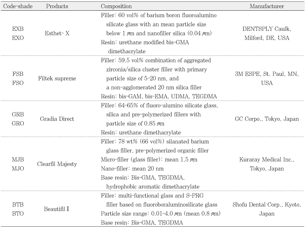
Table 1) selected for this study were five resin composites: Esthet-X (Dentsply, USA), Filtek supreme (3M ESPE, USA), Gradia Direct (GC Corporation, Japan), Clearfil Majesty Esthetic (Kuraray, Japan), and Beautifil II(Shofu, Japan). As the name of the same opaque shades vary in different products, for simplicity, the A2O, OA2, AO2 and A2D shades were given the generic name "A2O" in this study. Similarly, the A2 and A2B for body shades was described as" A2B".
Translucent acrylic plates (1-mm thick) with holes 10-mm in diameter were used as molds for making standardized disk-shaped specimens. The number of specimens for each group was 5. To determine the inherent color of each material, a measurement was made using a disk, 10-mm in diameter and 4-mm thick.
12 Each mold was filled with resin composite material, covered with clear celluloid strips on the top and bottom of the hole, and, with the acrylic plate, was pressed between two glass slides under a weight to achieve uniform thickness of the disk specimens. After removing the glass slides, the color of the materials was measured separately using a colorimeter: NF999 (Nippon Denshoku IND., Japan) against 2 backings: a black tile and a white tile. Calibration of the equipment was performed immediately before the series of measurements using a white tile supplied by the manufacturer. For each color measurement, the values obtained were expressed as CIELAB parameters (L
*, a
* and b
*). L
* refers to lightness, where 100 is white and 0 is black, a
* and b
* are the red-green and yellow-blue chromatic coordinates and a positive a
* or b
* indicates a red or a yellow shade, respectively. The white and black backings employed in this study were a white ceramic tile (L
* = 90.43, a
* = -0.3, b
* = 0.04) and a black ceramic tile (L
* = 10.86, a
* = -2.03, b
* =2.42).
After the initial series of color measurements of each uncured disk, light curing was performed through thin plastic film, using a Spectrum 800 (Dentsply, USA) at 600 mW/cm2 for 60 seconds each side. The color measurements were then repeated for each cured disk.
Calculation of color change (ΔE*) before and after light curing
The color change of each specimen (ΔE*) before and after light curing was calculated using the equation: ΔE* = [(L*after - L*before)2 + (a*after - a*before)2 + (b*after - b*before)2]1/2, where L*after, a*after, and b*after were CIELAB values of each specimen, the material itself, evaluated after light curing and L*before, a*before, and b*before were those values of each specimen, the material itself, evaluated before light curing.
Calculation of translucency parameter
The translucency of the materials 1-mm thick before and after light curing was calculated using the translucency parameter (TP) formula: TP
(D = 1 mm) = [(L
W* - L
B*)
2 + (a
W* - a
B*)
2 + (b
W* - b
B*)
2]
1/2, where the subscript "W"refers to the CIELAB values for each 1-mm thick specimen on a white backing and the subscript "B"refers to the values for specimens on a black backing.
6,
10,
19,
20
In order to extract the organic portion of the resins, 2 g of each material were weighed, using an analytical balance, and inserted into centrifuge tube. In each tube, 4 mL of HPLC-degree methanol were added and the sets were then taken to the ultrasonic appliance for 50 minutes so as to facilitate extraction.
Afterwards, the 1.5 mL polypropylene tubes were centrifuged in a microcentrifuge at a speed of 13,000 rpm for 200 seconds in order to increase the efficiency of filler sedimentation. After centrifuging, 3 mL of the supernatant portion in each polypropylene tube were collected with a pipette and transferred to individual vials.
The samples were injected into the gaseous chromatography appliance (HP 6890 plus GC) coupled to the mass spectrometer (HP 5973 MSD). A DB-5 column and a temperature slope from 40 to 280℃ in 25 minutes were used for the test.
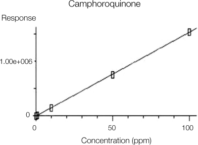
In order to create a calibration curve, 97% CQ (Sigma-Aldrich), containing a high level of pureness and no additional purification was used. The CQ was dissolved in methanol, HPLC level, so as to obtain solutions at concentrations of 0.1, 1, 10, 50, 100 ppm. These solutions were injected into the chromatograph under the same analysis conditions previously described for the samples, so that the calibration curve could be obtained, as shown in
Figure 1.
Refractive indices of resin composite samples were determined by spectroscopic ellipsometer (M-2000, J.A.Woollam Co., USA) at a 70°angle of incidence before and after light curing at 380-770 nm wave-length, where visible light is located.
Statistic Analysis
One-way ANOVA, two-way ANOVA, Tukey's post-hoc test and t-test were used for statistic analysis. The L*, a* b* changes and ΔE* before and after light curing were evaluated using one-way ANOVA and Tukey's post-hoc tests to detect statistically significant differences between groups (significance level 0.05). T-test was employed to detect statistically significant differences in a group before and after light curing (significance level 0.05). To detect any statistical changes in TP and refractive index before and after light curing, as primary factors, two-way ANOVA and Tukey's post-hoc test were carried out regarding products/shades and before or after light curing. When this analysis revealed interaction of any of the primary factors, one-way ANOVA and Tukey's post-hoc tests were employed to detect statistically significant differences between groups (significance level 0.05). T-test was employed to detect statistically significant differences in a group before and after light curing (significance level 0.05).
RESULTS
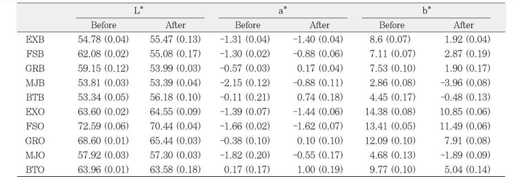
L*, a*, b* before and after light curing
The L
*, a
*, b
* before and after light curing are indicated in
Table 2. Comparing the L
*, a
*, b
* before and after light curing, significant differences were detected in all materials except for a
* of Esthet-X opaque and Filtek supreme opaque. The L
* of opaque shades are higher than that of body shades in all resin composites after curing. The L
* and b
* of FSO after curing is highest, it means FSO is the most bright and yellow material.
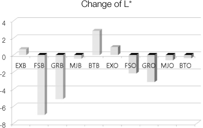
Changes of L
* after curing are indicated in
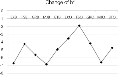
Figure 2. Body shade and opaque shade are divided into color of histogram. Changes of b
* are indicated in
Figure 3. b
* was decreased after curing, it means yellowness was reduced and blueness was increased in all resin composites. Significant difference was detected in Δb
* between body shades and opaque shades.
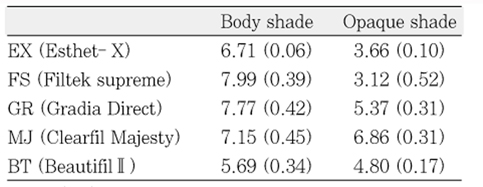
Table 3 summarizes the results in color differences (ΔE
*) before and after light curing. The ΔE
* of body shade was significantly greater compared with the opaque shade except for Clearfil Majesty.
Translucency parameter before and after light curing
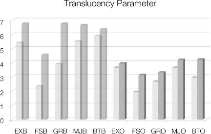
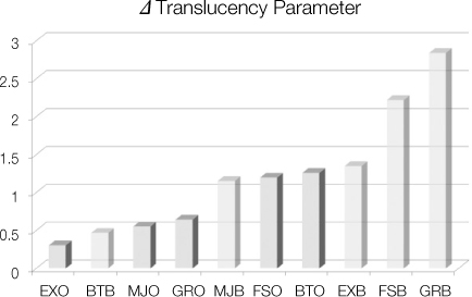
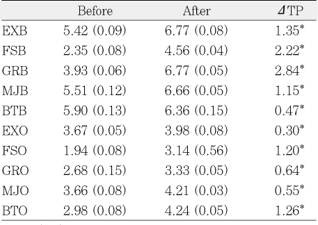
Table 4,
Figures 4 and
5 are about translucency parameter. All resin composites showed a statistically significant increase in TP after curing. Generally, ΔTP of body shades are greater than that of opaque shades. Specially, ΔTP of Gradia Direct body shade and Filtek supreme body shade are great.
Camphorquinone content
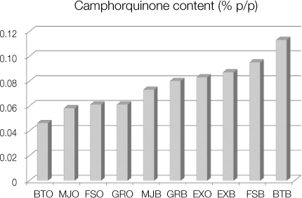
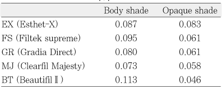
CQ contents are indicated in
Table 5 and
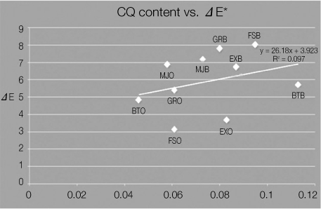
Figure 6. It showed from 0.046%p/p to 0.113 %p/p. The content of body shades are higher than that of opaque shades in all resin composites. The CQ content and ΔE
* showed a proportional trend (γ= 0.311) in
Figure 7.
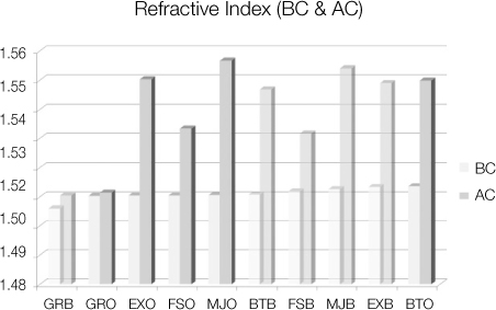
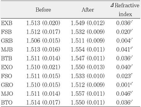
Table 6 summarized the means of refractive indices between 380-770 nm before and after light curing. After curing, the refractive indices were increased in all materials in
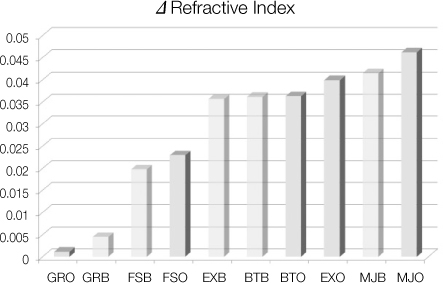
Figure 8. The growth of refractive indicesare indicated in
Figure 9. ΔRefractive index of Clearfil Majesty is the greatest and that of Gradia Direct is the smallest in body and opaque shade both.
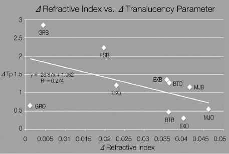
Figure 10 showed correlation between Δrefractive index and Δtranslucency parameter (γ= -0.523). In all resin composites except for Gradia Direct opaque, Δrefractive index and Δtranslucency parameter showed a inverse proportional trend.
DISCUSSION
In terms of the L
*, a
* and b
* changes after light curing, statistical change was indicated, except for the opaque A2 of Esthet X and Filtek supreme in a
* change. All the products/shades showed a significant decrease in b
*. The same phenomenon, in terms of a decrease in b
*, was also reported by Seghi and others.
7 These authors explained that the reason for the color change was due to a decrease in absorption of blue light by degradation of CQ after light curing.
7 Although Johnston and others attached importance to the L
* as an indicator of color change, the b
* appeared to reflect color change in this study, which agrees with Seghi and others.
7,
20
As for the color change (ΔE
*) before and after light curing, a significantly smaller ΔE
* was observed in Filtek supreme opaque and Esthet-X opaque. However, all the ΔE
* except for FSO were above 3.3, which was considered a clinically perceptable color difference.
21 Therefore, direct shade matching using uncured resin paste seemed inadequate for these products. This study has highlighted the fact that, in order to get a precise shade match, direct shade matching of these materials should be performed by using the cured composite.
The inherent translucency of resin composites may contribute to shade matching with a tooth by allowing the shade of the adjacent and underlying tooth structure to shine through. Clinicians have commonly observed this "chameleon"effect of composite restoration adjacent tooth substrate.
6,
22
However, in situations where there is no tooth structure to provide a backing for the restoration, such as in a large Class III or IV cavities, translucent materials may provide relatively poor color matches.
10,
23,
24 More specifically, a grayish or bluish shade is often seen in comparison with the surrounding tooth structure, as relatively translucent materials are probably affected by the darkness of the oral cavity. In such situations, opaque shade resin composites have been utilized.
Regarding the opaque shade, larger L* and b* were observed when compared to the body shade. Hence, the opaque shade may serve to add brightness and yellowish characteristics alongside opacity to a layered resin composite restoration. However, the effect of the underlying opaque shade on the resultant color of a layered restoration remains to be known. Therefore, more detailed studies should be conducted to clarify how the opaque shade affects the color of the layered restorations.
The technique employed in this study generated considerable data to evaluate color and translucency change before and after light curing. One of the advantages of the technique is that the CIELAB parameters on the 2 backings before and after light curing can be obtained from the same specimen in order to make the paired data. As a result, in this study, statistical analysis of color and TP change before and after light curing became possible. Since translucency of resin composite can be influenced by the thickness of specimens, the inherent colors of the resin composites were evaluated by using 4 mm thick specimens. For all the 4 mm thick specimens, their TP values were always below 1.1, regardless of shade. These TP values fall within the imperceptible range by human visual sense.
12,
25 Therefore, the colors of the 4 mm thick specimens could be considered as the inherent colors of the resin composites not affected by the background color.
The most used photoinitiator in dental polymers is CQ.
13,
18,
26-
30 CQ is a αdiketone that features a conjugated dicarbonilic group and also good absorption within the visible range of the spectrum, with a peak at the wavelength of 470 nm. CQ is featured as an intense yellow powder.
16,
18,
29,
31 Its peculiar color is due to its chromophore; it absorbs light in the region of 470 nm, thus, it reflects a resulting shade ranging between yellow and orange. Some photoinitiators break the chromophore after irradiation, loosing the yellow color, in a process called photobleaching. However, CQ shows poor photobleaching maintaining the same color after free radical generation. This feature limits the addition of CQ to the composites, since it can turn them excessively yellow, hazarding the final esthetic result.
13,
29
The content of CQ evaluated in this present study, among the resins that use this photoinitiator, varied from 0.046 to 0.113 %p/p. These results are similar to those obtained by Shintani et al. who found concentrations of CQ from 0.032 to 0.095 %p/p.28 These authors evaluated different brands of resin composites, comparing the results between microfilled and conventional filled composite, with no reference to the resin shade used. The same happened in the study performed by Taira et al. where only brands were compared, while the shades tested were not quoted.
29 The results found by these latter authors may seem at odds when compared to the others, since the amount of CQ varied from 0.17 to 1.03 %p/p. However, they calculated the amount of CQ related only to the resinous phase. When considering this feature of resins, with on average from 60 to 80% of inorganic filler, the results tend to be similar to those found in the present study and that carried out by Shintani et al. (approximately 0.04-0.275 %p/p of CQ).
28
The CQ contents of body shades are higher than that of opaque shades in all resin composites in this study. The CQ content and ΔE* showed a proportional trend. With the breakdown of CQ, color difference (ΔE*) is created. So, CQ content is less in opaque shade.
There are many reports about factors that contribute to the opacity of resin composites. Inokoshi and others
6,
32 stated that, the greater the difference between the refractive indices of inorganic particles and the matrix phase of resin composites, the greater the opacity of the materials, due to multiple reflection and refraction at the matrix particle interfaces. Campbell et al. stated that, in experimental PMMA resin composites, the efficiency of light scattering for a quartz filler decreased as the size of the filler increased.
6,
33 Kawaguchi et al. mentioned that certain types of hybrid resin composites could show smaller transmission coefficients because of the wide range of particle size.
24 Johnston and Reisbick insisted that the color and translucency of esthetic restorative materials is determined not only by more macroscopic phenomena, such as matrix and filler composition as well as filler content, but also by relatively minor pigment additions and potentially by all other chemical components of these materials.
20
In this study, the refractive indices increased in all materials after polymerization. ΔRefractive index of Clearfil Majesty is the greatest and that of Gradia Direct is the smallest in body and opaque shade both. In all resin composites except for Gradia Direct opaque, Δrefractive index and Δtranslucency parameter showed a inverse proportional trend.
CONCLUSION
It is important to be able to predict the amount of change in color and translucency of resin composite before and after curing for the successful restoration. The CQ content and refractive index influence on color and translucency of resin composite.
Resin composites used in this study were A2 body and A2 opaque shade of Esthet-X, Filtek supreme, Gradia Direct, Clearfil Majesty and Beautifil II. Color and translucency changes before and after light curing were evaluated by colorimeter, the CQ was quantified by GC-MS and refractive index changes were measured by spectroscopic ellipsometer. One-way ANOVA, two-way ANOVA, Tukey's post-hoc test and t-test were used for statistic analysis. Within the limit of this study, following conclusion can be obtained.
When comparing the L* and b* before and after light curing, significant differences were detected and b* showed a significant decrease in all materials. Significant difference was detected in Δb* between body shades and opaque shades.
The ΔE* of body shade was significantly greater compared to that of opaque shade except for the Clearfil Majesty. All the ΔE* except for Filtek supreme opaque were above 3.3, which was considered a clinically perceptable color difference.
Translucency parameter (TP) was significantly increased after curing. The ΔTP of body shades are greater than that of opaque shades.
CQ content (%p/p) varied between 0.046 - 0.113. The CQ content of body shades are higher than that of opaque shades in all resin composites. The CQ content and ΔE* showed a proportional trend.
Refractive index increased after polymerization in all materials and significant difference in Δ refractive index was found between body and opaque shade (p < 0.05). In all resin composites except for Gradia Direct opaque, Δrefractive index and Δtranslucency parameter showed a inverse proportional trend.
For an accurate shade match, direct shade matching of resin composite should be performed by using the cured material. From the viewpoint of different color and translucency between the products of the same shade, the using of custom shade guides made from the material itself is suggested.
REFERENCES
- 1. Yap AU, Tan KB, Bhole S. Comparison of aesthetic properties of tooth-colored restorative materials. Oper Dent. 1997;22: 167-172.PubMed
- 2. Park SJ, Lee HY, Na MY, Chang HS, Hwang YC, Oh WM, Hwang IN. The evaluation of color and color difference according to the layering placement of Incisal shade composites on the body composites of the indirect resin restoration. J Korean Acad Conserv Dent. 2011;36: 37-49.Article
- 3. Yap AU, Bhole S, Tan KB. Shade match of tooth-colored restorative materials based on a commercial shade guide. Quintessence Int. 1995;26: 697-702.PubMed
- 4. Kim HS, Um CM. Color differences between resin composites and shade guides. Quintessence Int. 1996;27: 559-567.PubMed
- 5. Park SJ, Hwang YC, Oh WM, Hwang IN. Opacity and mashing effect of the opaque shade composite resins. J Korean Acad Conserv Dent. 2007;32: 356-364.Article
- 6. Sidhu SK, Ikeda T, Omata Y, Fujita M, Sano H. Change of color and translucency by light curing in resin composites. Oper Dent. 2006;31: 598-603.ArticlePubMedPDF
- 7. Seghi RR, Gritz MD, Kim J. Colorimetric changes in composites resulting from visible-light-initiated polymerization. Dent Mater. 1990;6: 133-137.ArticlePubMed
- 8. Cho KJ, Park SJ, Cho HG, Kim DJ, Hwang YC, Oh WM, Hwang IN. Influence of the surface roughness on translucency and surface color of the dental composite resins. J Korean Acad Conserv Dent. 2006;31: 312-322.Article
- 9. Ikeda T, Nakanishi A, Yamamoto T, Sano H. Color differences and color changes in Vita Shade tooth-colored restorative materials. Am J Dent. 2003;16: 381-384.PubMed
- 10. Ikeda T, Murata Y, Sano H. Translucency of opaqueshade resin composites. Am J Dent. 2004;17: 127-130.PubMed
- 11. Ikeda T, Sidhu SK, Omata Y, Fujita M, Sano H. Colour and translucency of opaque-shades and bodyshades of resin composites. Eur J Oral Sci. 2005;113: 170-173.ArticlePubMed
- 12. Kamishima N, Ikeda T, Sano H. Color and translucency of resin composites for layering techniques. Dent Mater J. 2005;24: 428-432.ArticlePubMed
- 13. Alvim HH, Alecio AC, Vasconcellos WA, Furlan M, de Oliveira JE, Saad JR. Analysis of camphorquinone in composite resins as a function of shade. Dent Mater. 2007;23: 1245-1249.ArticlePubMed
- 14. Peutzfeldt A, Asmussen E. Hardness of restorative resins: effect of camphorquinone, amine, and inhibitor. Acta Odontol Scand. 1989;47: 229-231.ArticlePubMed
- 15. Rueggeberg FA, Ergle JW, Lockwood PE. Effect of photoinitiator level on properties of a light-cured and postcure heated model resin system. Dent Mater. 1997;13: 360-364.ArticlePubMed
- 16. Yoshida K, Greener EH. Effect of photoinitiator on degree of conversion of unfilled light-cured resin. J Dent. 1994;22: 296-299.ArticlePubMed
- 17. Yoshida K, Greener EH. Effects of two amine reducing agents on the degree of conversion and physical properties of an unfilled light-cured resin. Dent Mater. 1993;9: 246-251.ArticlePubMed
- 18. Park YJ, Chae KH, Rawls HR. Development of a new photoinitiation system for dental light-cure composite resins. Dent Mater. 1999;15: 120-127.ArticlePubMed
- 19. Johnston WM, Ma T, Kienle BH. Translucency parameter of colorants for maxillofacial prostheses. Int J Prosthodont. 1995;8: 79-86.PubMed
- 20. Johnston WM, Reisbick MH. Color and translucency changes during and after curing of esthetic restorative materials. Dent Mater. 1997;13: 89-97.ArticlePubMed
- 21. Ruyter IE, Nilner K, Moller B. Color stability of dental composite resin materials for crown and bridge veneers. Dent Mater. 1987;3: 246-251.ArticlePubMed
- 22. Swift EJ Jr, Hammel SA, Lund PS. Colorimetric evaluation of vita shade resin composites. Int J Prosthodont. 1994;7: 356-361.PubMed
- 23. Powers JM, Yeh CL, Miyagawa Y. Optical properties of composites of selected shades in white light. J Oral Rehabil. 1983;10: 319-324.ArticlePubMed
- 24. Kawaguchi M, Fukushima T, Miyazaki K. The relationship between cure depth and transmission coefficient of visible-light-activated resin composites. J Dent Res. 1994;73: 516-521.ArticlePubMedPDF
- 25. Gross MD, Moser JB. A colorimetric study of coffee and tea staining of four composite resins. J Oral Rehabil. 1977;4: 311-322.ArticlePubMed
- 26. Moin Jan C, Nomura Y, Urabe H, Okazaki M, Shintani H. The relationship between leachability of polymerization initiator and degree of conversion of visible light-cured resin. J Biomed Mater Res. 2001;58: 42-46.ArticlePubMedPDF
- 27. Michelsen VB, Lygre H, Skålevik R, Tveit AB, Solheim E. Identification of organic eluates from four polymer-based dental filling materials. Eur J Oral Sci. 2003;111: 263-271.ArticlePubMedPDF
- 28. Shintani H, Inoue T, Yamaki M. Analysis of camphorquinone in visible light-cured composite resins. Dent Mater. 1985;1: 124-126.ArticlePubMed
- 29. Taira M, Urabe H, Hirose T, Wakasa K, Yamaki M. Analysis of photo-initiators in visible-light-cured dental composite resins. J Dent Res. 1988;67: 24-28.ArticlePubMedPDF
- 30. Teshima W, Nomura Y, Tanaka N, Urabe H, Okazaki M, Nahara Y. ESR study of camphorquinone/amine photoinitiator systems using blue light-emitting diodes. Biomaterials. 2003;24: 2097-2103.ArticlePubMed
- 31. Neumann MG, Miranda WG Jr, Schmitt CC, Rueggeberg FA, Correa IC. Molar extinction coefficients and the photon absorption efficiency of dental photoinitiators and light curing units. J Dent. 2005;33: 525-532.ArticlePubMed
- 32. Inokoshi S, Burrow MF, Kataumi M, Yamada T, Takatsu T. Opacity and color changes of tooth-colored restorative materials. Oper Dent. 1996;21: 73-80.PubMed
- 33. Campbell PM, Johnston WM, O'Brien WJ. Light scattering and gloss of an experimental quartz-filled composite. J Dent Res. 1986;65: 892-894.ArticlePubMedPDF
- 34. Arimoto A, Nakajima M, Hosaka K, Nishimura K, Ikeda M, Foxton RM, Tagami J. Translucency, opalescence and light transmission characteristics of light-cured resin composites. Dent Mater. 2010;26: 1090-1097.ArticlePubMed
- 35. Lehtinen J, Laurila T, Lassila LV, Vallittu PK, Räty J, Hernberg R. Optical characterization of bisphenol-A-glycidyldimethacrylate-triethyleneglycoldimethacrylate (BisGMA/TEGDMA) monomers and copolymer. Dent Mater. 2008;24: 1324-1328.ArticlePubMed
- 36. Emami N, Sjödahl M, Söderholm KJ. How filler properties, filler fraction, sample thickness and light source affect light attenuation in particulate filled resin composites. Dent Mater. 2005;21: 721-730.ArticlePubMed
- 37. Shortall AC, Palin WM, Burtscher P. Refractive index mismatch and monomer reactivity influence composite curing depth. J Dent Res. 2008;87: 84-88.ArticlePubMedPDF
- 38. Westland S. Review of the CIE system of colorimetry and its use in dentistry. J Esthet Restor Dent. 2003;15: S5-S12.ArticlePubMed
- 39. Howard B, Wilson ND, Newman SM, Pfeifer CS, Stansbury JW. Relationships between conversion, temperature and optical properties during composite photopolymerization. Acta Biomater. 2010;6: 2053-2059.ArticlePubMed


 KACD
KACD


 ePub Link
 Cite
Cite

