
Search
- Page Path
- HOME > Search
Research Article
- Structural and morphological characterization of silver nanoparticles intruded mineral trioxide aggregate admixture as a chair-side restorative medicament: an in vitro experimental study
- H. Murali Rao, Rajkumar Krishnan, Chitra Shivalingam, Ramya Ramadoss
- Restor Dent Endod 2025;50(3):e30. Published online August 8, 2025
- DOI: https://doi.org/10.5395/rde.2025.50.e30
-
 Abstract
 PDF
PDF PubReader
 ePub
ePub - Objectives
The aim of this study was to create a rapid admixture of mineral trioxide aggregate (MTA) and silver nanoparticles (AgNPs) for chairside use in clinical settings to remediate the challenges associated with root canal treatment and pulp capping.
Methods
Synthesized AgNPs at ratios of 10 and 25% were added to commercially available MTA to create an admixture. The admixture was subjected to structural and morphological assessment using X-ray diffraction analysis (XRD), Fourier transform infrared (FT-IR) analysis, Raman spectroscopy, and scanning electron microscopy. Antioxidant activity was measured using the hydroxyl radical scavenging assay. A significance level of 0.05 was applied to determine statistical differences.
Results
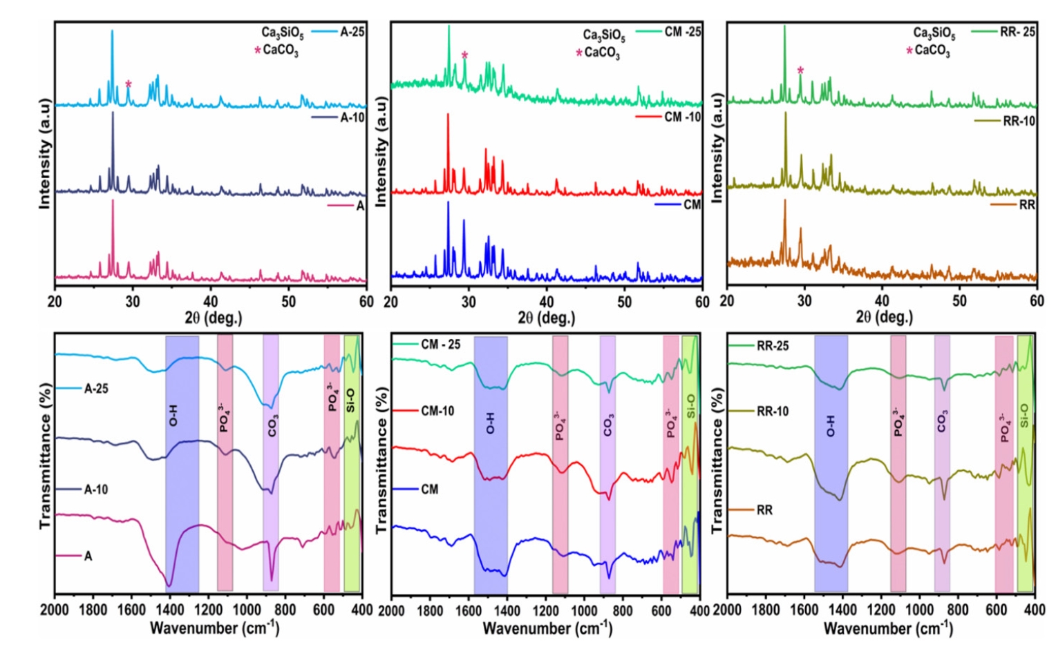
The addition of AgNPs decreased the carbonate peak intensity in XRD and FT-IR. The rod-like morphology of MTA was changed to a flake-like morphology with the addition of AgNPs. Antibacterial efficacy enhanced proportionally with the augmentation of AgNPs concentration.
Conclusions
The creation of rapid admixture of MTA and AgNPs during chairside use in clinical settings can deliver beneficial characteristics of enhanced morphological features favoring mineralization and profound antibacterial effects to overcome the challenges associated with root canal treatment and pulp capping.
- 2,648 View
- 95 Download

Review Article
- Stem cell-derived exosomes for dentin-pulp complex regeneration: a mini-review
- Dina A. Hammouda, Alaa M Mansour, Mahmoud A. Saeed, Ahmed R. Zaher, Mohammed E. Grawish
- Restor Dent Endod 2023;48(2):e20. Published online May 3, 2023
- DOI: https://doi.org/10.5395/rde.2023.48.e20
-
Abstract
PDFPubReaderePub
This mini-review was conducted to present an overview of the use of exosomes in regenerating the dentin-pulp complex (DPC). The PubMed and Scopus databases were searched for relevant articles published between January 1, 2013 and January 1, 2023. The findings of basic
in vitro studies indicated that exosomes enhance the proliferation and migration of mesenchymal cells, as human dental pulp stem cells, via mitogen-activated protein kinases and Wingless-Int signaling pathways. In addition, they possess proangiogenic potential and contribute to neovascularization and capillary tube formation by promoting endothelial cell proliferation and migration of human umbilical vein endothelial cells. Likewise, they regulate the migration and differentiation of Schwann cells, facilitate the conversion of M1 pro-inflammatory macrophages to M2 anti-inflammatory phenotypes, and mediate immune suppression as they promote regulatory T cell conversion. Basicin vivo studies have indicated that exosomes triggered the regeneration of dentin-pulp–like tissue, and exosomes isolated under odontogenic circumstances are particularly strong inducers of tissue regeneration and stem cell differentiation. Exosomes are a promising regenerative tool for DPC in cases of small pulp exposure or for whole-pulp tissue regeneration.-
Citations
Citations to this article as recorded by
- Extracellular vesicles derived from dental mesenchymal stem cells for regenerative medicine: a scoping review
Maria Emília Mota, Márcia Martins Marques, Thaís Gimenez, Suely Kunimi Kubo Ariga, Tiago Góss dos Santos, Fábio Abreu Alves, Maria Stella Moreira
Molecular Biology Reports.2026;[Epub] CrossRef - Stem cell-derived exosomes in tissue regeneration of oral and maxillofacial region: A systematic review
Qianlei Dai, Qianfen Dai
Medicine.2026; 105(1): e46948. CrossRef - Stem Cell-Derived Exosomes in Wound Healing and Skin Regeneration: Emerging Therapeutic Strategies and Mechanisms
Nithin Vidiyala, Pavani Sunkishala, Prashanth Reddy Parupathi, Dinesh Nyavanandi
Cells.2026; 15(10): 872. CrossRef - Cell Homing Strategies in Regenerative Endodontic Therapy
David Kim, Sahng G. Kim
Cells.2025; 14(3): 201. CrossRef - Impact of dental pulp cells-derived small extracellular vesicles on the properties and behavior of dental pulp cells: an in-vitro study
Dina A. Hammouda, Alaa M. Mansour, Ahmed R. Zaher, Mohammed E. Grawish
BMC Oral Health.2025;[Epub] CrossRef - Methodological Approaches for Economic Comparison of Mesenchymal Stem Cell and Exosome-based Therapies with Conventional Endodontic Treatments in Regenerative Endodontics
Madina A. Kurmanalina Kurmanalina, Nadiar M. Mussin, Aigul M. Sumanova, Violetta R. Detochkina, Maryam Mardani, Nader Tanideh, Amin Tamadon
West Kazakhstan Medical Journal.2025; 67(2): 188. CrossRef - Exosomal circ_0003057 promotes osteo/odontogenic differentiation of hDPSCs by binding with EIF4A3 through upregulated parental gene ANKH
Bingtao Wang, Yuanyuan Kong, Huixian Dong, Feng Lai, Zixin Guo, Liecong Lin, Jingyi Xu, Jingkun Zhang, Yiguo Jiang, Qianzhou Jiang
International Endodontic Journal.2025; 58(9): 1433. CrossRef - Mechanistic insights into dental stem cells‐derived exosomes in regenerative endodontics
Paras Ahmad, Nathan Estrin, Nima Farshidfar, Yufeng Zhang, Richard J. Miron
International Endodontic Journal.2025; 58(9): 1384. CrossRef - Development and characterization of an exosome-loaded biomimetic hydroxyapatite/gelatin scaffold for enhanced dental pulp regeneration
Yuen-Shan Tsai, Shih-Jung Cheng, Tsao-Li Chuang, Shu-Fang Chang, Feng-Huei Lin, Chun-Pin Lin
Journal of Dental Sciences.2025;[Epub] CrossRef - Exosomes as Promising Therapeutic Tools for Regenerative Endodontic Therapy
Qingyue Kong, Yujie Wang, Nan Jiang, Yifan Wang, Rui Wang, Xiaohan Hu, Jing Mao, Xin Shi
Biomolecules.2024; 14(3): 330. CrossRef - Role and Molecular Mechanism of miR-586 in the Differentiation of Dental Pulp Stem Cells into Odontoblast-like Cells
Gang Pan, Qianwen Zhou, Chenhua Pan, Yingxue Zhang
Cell Biochemistry and Biophysics.2024; 83(1): 507. CrossRef
- Extracellular vesicles derived from dental mesenchymal stem cells for regenerative medicine: a scoping review
- 8,237 View
- 141 Download
- 9 Web of Science
- 11 Crossref
Research Articles
- Bioactivity of endodontic biomaterials on dental pulp stem cells through dentin
- Bahar Javid, Narges Panahandeh, Hassan Torabzadeh, Hamid Nazarian, Ardavan Parhizkar, Saeed Asgary
- Restor Dent Endod 2020;45(1):e3. Published online November 4, 2019
- DOI: https://doi.org/10.5395/rde.2020.45.e3
-
Abstract
PDFPubReaderePub
Objectives This study investigated the indirect effect of calcium-enriched mixture (CEM) cement and mineral trioxide aggregate (MTA), as 2 calcium silicate-based hydraulic cements, on human dental pulp stem cells (hDPSCs) through different dentin thicknesses.
Materials and Methods Two-chamber setups were designed to simulate indirect pulp capping (IPC). Human molars were sectioned to obtain 0.1-, 0.3-, and 0.5-mm-thick dentin discs, which were placed between the 2 chambers to simulate an IPC procedure. Then, MTA and CEM were applied on one side of the discs, while hDPSCs were cultured on the other side. After 2 weeks of incubation, the cells were removed, and cell proliferation, morphology, and attachment to the discs were evaluated under scanning electron microscopy (SEM). Energy-dispersive X-ray (EDXA) spectroscopy was performed for elemental analysis. Alkaline phosphatase (ALP) activity was assessed quantitatively. The data were analyzed using the Kruskal-Wallis and Mann-Whitney tests.
Results SEM micrographs revealed elongated cells, collagen fibers, and calcified nucleations in all samples. EDXA verified that the calcified nucleations consisted of calcium phosphate. The largest calcifications were seen in the 0.1-mm-thick dentin subgroups. There was no significant difference in ALP activity across the CEM subgroups; however, ALP activity was significantly lower in the 0.1-mm-thick dentin subgroup than in the other MTA subgroups (
p < 0.05).Conclusions The employed capping biomaterials exerted biological activity on hDPSCs, as shown by cell proliferation, morphology, and attachment and calcific precipitations, through 0.1- to 0.5-mm-thick layers of dentin. In IPC, the bioactivity of these endodontic biomaterials is probably beneficial.
-
Citations
Citations to this article as recorded by- Dental pulp capping materials: modulators of stem cell behavior and regenerative potential
Ali Cheayto, Sara Ayoub, Sarah Ayad Al-Tameemi, Mohammad Fayyad-Kazan
Biomedical Physics & Engineering Express.2025; 11(6): 062004. CrossRef - Effect of pulp capping materials on odontogenic differentiation of human dental pulp stem cells: An in vitro study
Mahmoud M. Bakr, Mohamed Shamel, Shereen N. Raafat, Robert M. Love, Mahmoud M. Al‐Ankily
Clinical and Experimental Dental Research.2024;[Epub] CrossRef - Effects of Growth Factors on the Differentiation of Dental Stem Cells: A
Systematic Review and Meta-analysis (Part I)
Sayna Shamszadeh, Armin Shirvani, Hassan Torabzadeh, Saeed Asgary
Current Stem Cell Research & Therapy.2024; 19(4): 523. CrossRef - The Role of Growth Factor Delivery Systems on Cellular Activities of Dental
Stem Cells: A Systematic Review (Part II)
Sayna Shamszadeh, Armin Shirvani, Saeed Asgary
Current Stem Cell Research & Therapy.2024; 19(4): 587. CrossRef - Comprehensive review of composition, properties, clinical applications, and future perspectives of calcium-enriched mixture (CEM) cement: a systematic analysis
Saeed Asgary, Mahtab Aram, Mahta Fazlyab
BioMedical Engineering OnLine.2024;[Epub] CrossRef - Evaluation of dental pulp stem cells behavior after odontogenic differentiation induction by three different bioactive materials on two different scaffolds
Basma Ahmed, Mai H. Ragab, Rania A. Galhom, Hayam Y. Hassan
BMC Oral Health.2023;[Epub] CrossRef - Characterization of Dental Pulp Stem Cell Responses to Functional Biomaterials Including Mineralized Trioxide Aggregates
Sejin Bae, Bueonguk Kang, Hyungbin Lee, Harrison Luu, Eric Mullins, Karl Kingsley
Journal of Functional Biomaterials.2021; 12(1): 15. CrossRef - Incorporation of amoxicillin-loaded microspheres in mineral trioxide aggregate cement: an in vitro study
Fábio Rocha Bohns, Vicente Castelo Branco Leitune, Isadora Martini Garcia, Bruna Genari, Nélio Bairros Dornelles, Silvia Stanisçuaski Guterres, Fabrício Aulo Ogliari, Mary Anne Sampaio de Melo, Fabrício Mezzomo Collares
Restorative Dentistry & Endodontics.2020;[Epub] CrossRef
- Dental pulp capping materials: modulators of stem cell behavior and regenerative potential
- 2,307 View
- 17 Download
- 8 Crossref
- Effects of the exposure site on histological pulpal responses after direct capping with 2 calcium-silicate based cements in a rat model
- Panruethai Trongkij, Supachai Sutimuntanakul, Puangwan Lapthanasupkul, Chitpol Chaimanakarn, Rebecca Wong, Danuchit Banomyong
- Restor Dent Endod 2018;43(4):e36. Published online August 22, 2018
- DOI: https://doi.org/10.5395/rde.2018.43.e36

-
Abstract
PDFPubReaderePub
Objectives Direct pulp capping is a treatment for mechanically exposed pulp in which a biocompatible capping material is used to preserve pulpal vitality. Biocompatibility tests in animal studies have used a variety of experimental protocols, particularly with regard to the exposure site. In this study, pulp exposure on the occlusal and mesial surfaces of molar teeth was investigated in a rat model.
Materials and Methods A total of 58 maxillary first molars of Wistar rats were used. Forty molars were mechanically exposed and randomly assigned according to 3 factors: 1) the exposure site (occlusal or mesial), 2) the pulp-capping material (ProRoot White MTA or Bio-MA), and 3) 2 follow-up periods (1 day or 7 days) (
n = 5 each). The pulp of 6 intact molars served as negative controls. The pulp of 12 molars was exposed without a capping material (n = 3 per exposure site for each period) and served as positive controls. Inflammatory cell infiltration and reparative dentin formation were histologically evaluated at 1 and 7 days using grading scores.Results At 1 day, localized mild inflammation was detected in most teeth in all experimental groups. At 7 days, continuous/discontinuous calcified bridges were formed at exposure sites with no or few inflammatory cells. No significant differences in pulpal response according to the exposure site or calcium-silicate cement were observed.
Conclusions The location of the exposure site had no effect on rat pulpal healing. However, mesial exposures could be performed easily, with more consistent results. The pulpal responses were not significantly different between the 2 capping materials.
-
Citations
Citations to this article as recorded by- Bioactivity and biocompatibility of bioceramic-based pulp capping materials in laboratory and animal models
Rafiqul Islam, Md. Refat Readul Islam, Kenta Tsuchiya, Yu Toida, Hidehiko Sano, Monica Yamauti, Hany Mohamed Aly Ahmed, Atsushi Tomokiyo
Journal of Materials Science: Materials in Medicine.2025;[Epub] CrossRef - The road map to proper dental pulp experiments in animal models
Nuha A Elmubarak
International Dental Journal of Student's Research.2024; 11(4): 163. CrossRef - Treatment outcomes of root perforations repaired by calcium silicate-based cements with or without an accelerator: A randomized controlled trial
Kanyarat Tungputsa, Danuchit Banomyong, Sittichoke Osiri, Supachai Sutimuntanakul
Endodontology.2024; 36(4): 315. CrossRef - Biological evaluation of novel phosphorylated pullulan‐based calcium hydroxide formulations as direct pulp capping materials: An in vivo study on a rat model
Md Refat Readul Islam, Rafiqul Islam, Yunqing Liu, Yu Toida, Yasuhiro Yoshida, Hidehiko Sano, Hany Mohamed Aly Ahmed, Atsushi Tomokiyo
International Endodontic Journal.2024; 57(9): 1247. CrossRef - 3D-printed microgels supplemented with dentin matrix molecules as a novel biomaterial for direct pulp capping
Diana Cunha, Nayara Souza, Manuela Moreira, Nara Rodrigues, Paulo Silva, Cristiane Franca, Sivaporn Horsophonphong, Ashley Sercia, Ramesh Subbiah, Anthony Tahayeri, Jack Ferracane, Pamela Yelick, Vicente Saboia, Luiz Bertassoni
Clinical Oral Investigations.2022; 27(3): 1215. CrossRef - Calcium silicate and calcium aluminate cements for dentistry reviewed
Carolyn Primus, James L. Gutmann, Franklin R. Tay, Anna B. Fuks
Journal of the American Ceramic Society.2022; 105(3): 1841. CrossRef - Pulpal response to mineral trioxide aggregate containing phosphorylated pullulan-based capping material
Yu TOIDA, Shimpei KAWANO, Rafiqul ISLAM, Fu JIALE, AFM A CHOWDHURY, Shuhei HOSHIKA, Yasushi SHIMADA, Junji TAGAMI, Masahiro YOSHIYAMA, Satoshi INOUE, Ricardo M. CARVALHO, Yasuhiro YOSHIDA, Hidehiko SANO
Dental Materials Journal.2022; 41(1): 126. CrossRef - The Effect of Calcium-Silicate Cements on Reparative Dentinogenesis Following Direct Pulp Capping on Animal Models
Mihai Andrei, Raluca Paula Vacaru, Anca Coricovac, Radu Ilinca, Andreea Cristiana Didilescu, Ioana Demetrescu
Molecules.2021; 26(9): 2725. CrossRef - Histological evaluation of a novel phosphorylated pullulan‐based pulp capping material: An in vivo study on rat molars
Rafiqul Islam, Yu Toida, Fei Chen, Toru Tanaka, Satoshi Inoue, Tetsuya Kitamura, Yasuhiro Yoshida, Abu Faem Mohammad Almas Chowdhury, Hany Mohamed Aly Ahmed, Hidehiko Sano
International Endodontic Journal.2021; 54(10): 1902. CrossRef - Effectiveness of Direct Pulp Capping Bioactive Materials in Dentin Regeneration: A Systematic Review
Ermin Nie, Jiali Yu, Rui Jiang, Xiangzhen Liu, Xiang Li, Rafiqul Islam, Mohammad Khursheed Alam
Materials.2021; 14(22): 6811. CrossRef - A strontium and amorphous calcium phosphate dipped premixed injectable calcium silicate-based ceramic for dental root canal sealing
Huimin Jin, Yuzhu Li, Qingqing Wang, Menglu Dong, Mengmeng Yang, Wendy Chen, Shengrui Wang, Heng Zhang, Shunli Zheng, Chris Ying Cao, Zheng Zhou, Quan-Li Li
Ceramics International.2021; 47(23): 33738. CrossRef - Bioactive tri/dicalcium silicate cements for treatment of pulpal and periapical tissues
Carolyn M. Primus, Franklin R. Tay, Li-na Niu
Acta Biomaterialia.2019; 96: 35. CrossRef
- Bioactivity and biocompatibility of bioceramic-based pulp capping materials in laboratory and animal models
- 2,957 View
- 27 Download
- 12 Crossref
Basic Research
- Gene expression profiling in human dental pulp cells treated with mineral trioxide aggregate
- Yong-Beom Kim, Won-Jun Shon, WooCheol Lee, Kee-Yeon Kum, Seung-Ho Baek, Kwang-Shik Bae
- J Korean Acad Conserv Dent 2010;35(3):152-163. Published online May 31, 2010
- DOI: https://doi.org/10.5395/JKACD.2010.35.3.152
-
Abstract
PDFPubReaderePub
This study investigated the changes in gene expression when mineral trioxide aggregate (MTA) was applied
in vitro to human dental pulp cells (HDPCs). MTA in a teflon tube (diameter 10 mm, height 2 mm) was applied to HDPCs. Empty tube-applied HDPCs were used as negative control. For microarray analysis, total RNA was extracted at 6, 24, and 72 hrs after MTA application. The results were confirmed selectively by performing reverse transcriptase polymerase chain reaction for genes that showed changes of more than two-fold or less than half. Of the 24,546 genes, 109 genes were up-regulated greater than two-fold (e.g., FOSB, THBS1, BHLHB2, EDN1, IL11, FN1, COL10A1, and TUFT1) and 69 genes were down-regulated below 50% (e.g., SMAD6 and DCN). These results suggest that MTA, rather than being a bio-inert material, may have potential to affect the proliferation and differentiation of pulp cells in various ways.-
Citations
Citations to this article as recorded by- Analysis of gene expression during odontogenic differentiation of cultured human dental pulp cells
Min-Seock Seo, Kyung-Gyun Hwang, Hyongbum Kim, Seung-Ho Baek
Restorative Dentistry & Endodontics.2012; 37(3): 142. CrossRef - Comparison of gene expression profiles of human dental pulp cells treated with mineral trioxide aggregate and calcium hydroxide
Yong-Beom Kim, Won-Jun Shon, Woocheol Lee, Kee-Yeon Kum, Seung-Ho Baek, Kwang-Shik Bae
Journal of Korean Academy of Conservative Dentistry.2011; 36(5): 397. CrossRef
- Analysis of gene expression during odontogenic differentiation of cultured human dental pulp cells
- 1,728 View
- 5 Download
- 2 Crossref
 First
First Prev
Prev

