Search
- Page Path
- HOME > Search
- Evaluation of the effects of different file systems and apical functions of integrated endodontic motors on debris extrusion: an ex vivo experimental study
- Sıla Nur Usta, Antonio Magan-Fernandez, Cumhur Aydın
- Restor Dent Endod 2025;50(2):e14. Published online April 14, 2025
- DOI: https://doi.org/10.5395/rde.2025.50.e14

-
 Abstract
 PDF
PDF PubReader
 ePub
ePub - Objectives
This study aimed to evaluate the effects of two different file systems operated with three apical functions of an endodontic motor integrated with an electronic apex locator on debris extrusion.
Methods
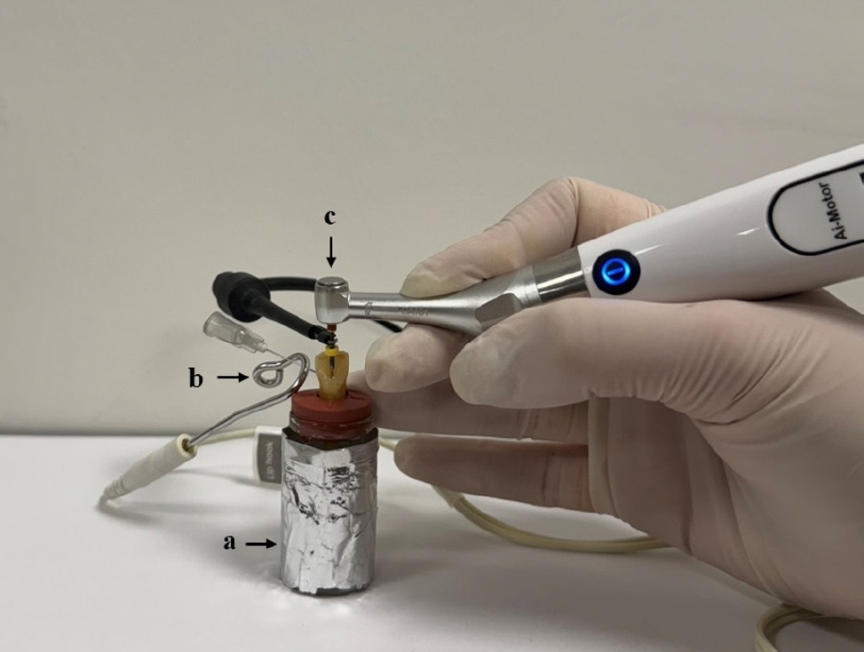
Sixty single-rooted teeth were prepared and divided into two main groups and three subgroups based on the file system (OneShape [Micro-Mega SA] and WaveOne [Dentsply Maillefer]) and apical function of the endodontic motor used (auto apical stop [AAS], auto apical reverse [AAR], and auto apical slowdown [ASD]). The teeth were mounted in pre-weighed glass tubes filled with 0.9% sodium chloride to complete the circuit with the apex locator. Files were advanced until the respective apical function (stop, reverse, or slowdown) was activated. The extruded debris was collected, dried, and weighed by subtracting pre-weighed values from post-weighed values. Preparation time was also recorded. Statistical analyses were performed to compare the groups.
Results
OneShape was associated with significantly less debris extrusion compared to WaveOne, regardless of the apical function (p < 0.05). The ASD function resulted in the least debris extrusion compared to AAS and AAR (p < 0.05). Preparation time was significantly longer in the ASD function (p < 0.05), while no differences were observed between the file systems (p > 0.05).
Conclusions
The OneShape file system and the ASD function produced the least amount of apical debris. While the ASD function requires more preparation time, its potential to minimize debris extrusion suggests it may reduce postoperative symptoms. -
Citations
Citations to this article as recorded by- Inflammatory Mediator Levels and Postoperative Pain Following Root Canal Shaping with Different Apical Actions: A Randomized Controlled Trial
Mustafa Mert Tulgar, Yağmur Kılıç, Oğuz Karalar, Huriye Erbak Yılmaz, Emrah Karataşlıoğlu
Journal of Endodontics.2025;[Epub] CrossRef
- Inflammatory Mediator Levels and Postoperative Pain Following Root Canal Shaping with Different Apical Actions: A Randomized Controlled Trial
- 4,065 View
- 214 Download
- 1 Crossref

- Effects of calcium silicate cements on neuronal conductivity
- Derya Deniz-Sungur, Mehmet Ali Onur, Esin Akbay, Gamze Tan, Fügen Daglı-Comert, Taner Cem Sayın
- Restor Dent Endod 2022;47(2):e18. Published online March 7, 2022
- DOI: https://doi.org/10.5395/rde.2022.47.e18

-
 Abstract
 PDF
PDF PubReader
 ePub
ePub Objectives This study evaluated alterations in neuronal conductivity related to calcium silicate cements (CSCs) by investigating compound action potentials (cAPs) in rat sciatic nerves.
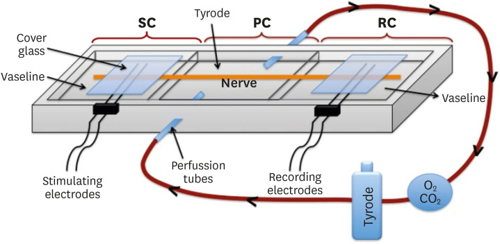
Materials and Methods Sciatic nerves were placed in a Tyrode bath and cAPs were recorded before, during, and after the application of test materials for 60-minute control, application, and recovery measurements, respectively. Freshly prepared ProRoot MTA, MTA Angelus, Biodentine, Endosequence RRM-Putty, BioAggregate, and RetroMTA were directly applied onto the nerves. Biopac LabPro version 3.7 was used to record and analyze cAPs. The data were statistically analyzed.
Results None of the CSCs totally blocked cAPs. RetroMTA, Biodentine, and MTA Angelus caused no significant alteration in cAPs (
p > 0.05). Significantly lower cAPs were observed in recovery measurements for BioAggregate than in the control condition (p < 0.05). ProRoot MTA significantly but transiently reduced cAPs in the application period compared to the control period (p < 0.05). Endosequence RRM-Putty significantly reduced cAPs.Conclusions Various CSCs may alter cAPs to some extent, but none of the CSCs irreversibly blocked them. The usage of fast-setting CSCs during apexification or regeneration of immature teeth seems safer than slow-setting CSCs due to their more favorable neuronal effects.
-
Citations
Citations to this article as recorded by- Endodontic Sealers and Innovations to Enhance Their Properties: A Current Review
Anna Błaszczyk-Pośpiech, Natalia Struzik, Maria Szymonowicz, Przemysław Sareło, Maria Wiśniewska-Wrona, Kamila Wiśniewska, Maciej Dobrzyński, Magdalena Wawrzyńska
Materials.2025; 18(18): 4259. CrossRef
- Endodontic Sealers and Innovations to Enhance Their Properties: A Current Review
- 1,370 View
- 19 Download
- 1 Web of Science
- 1 Crossref

- Shaping ability and apical debris extrusion after root canal preparation with rotary or reciprocating instruments: a micro-CT study
- Emmanuel João Nogueira Leal da Silva, Sara Gomes de Moura, Carolina Oliveira de Lima, Ana Flávia Almeida Barbosa, Waleska Florentino Misael, Mariane Floriano Lopes Santos Lacerda, Luciana Moura Sassone
- Restor Dent Endod 2021;46(2):e16. Published online February 25, 2021
- DOI: https://doi.org/10.5395/rde.2021.46.e16

-
 Abstract
 PDF
PDF PubReader
 ePub
ePub Objectives The aim of this study was to evaluate the shaping ability of the TruShape and Reciproc Blue systems and the apical extrusion of debris after root canal instrumentation. The ProTaper Universal system was used as a reference for comparison.
Materials and Methods Thirty-three mandibular premolars with a single canal were scanned using micro-computed tomography and were matched into 3 groups (
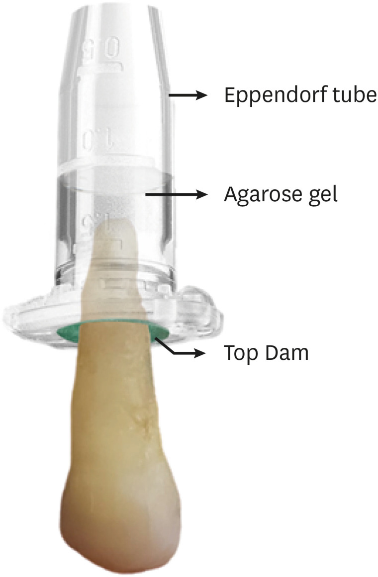
n = 11) according to the instrumentation system: TruShape, Reciproc Blue and ProTaper Universal. The teeth were accessed and mounted in an apparatus with agarose gel, which simulated apical resistance provided by the periapical tissue and enabled the collection of apically extruded debris. During root canal preparation, 2.5% sodium hypochlorite was used as an irrigant. The samples were scanned again after instrumentation. The percentage of unprepared area, removed dentin, and volume of apically extruded debris were analyzed. The data were analyzed using 1-way analysis of variance and the Tukey test for multiple comparisons at a 5% significance level.Results No significant differences in the percentage of unprepared area were observed among the systems (
p > 0.05). ProTaper Universal presented a higher percentage of dentin removal than the TruShape and Reciproc Blue systems (p < 0.05). The systems produced similar volumes of apically extruded debris (p > 0.05).Conclusions All systems caused apically extruded debris, without any significant differences among them. TruShape, Reciproc Blue, and ProTaper Universal presented similar percentages of unprepared area after root canal instrumentation; however, ProTaper Universal was associated with higher dentin removal than the other systems.
-
Citations
Citations to this article as recorded by- Evaluation of Silver-Ion-Coated Rotary Nickel Titanium Files - An In Vitro Study
Jhanvi H. Sadaria, Kondas V. Venkatesh, Dhanasekaran Sihivahanan
Indian Journal of Dental Research.2026;[Epub] CrossRef - Comparison of post-operative pain prevalence after single visit endodontic treatment with two NiTi rotary files - a randomized clinical trial
M. E. Khallaf, Yousra Aly, Amira Ibrahim Mohamed
Scientific Reports.2025;[Epub] CrossRef - A quantitative comparison of apically extruded debris during root canal preparation using NiTi full-sequence rotary and single-file rotary systems: An in vitro study
Pallavi Goel, R. Vikram, R. Anithakumari, M. S. Adarsha, M. E. Sudhanva
Endodontology.2024; 36(3): 235. CrossRef - Extrusion of Sodium Hypochlorite in Oval-Shaped Canals: A Comparative Study of the Potential of Four Final Agitation Approaches Employing Agarose-Embedded Mandibular First Premolars
Aalisha Parkar, Kulvinder Singh Banga, Ajinkya M. Pawar, Alexander Maniangat Luke
Journal of Clinical Medicine.2024; 13(10): 2748. CrossRef - Shaping Efficiency of Rotary and Reciprocating Kinematics of Engine-driven Nickel-Titanium Instruments in Moderate and Severely curved Root Canals Using Microcomputed Tomography: A Systematic Review of Ex Vivo Studies
Claudiu Călin, Ana-Maria Focșăneanu, Friedrich Paulsen, Andreea C. Didilescu, Tiberiu Niță
Journal of Endodontics.2024; 50(7): 907. CrossRef - Intracanal removal and apical extrusion of filling material after retreatment using rotary or reciprocating instruments: A new approach using human cadavers
Thamyres M. Monteiro, Victor O. Cortes‐Cid, Marilia F. V. Marceliano‐Alves, Andrea F. Campello, Luan F. Bastos, Ricardo T. Lopes, José F. Siqueira, Flávio R. F. Alves
International Endodontic Journal.2024; 57(1): 100. CrossRef - Assessment of debris extrusion on using automated irrigation device with conventional needle irrigation – An ex vivo study
Sahil Choudhari, Kavalipurapu Venkata Teja, Raja Kumar, Sindhu Ramesh
Saudi Endodontic Journal.2023; 13(3): 263. CrossRef - Postoperative pain perception and associated risk factors in children after continuous rotation versus reciprocating kinematics: A randomised prospective clinical trial
Ahmad Abdel Hamid Elheeny, Dania Ibrahem Sermani, Mahmoud Ahmed Abdelmotelb
Australian Endodontic Journal.2023; 49(S1): 345. CrossRef - A critical analysis of research methods and experimental models to study apical extrusion of debris and irrigants
Jale Tanalp
International Endodontic Journal.2022; 55(S1): 153. CrossRef - Quantitative evaluation of apically extruded debris using TRUShape, TruNatomy, and WaveOne Gold in curved canals
Nehal Nabil Roshdy, Reham Hassan
BDJ Open.2022;[Epub] CrossRef - Shaping ability of new reciprocating or rotary instruments with two cross‐sectional designs: An ex vivo study
Isabela G. Guedes, Renata C. V. Rodrigues, Marília F. Marceliano‐Alves, Flávio R. F. Alves, Isabela N. Rôças, José F. Siqueira
International Endodontic Journal.2022; 55(12): 1385. CrossRef
- Evaluation of Silver-Ion-Coated Rotary Nickel Titanium Files - An In Vitro Study
- 2,522 View
- 49 Download
- 8 Web of Science
- 11 Crossref

- Effects of the endodontic access cavity on apical debris extrusion during root canal preparation using different single-file systems
- Pelin Tüfenkçi, Koray Yılmaz, Mehmet Adigüzel
- Restor Dent Endod 2020;45(3):e33. Published online June 4, 2020
- DOI: https://doi.org/10.5395/rde.2020.45.e33

-
 Abstract
 PDF
PDF PubReader
 ePub
ePub Objectives This study was conducted to evaluate the effects of traditional and contracted endodontic cavity (TEC and CEC) preparation with the use of Reciproc Blue (RPC B) and One Curve (OC) single-file systems on the amount of apical debris extrusion in mandibular first molar root canals.
Materials and Methods Eighty extracted mandibular first molar teeth were randomly assigned to 4 groups (
n = 20) according to the endodontic access cavity shape and the single file system used for root canal preparation (reciprocating motion with the RCP B and rotary motion with the OC): TEC-RPC B, TEC-OC, CEC-RPC B, and CEC-OC. The apically extruded debris during preparation was collected in Eppendorf tubes. The amount of extruded debris was quantified by subtracting the weight of the empty tubes from the weight of the Eppendorf tubes containing the debris. Data were analyzed using 1-way analysis of variance with the Tukeypost hoc test. The level of significance was set atp < 0.05.Results The CEC-RPC B group showed more apical debris extrusion than the TEC-OC and CEC-OC groups (
p < 0.05). There were no statistically significant differences in the amount of apical debris extrusion among the TEC-OC, CEC-OC, and TEC-RPC B groups.Conclusions RPC B caused more apical debris extrusion in the CEC groups than did the OC single-file system. Therefore, it is suggested that the RPC B file should be used carefully in teeth with a CEC.
-
Citations
Citations to this article as recorded by- Comparative Evaluation of Periapical Expulsion Using Manual, Rotary, and Reciprocating Instrumentation With EndoVac Irrigation: An In Vitro Study
Sachin Metkari, Sanpreet S Sachdev, Pravin Patil, Manoj Ramugade, Kishor D Sapkale, Kulvinder S Banga, Dinesh Rao
Cureus.2025;[Epub] CrossRef - Comparison of Debris Extrusion and Preparation Time by Traverse, R‐Motion Glider C, and Other Glide Path Systems in Severely Curved Canals
Taher Al Omari, Layla Hassouneh, Khawlah Albashaireh, Alaa Dkmak, Rami Albanna, Ali Al-Mohammed, Ahmed Jamleh, Lucas da Fonseca Roberti Garcia
International Journal of Dentistry.2025;[Epub] CrossRef - Minimal İnvaziv Giriş Kavitelerinin Alt Kesici Dişlerdeki Apikal Ekstrüzyona Etkisi
İrem Haskarabağ, Cangül Keskin
Türk Diş Hekimliği Araştırma Dergisi.2025; 4(2): 75. CrossRef - Evaluation of apically extruded debris from root canal filling removal of the mesiobuccal canal of maxillary molars using XP shaper and protaper with two different irrigation
Sanaz Mirsattari, Maryam Zare Jahromi, Masoud Khabiri
Dental Research Journal.2024;[Epub] CrossRef - The Impact of Minimum Invasive Access Cavity Design on the Quality of Instrumentation of Root Canals of Maxillary Molars Using Cone-Beam Computed Tomography: An in Vitro Study
Fahad H Baabdullah, Samia M Elsherief , Rayan A Hawsawi, Hetaf S Redwan
Cureus.2024;[Epub] CrossRef - Assessment of Bacterial Load and Post-Endodontic Pain after One-Visit Root Canal Treatment Using Two Types of Endodontic Access Openings: A Randomized Controlled Clinical Trial
Ahmed M. Al-Ani, Ahmed H. Ali, Garrit Koller
Dentistry Journal.2024; 12(4): 88. CrossRef - The effect of different kinematics on apical debris extrusion with a single-file system
Taher M. N. Al Omari, Giusy Rita Maria La Rosa, Rami Haitham Issa Albanna, Abedelmalek Tabnjh, Flavia Papale, Eugenio Pedullà
Odontology.2023; 111(4): 910. CrossRef - The effects of laser and ultrasonic irrigation activation methods on smear and debris removal in traditional and conservative endodontic access cavities
Hüseyin Gündüz, Esin Özlek
Lasers in Medical Science.2023;[Epub] CrossRef - Influence of access cavity design, sodium hypochlorite formulation and XP‐endo Shaper usage on apical debris extrusion – A laboratory investigation
Jerry Jose, Aishuwariya Thamilselvan, Kavalipurapu Venkata Teja, Giampiero Rossi–Fedele
Australian Endodontic Journal.2023; 49(1): 6. CrossRef - Apically extruded debris, canal transportation, and shaping ability of nickel-titanium instruments on contracted endodontic cavities in molar teeth
Qinqin Zhang, Jingyi Gu, Jiadi Shen, Ming Ma, Ying Lv, Xin Wei
Journal of Oral Science.2023; 65(4): 203. CrossRef - Impact of contracted endodontic cavities on instrumentation efficacy—A systematic review
Manan Shroff, Karkala Venkappa Kishan, Nimisha Shah, Purnima Saklecha
Australian Endodontic Journal.2023; 49(1): 202. CrossRef - Present status and future directions – Minimal endodontic access cavities
Emmanuel João Nogueira Leal Silva, Gustavo De‐Deus, Erick Miranda Souza, Felipe Gonçalves Belladonna, Daniele Moreira Cavalcante, Marco Simões‐Carvalho, Marco Aurélio Versiani
International Endodontic Journal.2022; 55(S3): 531. CrossRef - Effect of guided conservative endodontic access and different file kinematics on debris extrusion in mesial root of the mandibular molars: An in vitro study
Sathish Sundar, Aswathi Varghese, KrithikaJ Datta, Velmurugan Natanasabapathy
Journal of Conservative Dentistry.2022; 25(5): 547. CrossRef - A critical analysis of research methods and experimental models to study apical extrusion of debris and irrigants
Jale Tanalp
International Endodontic Journal.2022; 55(S1): 153. CrossRef - Current strategies for conservative endodontic access cavity preparation techniques—systematic review, meta-analysis, and decision-making protocol
Benoit Ballester, Thomas Giraud, Hany Mohamed Aly Ahmed, Mohamed Shady Nabhan, Frédéric Bukiet, Maud Guivarc’h
Clinical Oral Investigations.2021; 25(11): 6027. CrossRef - Extrusion of debris with and without intentional foraminal enlargement – A systematic review and meta‐analysis
Ricardo Machado, Gislayne Vigarani, Tainara Macoppi, Ajinkya Pawar, Stella Maria Glaci Reinke, Ana Cristina Kovalik Gonçalves
Australian Endodontic Journal.2021; 47(3): 741. CrossRef - Apical debris extrusion of single-file systems in curved canals
Ecehan Hazar, Olcay Özdemir, Mustafa Murat Koçak, Baran Can Sağlam, Sibel Koçak
Endodontology.2021; 33(3): 128. CrossRef - Quantitative Evaluation of Apically Extruded Debris in Root Canals prepared by Single-file Reciprocating and Single File Rotary Instrumentation Systems: A Comparative In vitro Study
Sonal Sinha, Konark Singh, Anju Singh, Swati Priya, Avanindra Kumar, Sahil Kawle
Journal of Pharmacy and Bioallied Sciences.2021; 13(Suppl 2): S1398. CrossRef - THE INFLUENCE OF DIFFERENT PECKING DEPTH ON AMOUNT OF APICALLY EXTRUDED DEBRIS DURING ROOT CANAL PREPARATION
Fatih ÇAKICI, Busra UYSAL, Elif Bahar CAKİCİ, Adem GUNAYDIN
Atatürk Üniversitesi Diş Hekimliği Fakültesi Dergisi.2021; : 1. CrossRef
- Comparative Evaluation of Periapical Expulsion Using Manual, Rotary, and Reciprocating Instrumentation With EndoVac Irrigation: An In Vitro Study
- 2,407 View
- 23 Download
- 19 Crossref

- Influence of plugger penetration depth on the apical extrusion of root canal sealer in Continuous Wave of Condensation Technique
- Ho-Young So, Young-Mi Lee, Kwang-Keun Kim, Ki-Ok Kim, Young-Kyung Kim, Sung-Kyo Kim
- J Korean Acad Conserv Dent 2004;29(5):439-445. Published online January 14, 2004
- DOI: https://doi.org/10.5395/JKACD.2004.29.5.439
-
 Abstract
 PDF
PDF PubReader
 ePub
ePub ABSTRACT The purpose of this study was to evaluate the influence of plugger penetration depth on the apical extrusion of root canal sealer during root canal obturation with Continuous Wave of Condensation Technique.
Root canals of forty extracted human teeth were divided into four groups and were prepared up to size 40 of 0.06 taper with ProFile. After drying, canals of three groups were filled with Continuous Wave of Condensation Technique with System B™ and different plugger penetration depths of 3, 5, and 7 mm from the apex. Canals of one group were filled with cold lateral compaction technique as a control. Canals were filled with non-standardized master gutta-percha cones and 0.02 mL of Sealapex. Apical extruded sealer was collected in a container and weighed. Data was analyzed with one-way ANOVA and Duncan’s Multiple Range Test. 3 and 5 mm penetration depth groups in Continuous Wave of Condensation Technique showed significantly more extrusion of root canal sealer than 7 mm penetration depth group (
p < 0.05). However, there was no significant difference between 7 mm depth group in Continuous Wave of Condensation Technique and cold lateral compaction group (p < 0.05).The result of this study demonstrates that deeper plugger penetration depth causes more extrusion of root canal sealer in root canal obturation by Continuous Wave of Condensation Technique. Therefore, special caution is needed when plugger penetration is deeper in the canal in Continuous Wave of Condensation Technique to minimize the amount of sealer extrusion beyond apex.
-
Citations
Citations to this article as recorded by- Influence of plugger penetration depth on the area of the canal space occupied by gutta-percha
Young Mi Lee, Ho-young So, Young Kyung Kim, Sung Kyo Kim
Journal of Korean Academy of Conservative Dentistry.2006; 31(1): 66. CrossRef
- Influence of plugger penetration depth on the area of the canal space occupied by gutta-percha
- 1,246 View
- 7 Download
- 1 Crossref


 KACD
KACD

 First
First Prev
Prev


