-
Mineral content analysis of root canal dentin using laser-induced breakdown spectroscopy
-
Selen Küçükkaya Eren, Emel Uzunoğlu, Banu Sezer, Zeliha Yılmaz, İsmail Hakkı Boyacı
-
Restor Dent Endod 2018;43(1):e11. Published online February 4, 2018
-
DOI: https://doi.org/10.5395/rde.2018.43.e11
-
-
 Abstract Abstract
 PDF PDF PubReader PubReader ePub ePub
- Objectives
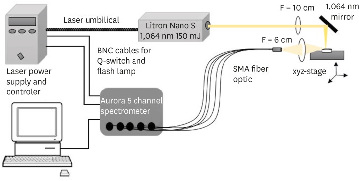
This study aimed to introduce the use of laser-induced breakdown spectroscopy (LIBS) for evaluation of the mineral content of root canal dentin, and to assess whether a correlation exists between LIBS and scanning electron microscopy/energy dispersive spectroscopy (SEM/EDS) methods by comparing the effects of irrigation solutions on the mineral content change of root canal dentin. Materials and MethodsForty teeth with a single root canal were decoronated and longitudinally sectioned to expose the canals. The root halves were divided into 4 groups (n = 10) according to the solution applied: group NaOCl, 5.25% sodium hypochlorite (NaOCl) for 1 hour; group EDTA, 17% ethylenediaminetetraacetic acid (EDTA) for 2 minutes; group NaOCl+EDTA, 5.25% NaOCl for 1 hour and 17% EDTA for 2 minutes; a control group. Each root half belonging to the same root was evaluated for mineral content with either LIBS or SEM/EDS methods. The data were analyzed statistically. ResultsIn groups NaOCl and NaOCl+EDTA, the calcium (Ca)/phosphorus (P) ratio decreased while the sodium (Na) level increased compared with the other groups (p < 0.05). The magnesium (Mg) level changes were not significant among the groups. A significant positive correlation was found between the results of LIBS and SEM/EDS analyses (r = 0.84, p < 0.001). ConclusionsTreatment with NaOCl for 1 hour altered the mineral content of dentin, while EDTA application for 2 minutes had no effect on the elemental composition. The LIBS method proved to be reliable while providing data for the elemental composition of root canal dentin.
-
Citations
Citations to this article as recorded by  - In vitro evaluation of antimicrobial photodynamic therapy with photosensitizers and calcium hydroxide on bond strength, chemical composition, and sealing of glass-fiber posts to root dentin
Thalya Fernanda Horsth Maltarollo, Paulo Henrique dos Santos, Henrique Augusto Banci, Mariana de Oliveira Bachega, Beatriz Melare de Oliveira, Marco Hungaro Antonio Duarte, Índia Olinta de Azevedo Queiroz, Rodrigo Rodrigues Amaral, Luciano Angelo Tavares
Lasers in Medical Science.2025;[Epub] CrossRef - Effect of Using 5% Apple Vinegar Irrigation Solution Adjunct to Diode Laser on Smear Layer Removal and Calcium/Phosphorus Ion Ratio during Root Canal Treatment
Tarek AA Salam, Haythem SA Kader, Elsayed E Abdallah
CODS - Journal of Dentistry.2024; 15(1): 3. CrossRef - Evaluation of chemical composition of root canal dentin between two age groups using different irrigating solutions: An in vitro sem-eds study
Naresh Kumar K, Abhijith Kallu, Surender L.R, Sravani Nirmala, Narender Reddy
International Dental Journal of Student's Research.2024; 12(1): 18. CrossRef - Minimally invasive management of vital teeth requiring root canal therapy
E. Karatas, M. Hadis, W. M. Palin, M. R. Milward, S. A. Kuehne, J. Camilleri
Scientific Reports.2023;[Epub] CrossRef - The Effects of a Novel Nanohydroxyapatite Gel and Er: YAG Laser Treatment on Dentin Hypersensitivity
Demet Sahin, Ceren Deger, Burcu Oglakci, Metehan Demirkol, Bedri Onur Kucukyildirim, Mehtikar Gursel, Evrim Eliguzeloglu Dalkilic
Materials.2023; 16(19): 6522. CrossRef - Chitosan Homogenizing Coffee Ring Effect for Soil Available Potassium Determination Using Laser-Induced Breakdown Spectroscopy
Xiaolong Li, Rongqin Chen, Zhengkai You, Tiantian Pan, Rui Yang, Jing Huang, Hui Fang, Wenwen Kong, Jiyu Peng, Fei Liu
Chemosensors.2022; 10(9): 374. CrossRef - Quantitative analysis of cadmium in rice roots based on LIBS and chemometrics methods
Wei Wang, Wenwen Kong, Tingting Shen, Zun Man, Wenjing Zhu, Yong He, Fei Liu
Environmental Sciences Europe.2021;[Epub] CrossRef
-
210
View
-
3
Download
-
7
Crossref
-
Influence of a glide path on the dentinal crack formation of ProTaper Next system
-
Sevinç Aktemur Türker, Emel Uzunoğlu
-
Restor Dent Endod 2015;40(4):286-289. Published online September 2, 2015
-
DOI: https://doi.org/10.5395/rde.2015.40.4.286
-
-
 Abstract Abstract
 PDF PDF PubReader PubReader ePub ePub
- Objectives
The aim was to evaluate dentinal crack formation after root canal preparation with ProTaper Next system (PTN) with and without a glide path. Materials and MethodsForty-five mesial roots of mandibular first molars were selected. Fifteen teeth were left unprepared and served as controls. The experimental groups consist of mesiobuccal and mesiolingual root canals of remaining 30 teeth, which were divided into 2 groups (n = 15): Group PG/PTN, glide path was created with ProGlider (PG) and then canals were shaped with PTN system; Group PTN, glide path was not prepared and canals were shaped with PTN system only. All roots were sectioned perpendicular to the long axis at 1, 2, 3, 4, 6, and 8 mm from the apex, and the sections were observed under a stereomicroscope. The presence/absence of cracks was recorded. Data were analyzed with chi-square tests with Yates correction. ResultsThere were no significant differences in crack formation between the PTN with and without glide path preparation. The incidence of cracks observed in PG/PTN and PTN groups was 17.8% and 28.9%, respectively. ConclusionsThe creation of a glide path with ProGlider before ProTaper Next rotary system did not influence dentinal crack formation in root canals.
-
Citations
Citations to this article as recorded by  - Glide Path in Endodontics: A Literature Review of Current Knowledge
Vlad Mircea Lup, Giulia Malvicini, Carlo Gaeta, Simone Grandini, Gabriela Ciavoi
Dentistry Journal.2024; 12(8): 257. CrossRef - Comparative Evaluation of the Shaping Ability of the Recent, Fifth-generation ProTaper Next and Revo-S NiTi Rotary Endodontic Files Using Three-dimensional Imaging: An Imaging-based Study
Prajna Pattanaik, Akilan Balasubramanian, P. Veeralakshmi, Gautam Singh, Vandana Sadananda, Hina Ahmed, J. Suresh Babu, C. Swarnalatha, Abhishek Singh Nayyar
Journal of Microscopy and Ultrastructure.2023;[Epub] CrossRef - Microscopic Assessment of Dentinal Defects Induced by ProTaper Universal, ProTaper Gold, and Hyflex Electric Discharge Machining Rotary File Systems – An in vitro Study
Takhellambam Premlata Devi, Amandeep Kaur, Shamurailatpam Priyadarshini, B. S. Deepak, Sumita Banerjee, Ng Sanjeeta
Contemporary Clinical Dentistry.2021; 12(3): 230. CrossRef - Influence of Negotiation, Glide Path, and Preflaring Procedures on Root Canal Shaping—Terminology, Basic Concepts, and a Systematic Review
Gianluca Plotino, Venkateshbabu Nagendrababu, Frederic Bukiet, Nicola M. Grande, Sajesh K. Veettil, Gustavo De-Deus, Hany Mohamed Aly Ahmed
Journal of Endodontics.2020; 46(6): 707. CrossRef
-
186
View
-
2
Download
-
4
Crossref
-
Calcium hydroxide dressing residues after different removal techniques affect the accuracy of Root-ZX apex locator
-
Emel Uzunoglu, Ayhan Eymirli, Mehmet Özgür Uyanik, Semra Çalt, Emre Nagas
-
Restor Dent Endod 2015;40(1):44-49. Published online November 5, 2014
-
DOI: https://doi.org/10.5395/rde.2015.40.1.44
-
-
 Abstract Abstract
 PDF PDF PubReader PubReader ePub ePub
- Objectives
This study compared the ability of several techniques to remove calcium hydroxide (CH) from the root canal and determined the influence of CH residues on the accuracy of the electronic apex locator. Materials and MethodsRoot canals of 90 human maxillary lateral incisors with confirmed true working length (TWL) were prepared and filled with CH. The teeth were randomly assigned to one of the experimental groups according to the CH removal technique (n = 14): 0.9% saline; 0.9% saline + master apical file (MAF); 17% ethylenediamine tetraacetic acid (EDTA); 17% EDTA + MAF; 5.25% sodium hypochlorite (NaOCl); 5.25% NaOCl + MAF. Six teeth were used as negative control. After CH removal, the electronic working length was measured using Root-ZX (Morita Corp.) and compared with TWL to evaluate Root-ZX accuracy. All specimens were sectioned longitudinally, and the area of remaining CH (CH) and total canal area were measured using imaging software. ResultsThe EDTA + MAF and NaOCl + MAF groups showed better CH removal than other groups (p < 0.05). Root-ZX reliability to prevent overestimated working length to be > 85% within a tolerance of ± 1.0 mm (p < 0.05). There was strong negative correlation between amount of CH residues and EAL accuracy (r = -0.800 for ± 0.5 mm; r = -0.940 for ± 1.0 mm). ConclusionsThe mechanical instrumentation improves the CH removal of irrigation solutions although none of the techniques removed the dressing completely. Residues of CH medication in root canals affected the accuracy of Root-ZX adversely.
-
Citations
Citations to this article as recorded by  - Evaluation of the Effect of Calcium Hydroxide Residues Including Different Vehicles on the Accuracy of Electronic Apex Locators
Simay Koç, Damla Erkal, Dide Tekinarslan, Kürs¸at Er
Journal of Advanced Oral Research.2025;[Epub] CrossRef - Comparative evaluation of the accuracy of electronic apex locators and cone-beam computed tomography in detection of root canal perforation and working length during endodontic retreatment
Simay Koç, Hatice Harorlı, Alper Kuştarcı
BMC Oral Health.2024;[Epub] CrossRef - Effects of Intracanal Medicaments on the Measurement Accuracy of Four Apex Locators: An In Vitro Study
Hamza Cudal, Tuğrul Aslan, Bertan Kesim
Meandros Medical and Dental Journal.2023; 24(3): 215. CrossRef - Electronic Apex Locators and their Implications in Contemporary Clinical Practice: A Review
Zainab Shirazi, Anas Al-Jadaa, Abdul Rahman Saleh
The Open Dentistry Journal.2023;[Epub] CrossRef - Influence of Apical Patency, Coronal Preflaring and Calcium Hydroxide on the Accuracy of Root ZX Apex Locator for Working Length Determination: An In Vitro Study
Mostafa Godiny, Reza Hatam, Roya Safari-Faramani, Atefeh Khavid, Mohammad Reza Rezaei
Journal of Advanced Oral Research.2022; 13(1): 38. CrossRef - Endodontic cement penetration after removal of calcium hydroxide dressing using XP-endo finisher
Alyssa Sales dos Santos, Maria Aparecida Barbosa de Sá, Marco Antônio Húngaro Duarte, Martinho Campolina Rebello Horta, Frank Ferreira Silveira, Eduardo Nunes
Brazilian Oral Research.2022;[Epub] CrossRef - Efficacy of glycolic acid for the removal of calcium hydroxide from simulated internal Resorption cavities
Cangül Keskin, Ali Keleş, Öznur Sarıyılmaz
Clinical Oral Investigations.2021; 25(7): 4407. CrossRef - Accuracy of electronic apex locator in the presence of different irrigating solutions
Padmanabh Jha, Vineeta Nikhil, Shalya Raj, Rohit Ravinder, Preeti Mishra
Endodontology.2021; 33(4): 232. CrossRef - Farklı Kanal İçi Ortamların Apeks Bulucuların Doğruluğu Üzerine Etkisi
Asena OKUR, Tuğrul ASLAN, Burak SAĞSEN
Selcuk Dental Journal.2021; 8(3): 859. CrossRef - Evaluation of the accuracy of different apex locators in determiningthe working length during root canal retreatment
Pelin Tufenkci, Aylin Kalaycı
Journal of Dental Research, Dental Clinics, Dental Prospects.2020; 14(2): 125. CrossRef - Influence of calcium hydroxide residues after using different irrigants on the accuracy of two electronic apex locators: An in vitro study
NooshinSadat Shojaee, Zahra Zaeri, MohammadMehdi Shokouhi, Fereshteh Sobhnamayan, Alireza Adl
Dental Research Journal.2020; 17(1): 48. CrossRef - The Effect of Calcium Hydroxide and File Sızes on the Accuracy of the Electronic Apex Locator in Simulated Immature Teeth
Leyla AYRANCİ, Ahmet ÇETİNKAYA, Serkan ÖZKAN
Middle Black Sea Journal of Health Science.2019; 5(3): 273. CrossRef - The Effect of File Size and Type and Irrigation Solutions on the Accuracy of Electronic Apex Locators: AnIn VitroStudy on Canine Teeth
Maciej Janeczek, Piotr Kosior, Dagmara Piesiak-Pańczyszyn, Krzysztof Dudek, Aleksander Chrószcz, Agnieszka Czajczyńska-Waszkiewicz, Małgorzata Kowalczyk-Zając, Aleksandra Gabren-Syller, Karol Kirstein, Aleksandra Skalec, Ewelina Bryła, Maciej Dobrzyński
BioMed Research International.2016; 2016: 1. CrossRef
-
222
View
-
2
Download
-
13
Crossref
-
Effects of dentin moisture on the push-out bond strength of a fiber post luted with different self-adhesive resin cements
-
Sevinç Aktemur Türker, Emel Uzunoğlu, Zeliha Yılmaz
-
Restor Dent Endod 2013;38(4):234-240. Published online November 12, 2013
-
DOI: https://doi.org/10.5395/rde.2013.38.4.234
-
-
 Abstract Abstract
 PDF PDF PubReader PubReader ePub ePub
- Objectives
This study evaluated the effects of intraradicular moisture on the pushout bond strength of a fibre post luted with several self-adhesive resin cements. Materials and MethodsEndodontically treated root canals were treated with one of three luting cements: (1) RelyX U100, (2) Clearfil SA, and (3) G-Cem. Roots were then divided into four subgroups according to the moisture condition tested: (I) dry: excess water removed with paper points followed by dehydration with 95% ethanol, (II) normal moisture: canals blot-dried with paper points until appearing dry, (III) moist: canals dried by low vacuum using a Luer adapter, and (IV) wet: canals remained totally flooded. Two 1-mm-thick slices were obtained from each root sample and bond strength was measured using a push-out test setup. The data were analysed using a two-way analysis of variance and the Bonferroni post hoc test with p = 0.05. ResultsStatistical analysis demonstrated that moisture levels had a significant effect on the bond strength of luting cements (p < 0.05), with the exception of G-Cem. RelyX U100 displayed the highest bond strength under moist conditions (III). Clearfil SA had the highest bond strength under normal moisture conditions (II). Statistical ranking of bond strength values was as follows: RelyX U100 > Clearfil SA > G-Cem. ConclusionsThe degree of residual moisture significantly affected the adhesion of luting cements to radicular dentine.
-
Citations
Citations to this article as recorded by  - Push-Out Bond Strength of Different Luting Cements Following Post Space Irrigation with 2% Chitosan: An In Vitro Study
Shimaa Rifaat, Ahmed Rahoma, Hind Muneer Alharbi, Sawsan Jamal Kazim, Shrouq Ali Aljuaid, Basmah Omar Alakloby, Faraz A. Farooqi, Noha Taymour
Prosthesis.2025; 7(1): 18. CrossRef - Dentin bond strength of resin luting agents under a simulated intra-oral environment
Takashi Washino, Hanemi Tsuruta, Masaomi Ikeda, Michael F. Burrow, Toru Nikaido
Asian Pacific Journal of Dentistry.2024; 24(2): 13. CrossRef - Effects of a relined fiberglass post with conventional and self-adhesive resin cement
Wilton Lima dos Santos Junior, Marina Rodrigues Santi, Rodrigo Barros Esteves Lins, Luís Roberto Marcondes Martins
Restorative Dentistry & Endodontics.2024;[Epub] CrossRef - Effect of dentin moisture on the adhesive properties of luting fiber posts using adhesive strategies
Renata Terumi JITUMORI, Rafaela Caroline RODRIGUES, Alessandra REIS, João Carlos GOMES, Giovana Mongruel GOMES
Brazilian Oral Research.2023;[Epub] CrossRef - Influence of different intraradicular chemical pretreatments on the bond strength of adhesive interface between dentine and fiber post cements: A systematic review and network meta‐analysis
Ana Luiza Barbosa Jurema, Ayla Macyelle de Oliveira Correia, Manuela da Silva Spinola, Eduardo Bresciani, Taciana Marco Ferraz Caneppele
European Journal of Oral Sciences.2022;[Epub] CrossRef - SELF ADEZİV REZİN SİMANLAR / SELF ADHESIVE RESIN CEMENTS
Kübra AMAÇ, Engin ESENTÜRK, Bilge TURHAN BAL
Atatürk Üniversitesi Diş Hekimliği Fakültesi Dergisi.2022; : 1. CrossRef - Postspace pretreatment with 17% ethylenediamine tetraacetic acid, 7% maleic acid, and 1% phytic acid on bond strength of fiber posts luted with a self-adhesive resin cement
PriyaC Yadav, Ramya Raghu, Ashish Shetty, Subhashini Rajasekhara
Journal of Conservative Dentistry.2021; 24(6): 558. CrossRef - Development and characterization of biological bovine dentin posts
Alice Gonçalves Penelas, Eduardo Moreira da Silva, Laiza Tatiana Poskus, Amanda Cypriano Alves, Isis Ingrid Nogueira Simões, Viviane Hass, José Guilherme Antunes Guimarães
Journal of the Mechanical Behavior of Biomedical Materials.2019; 92: 197. CrossRef - Evaluation of the influence of time and concentration of sodium hypochlorite on the bond strength of glass fibre post
Beau Knight, Robert M. Love, Roy George
Australian Endodontic Journal.2018; 44(3): 267. CrossRef - Test methods for bond strength of glass fiber posts to dentin: A review
F. C. Dos Santos, M. D. Banea, H. L. Carlo, S. De Barros
The Journal of Adhesion.2017; 93(1-2): 159. CrossRef - Is the bonding of self-adhesive cement sensitive to root region and curing mode?
Thaynara Faelly BOING, Giovana Mongruel GOMES, João Carlos GOMES, Alessandra REIS, Osnara Maria Mongruel GOMES
Journal of Applied Oral Science.2017; 25(1): 2. CrossRef - A Twofold Comparison between Dual Cure Resin Modified Cement and Glass Ionomer Cement for Orthodontic Band Cementation
Hanaa El Attar, Omnia Elhiny, Ghada Salem, Ahmed Abdelrahman, Mazen Attia
Open Access Macedonian Journal of Medical Sciences.2016; 4(4): 695. CrossRef - Shear bond strengths of various self-adhesive resin cements between bovine dentin and 4 types of adherends
Ah-Jin Kim, Da-Ryeong Park, Seunghan Oh, Ji-Myung Bae
Korean Journal of Dental Materials.2015; 42(4): 365. CrossRef
-
212
View
-
1
Download
-
13
Crossref
|


