Articles
- Page Path
- HOME > Restor Dent Endod > Volume 28(1); 2003 > Article
- Original Article Morphological patterns of self-etching primers and self-etching adhesive bonded to tooth structure
- Young-Gon Cho, Seok-Jong Lee, Jin-Ho Jeong, Young-Gon Lee, Soo-Mee Kim
-
2003;28(1):-33.
DOI: https://doi.org/10.5395/JKACD.2003.28.1.023
Published online: January 31, 2003
Department of Conservative Dentistry, College of Dentistry, Chosun University, Korea.
- Corresponding author (ygcho@mail.chosun.ac.kr)
Copyright © 2003 Korean Academy of Conservative Dentistry
- 932 Views
- 1 Download
- 1 Crossref
Abstract
-
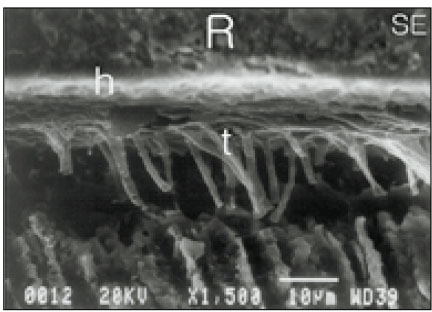
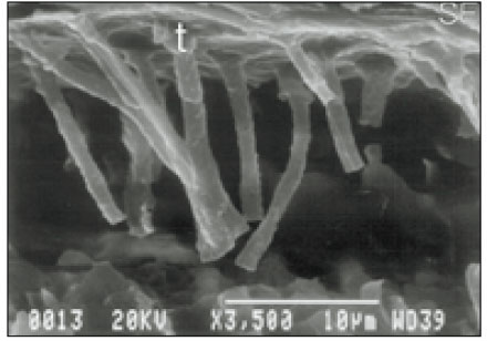
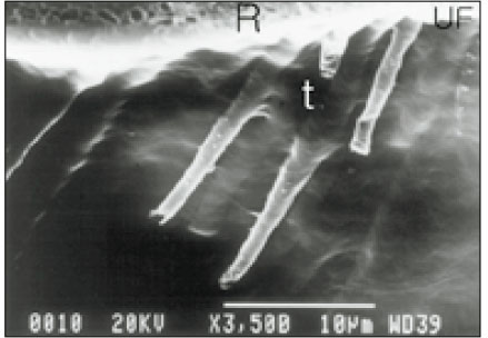
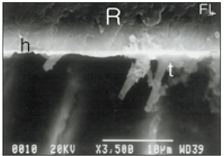
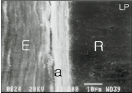
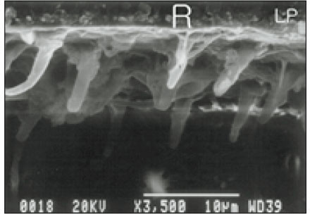
The purpose of this study was to compare in vitro interfacial relationship of restorations bonded with three self-etching primer adhesives and one self-etching adhesive.Class I cavity preparations were prepared on twenty extracted human molars. Prepared teeth were divided into four groups and restored with four adhesives and composites: Clearfil SE Bond/Clearfil™ AP-X (SE), UniFil Bond/UniFil® F (UF), FL Bond/Filtek™ Z 250 (FL) and Prompt L-Pop/Filtek™ Z 250 (LP)After storing in distilled water of room temperature for 24 hours, the specimens were vertically sectioned and decalcified. Morphological patterns between the enamel/dentin and adhesives were observed under SEM.The results of this study were as follows;1. They showed close adaptation between enamel and SE, UF and FL except for LP.2. The hybrid layer in dentin was 2 µm thick in SE, 1.5 µm thick in UF, and 0.4 µm in both FL and LP. So, the hybrid layers of SE and UF were slightly thicker than that of FL and LP.3. The lengths and diameters of resin tags in UF and FL were similar, but those of LP were slightly shorter and slenderer than those of SE.4. The resin tags were long rod shape in SE, and funnel shape in other groups.Within the limitations of this study, it was concluded that self-etching primer adhesives showed close adaptation on enamel. In addition, the thickness of hybrid layer ranged from 0.4-1.5 µm between adhesives and dentin. The resin tags were long rod or funnel shape, and dimension of them was similar or different among adhesives.
- 1. Besnault C, Attal JP. Influence of a simulated oral environment on microleakage of two adhesive systems in Class II composite restorations. J Dent. 2002;30: 1-6.ArticlePubMed
- 2. Cardoso PEC, Carrilho MRO, Francci CEF, Perdigao J. Microtensile bond strengths of one-bottle dentin adhesives. Am J Dent. 2001;14: 22-24.PubMed
- 3. Hashimoto M, Ohno H, Kaga M, Endo K, Sano H, Oguchi H. Resin-tooth adhesive interfaces after long-term function. Am J Dent. 2001;14: 211-215.PubMed
- 4. Shimada Y, Senawongse P, Harnirattisai C, Burrow MF, Nakaoki Y, Tagami J. Bone strength of two adhesive systems to primary and permanent enamel. Oper Dent. 2002;27: 403-409.PubMed
- 5. Nunes MF, Swift EJ Jr, Perdigao J. Effects of adhesive composition on microtensile bond strength to humam dentin. Am J Dent. 2001;14: 340-343.PubMed
- 6. Abdalla AI, Garcia-Godoy F. Morphological characterization of single bottle adhesives and vital dentin interface. Am J Dent. 2002;15: 31-34.PubMed
- 7. Pereira PNR, Okuda M, Nakajima M, Sano H, Tagami J, Pashley DH. Relationship between bond strengths and nanoleakage: Evaluation of a new assessment method. Am J Dent. 2001;14: 100-104.PubMed
- 8. Braga RR, Cesar PF, Gonzaga CC. Tensile bond strength of filled and unfilled adhesives to bovine dentin. Am J Dent. 2000;13: 73-76.PubMed
- 9. Toledano M, Osorio R, Leonardi GD, Rosales-Leal JI, Ceballos L, Cabrerizo-Vilchez MA. Influence of self-etching primer on the resin adhesion to enamel and dentin. Am J Dent. 2001;14: 205-210.PubMed
- 10. Hara AT, Amaral CM, Pimenta LAF, Sinhoreti MAC. Shear bond strength of hydrophilic adhesive systems to enamel. Am J Dent. 1999;12: 181-184.PubMed
- 11. Inoue S, Meerbeek BV, Vargas M, Yoshida Y, Lambrechts P, Vanherle G. Adhesion mechanism of self-etching adhesives. Advanced Adhesive Dentistry. 1999;3rd ed. Internaltional Kuraray symposium; 131-148.
- 12. Kubo S, Yokota H, Sata Y, Hayashi Y. Microleakage of self-etching primers after thermal and flexural load cycling. Am J Dent. 2001;14: 163-169.PubMed
- 13. Nakabayashi N. Resin reinforced denin due to infiltration of monomers into dentin at the adhesive interface. Dent Mater. 1982;1: 78-81.
- 14. Li H, Burrow MF, Tyas MJ. The effect of load cycling on the nanoleakage of dentin bonding systems. Dent Mater. 2002;18: 111-119.ArticlePubMed
- 15. Pontes DG, de Melo AT, Monnerat AF. Microleakage of new all-in-one adhesive systems on dentinal and enamel margins. Quintessence Int. 2002;33: 136-139.PubMed
- 16. Pradelle-Plasse N, Nechad S, Tavernier B, Colon P. Effect of dentin adhesives on the enamel-dentin/composite interfacial microleakage. Am J Dent. 2001;14: 344-347.PubMed
- 17. Rosa BT, Perdigão J. Bond strengths of nonrinsing adhesives. Quintessence Int. 2000;31: 353-358.PubMed
- 18. Perdigão J, Frankenberger R, Rosa BT. New trends in dentin/enamel adhesion. Am J Dent. 2000;13: 25D-30D.PubMed
- 19. Breschi L, Perdigao J, Mazzotti G. Ultramorphology and shear bond strengths of self-etching adhesives on enamel. J Dent Res. 1999;78: 475. (Abstract 2957).
- 20. Vargas MA. Interfacial ultrastructure of a self-etching primer/adhesive. J Dent Res. 1999;78: 224. (Abstract 950).
- 21. Nakajima M, Ogata M, Okuda M, Tagami J, Sano H, Pashley DH. Bonding to caries-affected dentin using self-etching primers. Am J Dent. 1999;12: 309-314.PubMed
- 22. Prati C, Chersoni S, Mongiorgi R, Pashley DH. Resin-infiltrated dentin layer formation of new bonding systems. Oper Dent. 1998;23: 185-194.PubMed
- 23. Prati I, Pashely DH, Chersoni S, Mongiorgi R. Marginal hybrid layer in Class V restorations. Oper Dent. 2000;25: 228-233.PubMed
- 24. Opdam NJM, Roeters FJM, Feilzer AJ, Verdonschot EH. Marginal integrity and postoperative sensitivity in Class 2 resin composite restorations in vivo. J Dent. 1998;26: 555-562.ArticlePubMed
- 25. Buonocore MG. A simple method of increasing the adhesion of acrylic filling materials to enamel surfaces. J Dent Res. 1955;34(6):349-853.
- 26. Ferrari M, Mason PN, Vichi A, Davidson CL. Role of hybridization on leakage and bond strength. Am J Dent. 2000;13: 329-336.PubMed
- 27. Hashimoto M, Ohno H, Kaga M, Endo K, Sano H, Oguchi H. Fractographical analysis of resin-dentin bonds. Am J Dent. 2001;14: 355-360.PubMed
- 28. Besnault C, Attal JP. Influence of a simulated oral environment on dentin bond strength of two adhesive systems. Am J Dent. 2001;14: 367-372.PubMed
- 29. Miyazaki M, Onose H, Moore BK. Effect of operator variability on dentin bond strength of two-step bonding systems. Am J Dent. 2000;13: 101-104.PubMed
- 30. Hannig M, Reihardt KJ, Bott B. Self-etching primer vs phosphoric acid: An alternative concept for composite-to-enamel bonding. Oper Dent. 1999;24: 172-180.PubMed
- 31. Ogata M, Nakajima M, Sano H, Tagami J. Effect of dentin primer application on regional bond strength to cervical wedge-shaped cavity walls. Oper Dent. 1999;24: 81-88.PubMed
- 32. Miyazaki M, Iwasaki K, Onose H, Moore BK. Enamel and dentin bond strengths of single application bonding systems. Am J Dent. 2001;14: 361-366.PubMed
- 33. Yoshiyama M, Matsuo T, Ebisu S, Pashley D. Regional bond strengths of self-etching/self-priming adhesive systems. J Dent. 1998;26: 609-616.PubMed
- 34. Cho YG, Cho KC. Marginal microleakage of self-etching primer adhesives and a self-etching adhesive. J Korean Acad Conserv Dent. 2002;27(5):493-501.
- 35. Spohr AM, Conceicao EN, Pacheco JFM. Tensile bond strength of four adhesive systems to dentin. Am J Dent. 2001;14: 247-251.PubMed
- 36. Yoshiyama M, Carvalho RM, Sano H, et al. Regional bond strengths of resins to human root dentine. J Dent. 1996;24: 435-442.ArticlePubMed
- 37. Chigira H, Yukitani W, Hasegawa T, et al. Self-etching dentin primers containing phenyl-P. J Dent Res. 1994;73: 1088-1095.PubMed
- 38. Watanabe I, Nakabayashi N, Pashley DH. Bonding to ground dentin by a Phenyl-P self-etching Primer. J Dent Res. 1994;73: 1212-1220.PubMed
- 39. Santini A, Plasschaert AJM, Mitchell S. Effect of composite resin placement techniques on the microleakage of two self-etching dentin-bonding agents. Am J Dent. 2001;14: 132-136.PubMed
- 40. Milia E, Lallai MR, Garcia-Godoy F. In vivo effect of a self-etching primer on dentin. Am J Dent. 1999;12: 167-171.PubMed
- 41. Nakabayashi N, Kojima K, Masuhara E. The promotion of adhesion by the infiltration of monomers into tooth substrates. J Biomed Mater Res. 1982;16: 265-273.PubMed
- 42. Ogata M, Harada N, Yamaguchi S, Nakajima M, Pereira PNR, Tagami J. Effects of different burs on dentin bond strengths of bonding systems. Oper Dent. 2001;26: 375-382.PubMed
- 43. Yoshiyama M, Urayama A, Kimochi T, Matsuo T, Pashley DH. Comparison of conventional vs self-etching adhesive bonds to caries-affected dentin. Oper Dent. 2000;25: 163-169.PubMed
- 44. Ogata M, Okuda M, Nakajima M, Pereira PNR, Sano H, Tagami J. Influence of the direction of tubules on bond strength to dentin. Oper Dent. 2001;26: 27-35.PubMed
- 45. Frankenberger R, Perdigão J, Rosa BT, Lopes M. "No-bottle" vs "multi-bottle" dentin adhesives--a microtensile bond strength and morphological study. Dent Mater. 2001;17: 373-380.PubMed
- 46. Ikemura K, Kouro Y, Endo T. Effect of 4-acryloxyethyltrimellitic acid in a self-etching primer on bonding to ground dentin. Dent Mater J. 1996;15: 132-143.PubMed
- 47. Ferrari M, Cagidiaco MC, Kugel G, et al. Dentin infiltration by three adhesive systems in clinical and laboratory conditions. Am J Dent. 1996;9: 240-244.PubMed
- 48. Ferrari M, Mannocci F, Kugel G, Garcia-Godoy F. Standardized microscopic evaluation of the bonding mechanism of NRC/Prime & Bond NT. Am J Dent. 1999;12: 77-83.PubMed
- 49. Mjör IA, Nordahl I. The density and branching of dentinal tubules in human teeth. J Dent Res. 1996;75: 346. (abstract 2628).
REFERENCES

Tables & Figures
REFERENCES
Citations

- Influence of appication time of self-etching primers on dentinal microtensile bond strength
Young-Gon Cho, Young-Gon Lee, Jong-Uk Kim, Byung-Cheul Park, Jong-Jin Kim, Hee-Young Choi, Cheul-Hee Jin, Sang-Hoon Yoo
Journal of Korean Academy of Conservative Dentistry.2004; 29(5): 430. CrossRef
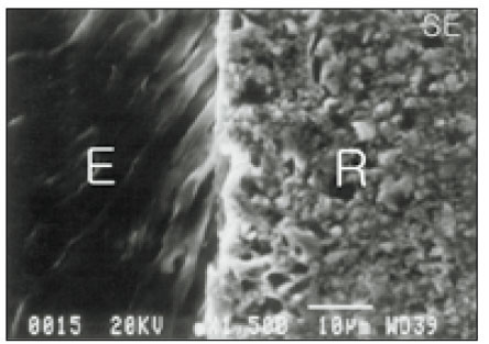


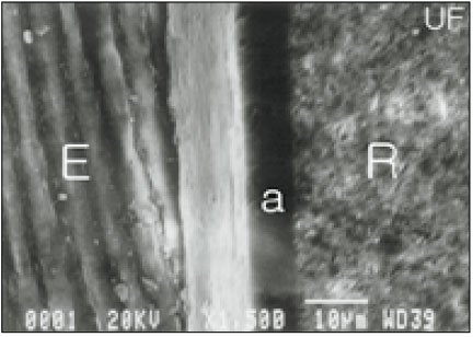
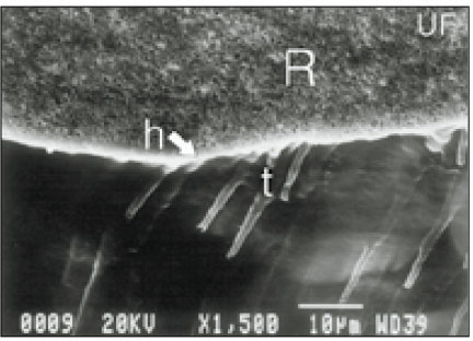

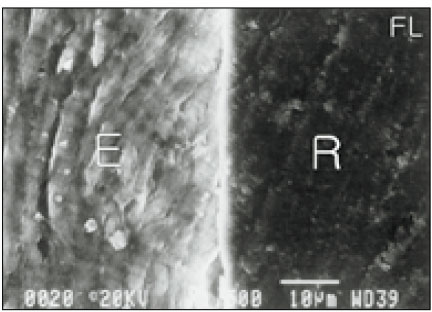
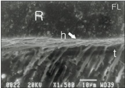


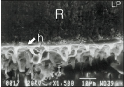

Fig. 1
Fig. 2
Fig. 3
Fig. 4
Fig. 5
Fig. 6
Fig. 7
Fig. 8
Fig. 9
Fig. 10
Fig. 11
Fig. 12
Group classification of three self-etching primer adhesives and one self-etching adhesive
Chemical formulations of four adhesive systems
MDP: 10-methacryloyloxydecyl dihydrogen phosphate,
HEMA: 2-hydroxyethylmethacylate, 4-MET: 4-methacryethyl trimettalic acid,
MFM: multi-functional methacrylate, 4-AET:4-acryloxyethyltrimellitic acid,
4-AETA: 4-acryloxyethyltrimellitate anhydride
Hybrid layer thickness (HLT), resin tags length (RTL), resin tags diameter (RTD) and resin tags shape (RTS) of the tested adhesives
*B: Base diameter of resin tags, E: End diameter of resin tags
MDP: 10-methacryloyloxydecyl dihydrogen phosphate, HEMA: 2-hydroxyethylmethacylate, 4-MET: 4-methacryethyl trimettalic acid, MFM: multi-functional methacrylate, 4-AET:4-acryloxyethyltrimellitic acid, 4-AETA: 4-acryloxyethyltrimellitate anhydride
*B: Base diameter of resin tags, E: End diameter of resin tags

 KACD
KACD


 ePub Link
 Cite
Cite

