Articles
- Page Path
- HOME > Restor Dent Endod > Volume 27(1); 2002 > Article
- Original Article Color changes in composites according to various light curing sources
- Young-Gon Cho, Myung-Cho Kim
-
2002;27(1):-94.
DOI: https://doi.org/10.5395/JKACD.2002.27.1.087
Published online: January 31, 2002
Department of Conservative Dentistry, College of dentistry, Chosun University, Korea.
Copyright © 2002 Korean Academy of Conservative Dentistry
- 958 Views
- 2 Download
- 3 Crossref
Abstract
-
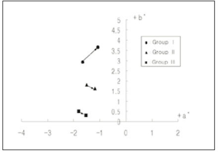
The purpose of this study was to evaluate the color changes of composite resin polymerized with three type of light curing units. Composite resin (Z100, shade A2) were applied in a cylindrical metal mold(2 mm thick, 7 mm diameter).Twenty specimens according to light curing units were made.Group1: the specimens were polymerized with Apollo 95E for 3seconds(1370 mW/cm2).Group2: the specimens were polymerized with XL 3000 for 40seconds(480 mW/cm2).Group3: the specimens were polymerized with Spectrum 800 for 10 seconds(250 mW/cm2) and 30 seconds(700 mW/cm2).The microhardness values(VHN) of upper and lower surfaces specimens after light polymerization were measured for the degree of polymerization. All specimens were stored in distilled water at 60℃ for 30 days.The color characteristics(L*, a*, b*) of the specimens before and after immersion were measured by spectrophotometer and the total color difference (ΔE*) was computed.The results obtained were as follows:1. The microhardness values of Group I showed significantly lower than those of Group II and III(p<0.05).2. In all groups the ΔE* values presented below 2.0.3. Group I showed the highest ΔE* values followed order from highest to lowest by Group II and III (p<0.05).
- 1. Ruyter IE. Conversion in different depths of ultraviolet and visible light activated composite materials. Acta Odontol Scand. 1982;40: 179-182.ArticlePubMed
- 2. Shintani H, Inoue T, Yamaki M. Analysis of camphoroquinone invisible light cured composite resins. Dent Mater. 1985;1: 124-126.PubMed
- 3. Leung R, Fan P, Johnson W. Post-irradiation polymerization of visible light activated composite resin. J Dent Res. 1983;62: 363-365.ArticlePDF
- 4. Friedman J. Care and maintenance of dental curing light. Dent Today. 1991;10: 40-41.
- 5. Carvalho RM, Pereira JC, Yoshiyama M, Pashley DH. A review of polymerization contraction: the incidence of stress development versus stress relief. Oper Dent. 1996;21(1):17-24.PubMed
- 6. Uno S, Asmussen E. Marginal adaptation of restorative resin polymerized at a reduced rate. Scand J Dent Res. 1991;99: 440-444.PubMed
- 7. Unterbrink GL, Muessner R. Influence of light intensity on two restorative systems. J Dent. 1995;23(3):183-189.ArticlePubMed
- 8. Mehl A, Hickel R, Kunzelmann KH. Physical properties and gap formation of light-cured composites with and without soft start-polymerization. J Dent. 1997;25(3):321-330.ArticlePubMed
- 9. Koran P, Kurschner R. Effect of sequential versus continuous irradiation of a light-cured resin composite on shrinkage, viscosity, adhesion and degree of polymerization. Am J Dent. 1998;11(1):17-22.PubMed
- 10. Peutzfeldt A, Sahafi A, Asmussen E. Characterization of resin composites polymerized with plasma arc curing units. Dent Mater. 2000;16(5):330-336.ArticlePubMed
- 11. Hanyang University Department of Physics. Plasma Application. 2000;Laboratory.
- 12. Eldiwany M, Komatsu S, Powers JM. Curing light intensityaffects mechanicalproperties of composites. J Dent Res. 1997;76: 73.
- 13. Caughman WF, Caughman GB, Shiflett RA, et al. Correlation of cytotoxicity, filler loading and curing time of dental composites. Biomaterials. 1991;12: 737-740.ArticlePubMed
- 14. Seghi RR, Gritz MD, Kim J. Colorimetric changes in composites resulting fromvisible-light-initiated polymerization. Dent Mater. 1990;6: 133-137.ArticlePubMed
- 15. Brauer GM. Color changes of composites on exposure to various energy sources. Dent Mater. 1988;4: 55-59.ArticlePubMed
- 16. Powers JM, Barakat MM, Ogura H. Color and optical properties of posteriorcomposites resin restoration. Dent Mater J. 1985;4: 62-67.PubMed
- 17. Noie F, O'Keeefe KL, Powers JM. Color stability of resin cements after accelerated aging. Int J Prosthodont. 1995;8: 51-55.PubMed
- 18. Swift EJ, Hammel SA, Lund PS. Colorimetric evaluation of vita shade resin composites. Int J Prosthodont. 1994;7: 356-361.PubMed
- 19. Asmussen E. An accelerated test for color stability of restorative resins. Acta Odontol Scand. 1981;39: 329-332.Article
- 20. Roh BD, Park SH, Lee CS. An experimental study of the degree of conversion and cytotoxicity of dual cure resin cements. J Korean Acad Conserv Dent. 1995;20(1):33-33.
- 21. DeWald JP, Ferracane JL. A comparison of four modes of evaluation depth of cure of light-activated composites. J Dent Res. 1982;66: 727-730.ArticlePDF
- 22. Vargas MA, Cobb DS, Schmit JL. Polymerization of composite resins:Argon laser vs conventional light. Oper Dent. 1998;23: 87-93.PubMed
- 23. Sakaguchi RL, Sasik CT, Bunczak MA, Douglas WH. Strain gauge method for mearsuring polymerization contraction of composite restoratives. J Dent. 1991;19(5):312-326.PubMed
- 24. Goracci G, et al. Curing light intensity and marginal leakage of resin composite restorations. Quintessence Int. 1996;27(5):355-362.
- 25. Martin FE. A survey of the efficiency of visible light curing units. J Dent. 1998;26(3):239-243.ArticlePubMed
- 26. Rueggeberg FA, Craig RC. Correlation of parameters used to estimate monomer conversion in a lightcured composite. J Dent Res. 1988;67: 932-937.ArticlePubMedPDF
- 27. Asmussen E. Restorative resins:hardness and strengthvsquality of remaining double bonds. Scand J Dent Res. 1982;90: 484-489.PubMed
- 28. Hansen EK. After-polymerization of visible light activated resins ; surface hardness vs light source. Scand J Dent Res. 1983;91: 406-410.ArticlePubMed
- 29. Backer J, Dermaut L, Bruynooghe W. The depth of polymerization of visible light-cured composite resins. Quintessence Int. 1985;10: 693-699.
- 30. Leung RL, Kahn RL, Fan PL. Comparison of depth of polymerization evaluation method for photoactivated composite. J Dent Res. 1982;61: 300. IADR Abstract # 1095.
- 31. Munksgaard EC, Peutzfeldt A, Asmussen E. Elution of TEGDMA and BisGMA from a resin and a resin composite cured with halogen or plasma light. Eur J Oral Sci. 2000;108(4):341-345.ArticlePubMedPDF
- 32. Silikas N, Eliades G, Watts DC. Light intensity effects on resin-composite degree of conversion and shrinkage strain. Dent Mater. 2000;16(4):292-296.ArticlePubMed
- 33. Asmussen E. Factor affecting the color stability of restorative resins. Acta Odontol Scand. 1983;41: 11-18.PubMed
- 34. Um CM, Ruyter IE. Staining of resin-based veneering materials with coffee and tea. Quintessence Int. 1991;22: 377-386.PubMed
- 35. Ruyter IE, Svendsen SA. Remaining methacrylate groups in composite restorative materials. Acta Odontol Scand. 1978;36: 75-82.ArticlePubMed
- 36. Hayshi H, Maejima K, Kezuka K, Ogushi K, Kono A, Fusayama TT. In vitro study of discoloration of composite resins. J Prosthet Dent. 1974;32: 66-69.ArticlePubMed
- 37. Dodge WW, Dale RA, Colley RL, Duke ES. Comparison of wet and dry finishing of resin composites with aluminium oxide discs. Dent Mater. 1991;7(1):18-20.PubMed
- 38. Han WS, Kum KY, Lee CY. The influence of fluoride on remineralization of artificial dental caries. J Korean Dent Assoc. 1977;15: 1009-1012.
- 39. Gross MD, Moser JB. A colorimetric study ofcoffeeandtea staining of four composite resins. J Oral Rehabil. 1977;4: 311-322.ArticlePubMed
- 40. Satou N, Khan AM, Mastsumae I, Satou J, Shintani H. In vitro color change of composite-based resins. Dent Mater. 1989;5: 384-389.ArticlePubMed
- 41. Wozniak WT, Muller TP, Silverman R, Moser JB. Photographic assessment of colour changes in cold and heat-cure resins. J Oral Rehabil. 1981;8: 333-339.ArticlePubMed
- 42. Raptis CN, Powers JM, Fan PL, Yu R. Staining of composite resins by cigarette smoke. J Oral Rehabil. 1982;9: 367-371.ArticlePubMed
- 43. Ruyter IE, Nilner K, Moller B. Color stability of dental composite resin materials for crown and bridge veneers. Dent Mater. 1987;13: 246-251.Article
- 44. Seghi RR, Jonston WM, O'Brien WJ. Performance assessment of colorimetric devices on dental porcelain. J Dent Res. 1989;68: 1755-1759.ArticlePubMedPDF
- 45. Dijken JWV. A clinical evaluation of anterior conventional microfilled and hybrid composite resin filling. Acta Odontol Scand. 1986;44: 357.PubMed
- 46. Kim CW, Lim BS, Moon HJ. Effect of Organic Solutions on the Surface Roughness and Color Stability of Dental Composite Resins. J Korea Res Soc Dent Mater. 1999;26(1):17-34.
- 47. Dietchi D, Campanile G, Holz J, Meyer JM. Comparison of the color stability of ten new-generation composites : An in vitro study. Dent Mater. 1994;10: 353-362.ArticlePubMed
REFERENCES
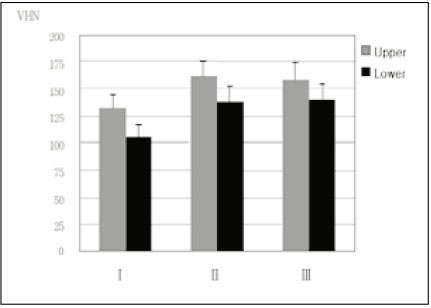
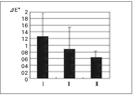
I: Apollo 95E , II: XL 3000, III: Spectrum 800
*: Statistically significant difference between groups (p < 0.05)

ΔL*, Δa*, Δb*, ΔC*: color difference, ΔE*: total color difference.
I: Apollo 95E, II: XL 3000, III: Spectrum 800
Standard deviations are in parentheses. *: significant differences (p<0.05)

Tables & Figures
REFERENCES
Citations

- Effects of the color components of light-cured composite resin before and after polymerization on degree of conversion and flexural strength
Ji-A Yoo, Byeong-Hoon Cho
Journal of Korean Academy of Conservative Dentistry.2011; 36(4): 324. CrossRef - Effect of the difference in spectral outputs of the single and dual-peak LEDs on the microhardness and the color stability of resin composites
Hye-Jung Park, Sung-Ae Son, Bock Hur, Hyeon-Cheol Kim, Yong-Hoon Kwon, Jeong-Kil Park
Journal of Korean Academy of Conservative Dentistry.2011; 36(2): 108. CrossRef - Color changes in composite resins exposed to xenon lamp
Young-Gon Cho, Jeong-Il Seo, Soo-Mee Kim, Jin-Ho Jeong, Young-Gon Lee
Journal of Korean Academy of Conservative Dentistry.2003; 28(3): 195. CrossRef
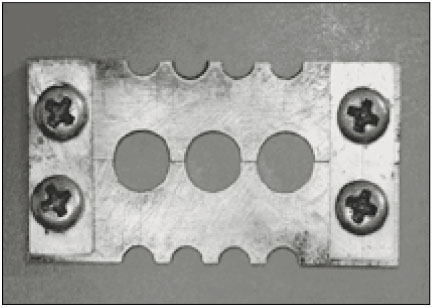
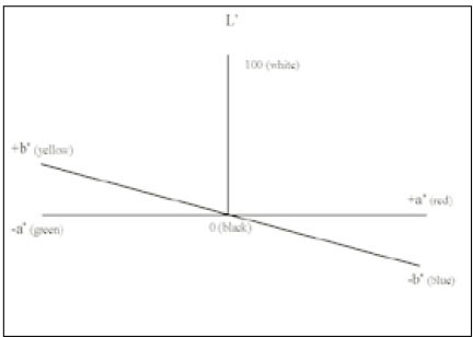


Fig. 1
Fig. 2
Fig. 3
Fig. 4
Fig. 5
Light curing units used in this study
Microhardness values(VHN) of upper surfaces and lower surfaces of each group(Mean±SD)
I: Apollo 95E , II: XL 3000, III: Spectrum 800
*: Statistically significant difference between groups (p < 0.05)
Result of color changes of group I, II and III after storing for 30 days in distilled water at 60℃ expressed as means
ΔL*, Δa*, Δb*, ΔC*: color difference, ΔE*: total color difference.
I: Apollo 95E, II: XL 3000, III: Spectrum 800
Standard deviations are in parentheses. *: significant differences (p<0.05)
I: Apollo 95E , II: XL 3000, III: Spectrum 800 *: Statistically significant difference between groups (p < 0.05)
ΔL*, Δa*, Δb*, ΔC*: color difference, ΔE*: total color difference. I: Apollo 95E, II: XL 3000, III: Spectrum 800 Standard deviations are in parentheses. *: significant differences (p<0.05)

 KACD
KACD


 ePub Link
 Cite
Cite

