Articles
- Page Path
- HOME > Restor Dent Endod > Volume 27(2); 2002 > Article
- Original Article Comparative study of digital and conventional radiography for the diagnostic ability of artificial proximal surface caries
- Young-Gon Cho, Si-Seung Park
-
2002;27(2):-121.
DOI: https://doi.org/10.5395/JKACD.2002.27.2.113
Published online: March 31, 2002
Department of Conservative Dentistry, College of dentistry, Chosun University, Korea.
Copyright © 2002 Korean Academy of Conservative Dentistry
- 1,955 Views
- 35 Download
Abstract
-
Conventional intraoral radiography continues to be the most widely used image modality for the diagnosis of dental caries. But, conventional intraoral radiography has several shortcomings, including the difficulty of exposing and processing intraoral film of consistently acceptable quality. In addition, radiographic retaking that was the result of processing errors, may result in increased discomfort and radiation dose to the patient.Recently, various digital radiographies substitute for conventional intraoral radiography to overcome these disadvantages. The advantages of digital radiography are numerous. One of advantages is the elimination of processing errors. In addition, the radiation dose for digital system is approximately 20% to 25% of that required for conventional intraoral radiography. Another potential advantage of digital imaging is the ability to perform image quality enhancements such as contrast and density modulation, which may increase diagnostic accuracy.The purpose of this study was to compare the diagnostic ability of artificial proximal defects to conventional intraoral radiography, direct digital image(CDX2000HQ®) and indirect digital image(Digora®).Artificial defects were made in proximal surfaces of 60 extracted human molars using #1/2, #1, #2 round bur. Five dentists assessed proximal defects on conventional intraoral radiography, direct digital image(CDX2000HQ®) and indirect digital image(Digora®). ROC(Receiver Operating Characteristic) analysis and Two-way ANOVA test were used for the evaluation of detectability, and following results were acquired.1. The mean ROC area of conventional intraoral radiography, direct digital image(CDX2000HQ®)and indirect digital image(Digora®) were 0.6766, 0.7538, 0.6791(Grade I), 0.7176, 0.7594, 0.7361(Grade II), and 0.7449, 0.7608, 0.7414(Grade III), respectively.2. Diagnostic ability of direct digital image was higher than other image modalities. But, there was no statistically significant difference among other imaging modalities for Grade I, II, III lesion(p>0.05).In conclusion, when direct and indirect digital system are comparable with conventional intraoral radiography, these systems may be considered an alternative of conventional intraoral radiography for the diagnosis of proximal surface caries.
- 1. Benz C, Mouyen F. Evaluation of the new RadioVisioGraphy system image quality. Oral Surg Oral Med Oral Pathol. 1991;72: 627-631.ArticlePubMed
- 2. Marthaler TM. Improvement of diagnostic methods in clinical caries trials. J Dent Res. 1984;63(Spec. Iss):746-750.PubMed
- 3. Eggertsson H, et al. Detection of Early interproximal caries in vitro using laser fluorescence, dye-enhanced laser fluorescence and direct visual examination. Caries Res. 1999;33: 227-233.ArticlePubMedPDF
- 4. Lee YS. Radiolographic densities diagnosis of periapical lesions using image enhancement techniques. 1993;Soongsil University, Department of Information Technology; 1-73 master's degree.
- 5. Furkart AJ, Dove SB, McDavid WD, et al. Direct digital radiography for the detection of periodontal bone lesions. Oral Surg Oral Med Oral Pathol. 1992;74: 652-660.ArticlePubMed
- 6. Wenzel A, Fejerskov O, Kidd E, et al. Depth of occlusal caries assessed clinically, by conventional film radiographs, and by digitized, processed radiographs. Caries Res. 1990;24: 327-333.ArticlePubMed
- 7. Kwon KJ, Hwang EH, Lee SR. A Study on the Artificial Interproximal Caries Detection with the Digital Radiography. J Korean Acad Oral Maxillofac Radiol. 1994;24: 85-94.
- 8. Cho HH, Kim EK. Experimental Study on Quantitative Evaluation of Film-based Digital Imaging System. J Korean Acad Oral Maxillofac Radiol. 1994;24: 137-150.
- 9. Welander U, et al. Resolution as defined by line spread and modulation transfer function four digital intraoral radiographic systems. Oral Surg Oral Med Oral Pathol. 1994;78: 109-115.PubMed
- 10. Oh KR, Choi EH, Kim JD. A Study on the Diagnostic Detection Ability of the Artificial Proximal Caries by Digora®. J Korean Acad Oral Maxillofac Radiol. 1998;28(2):415-433.
- 11. Gotfredsen E, Wenzel A, Grondahl HG. Observers' use of image enhancement in assessing caries in radiographs taken by four intraoral digital system. Dentomaxillofac Radiol. 1996;25: 34-38.PubMed
- 12. Pitts NB. Detection of approximal radiolucencies in enamel : A preliminary comparison between experienced clinicians and an image analysis method. J Dent. 1987;15: 191-197.ArticlePubMed
- 13. Kassebaum DK, Mcdavid WD, Dove SB, Waggener RG. Spatial resolution requirements for digitizing dental radiographs. Oral Surg Oral Med Oral Pathol. 1989;67: 760-769.ArticlePubMed
- 14. Fujita M, Kodera Y, Okawa M, Wada T, Doi K. Digital image processing of periapical radiographs. Oral Surg. 1988;65: 490-449.ArticlePubMed
- 15. White SC, Yoon DC. Comparative performance of digital and conventional images for detecting proximal surface caries. Dentomaxillofac Radiol. 1997;26: 32-38.ArticlePubMed
- 16. Swets JA. The relative operating characteristic in psychology. Science. 1973;182: 990-1000.ArticlePubMed
- 17. Wenzel A. Effect of image enhancement for detectability of bone lesion in digitized intraoral radiographs. Scand J Dent Res. 1988;96: 149-160.PubMed
- 18. Do JJ, Kim EK. Comparative Study of Direct Digital Radiographic System with Film-based Digital Imaging System Using Ektaspeed and Ektaspeed Plus Film. J Korean Acad Oral Maxillofac Radiol. 1995;25(1):51-66.
- 19. Lee K, Lee SR. A Study on the Readability of Periapical Radiograph with the Digital Radiography. J Korean Acad Oral Maxillofac Radiol. 1992;22: 117-127.
- 20. Farman AG, Scafe WC. Pixel perception and voxel vision: constructs for a new paradigm in maxillofacial imaging. Dentomaxillofac Radiol. 1994;23: 5-9.ArticlePubMed
- 21. McDonnell D, Price C. An evaluation of the Sens-A-Ray digital dental imaging system. Dentomaxillofac Radiol. 1993;2: 121-126.Article
- 22. Molteni R. Direct digital dental x-ray imaging with Visualix/Vixa. Oral Surg Oral Med Oral Pathol. 1993;76: 235-243.ArticlePubMed
- 23. Webber RL. Computers in dental radiography : A scenario for the future. J Am Dent Assoc. 1985;111: 419-424.ArticlePubMed
- 24. Welander U, Nelvig P, Tronje G, McDavid WD, Dove SB, Morner AC, Cederlund T. Basic technical properties of a system for direct acquisition of digital intraoral radiographs. Oral Surg Oral Med Oral Pathol. 1993;75: 506-516.ArticlePubMed
- 25. Wenzel A. New Caries Disgnostic Methods. J Dent Educ. 1993;57: 428-432.PubMed
- 26. Park KS, Lee SR. Digital Image Processing and Clinical Application of Videodensitometer. J Korean Acad Oral Maxillofac Radiol. 1992;22: 273-282.
- 27. Wenzel A. Tandlegebladt. Digital billedbehandling. Danish Dent J. 1991;95: 1-33.
- 28. Ado S, Nishioka T, Ozawa M, et al. Computer analysis of radiographic image. J Nihon Univ School Dent. 1968;10: 65-70.
- 29. Dove SB, Mcdavid WD, Tronje G, Wilcox CD. Design an implementation of an image management and communications system(IMACS) for dentomaxillofacial radiology. Dentomaxillofac Radiol. 1992;21: 216-221.PubMed
- 30. Ohki M, Okano T, Tamada N. A contrast correction method for digital subtraction radiography. J periodontal Res. 1988;23: 277-280.ArticlePubMed
- 31. Svanaes DB, Moystad A, Risnes S, Larheim TA, Gröndahl HG. Intraoral storage phosphor radiography for approximal caries detection and effect of image magnification comparison with conventional radiography. Oral Surg Oral Med Oral Pathol Oral Radiol Endod. 1996;82: 94-100.PubMed
- 32. Lusted LB. Logical analysis in roentgen diagnosis. Radiology. 1960;74: 178-193.ArticlePubMed
- 33. Douglass CW, McNeil BJ. Clinical decision analysis methods applied to diagnostic tests in dentistry. J Dent Educ. 1983;47: 708-712.ArticlePubMedPDF
- 34. Verdonschot EH, Kuijpers JMC, Polder BJ, et al. Effects of digital grey-scale modification on the diagnosis of small approximal carious lesion. J Dent. 1992;20: 44-49.PubMed
- 35. Verdonschot EH, Wenzel A, Bronkhorst EM. Applicability of Receiver Operating Charateristic (ROC) analysis on discrete caries depth ratings. Community Dent Oral Epidemiol. 1993;21: 269-272.PubMed
- 36. Wenzel A. Sensor noise in indirect digital imaging(the RadioVisioGraphy, Sense-A-Ray, and Visualix/vixa system) evaluated by subtraction radiography. Oral Surg Oral Med Oral Pathol. 1994;77: 70-74.PubMed
- 37. Benn DK. Radiographic caries diagnosis and monitoring : Review. Dentomaxillofac Radiol. 1994;23: 69-72.PubMed
- 38. Verdonschot EH, Wenzel A, Bronkhorst EM. Assessment of diagnostic accuracy in caries detection : An analysis of two methods. Community Dent Oral Epidemiol. 1993;21: 203-208.ArticlePubMed
- 39. Pitts NB. Detection and measurement of approximal radiolucencies by computer-aided image analysis. Oral Surg Oral Med Oral Pathol. 1984;58: 358-366.ArticlePubMed
- 40. Mol A, Van der Stelt PF. Locating the periapical legion in dental radiographs using digital image analysis. Oral Surg Oral Med Oral Pathol. 1993;75: 373-382.PubMed
REFERENCES
Fig. 2
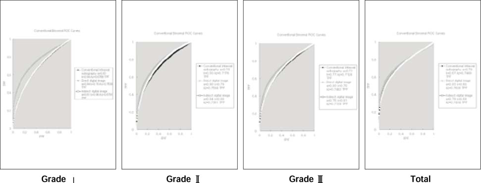
Receiver Operating Characteristic (ROC) curves obtained by five observers for detection of artificial proximal defects (Grade I,II,III) with three imaging modalities(conventional intraoral film, direct & indirect digital image).

Fig. 5
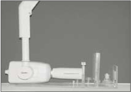
Geometrically standardized experiemental design. A: tube head of Gendex introral X-ray unit, B: acrylic resin plate to hole block of tooth, C: 2cm thick acrylic block simulating the soft tissue.

Fig. 6

Each image of same tooth in conventional intraoral radiograph (A), direct digital image (CDX2000HQ®) (B), indrectdigitalimage(Digora®) (C).

Table 2
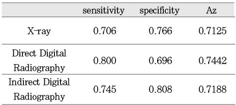
The mean values of sensitivity, specificity and area under ROC curve(Az) according to image modality.

Tables & Figures
REFERENCES
Citations
Citations to this article as recorded by 

Comparative study of digital and conventional radiography for the diagnostic ability of artificial proximal surface caries
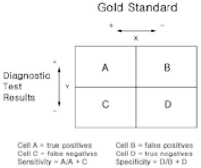

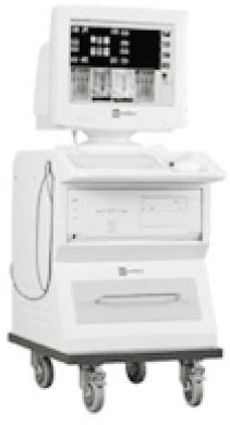
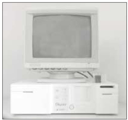


Fig. 1
Contingency table for interpretation of diagnostic tests.
Fig. 2
Receiver Operating Characteristic (ROC) curves obtained by five observers for detection of artificial proximal defects (Grade I,II,III) with three imaging modalities(conventional intraoral film, direct & indirect digital image).
Fig. 3
CDX2000HQ®: Direct digital image system
Fig. 4
Digora®: Indirect digital image system
Fig. 5
Geometrically standardized experiemental design. A: tube head of Gendex introral X-ray unit, B: acrylic resin plate to hole block of tooth, C: 2cm thick acrylic block simulating the soft tissue.
Fig. 6
Each image of same tooth in conventional intraoral radiograph (A), direct digital image (CDX2000HQ®) (B), indrectdigitalimage(Digora®) (C).
Fig. 1
Fig. 2
Fig. 3
Fig. 4
Fig. 5
Fig. 6
Comparative study of digital and conventional radiography for the diagnostic ability of artificial proximal surface caries
Classification of grade according to the depth of artificial defect.
The mean values of sensitivity, specificity and area under ROC curve(Az) according to image modality.
The mean values of the sensitivity and specificity according to the depth of artificial defect.
The mean values of area under ROC curve (Az) according to the depth of artificial defect.
* : statistically significant difference(p<0.05) by Two-way ANOVA test
Table 1
Classification of grade according to the depth of artificial defect.
Table 2
The mean values of sensitivity, specificity and area under ROC curve(Az) according to image modality.
Table 3
The mean values of the sensitivity and specificity according to the depth of artificial defect.
Table 4
The mean values of area under ROC curve (Az) according to the depth of artificial defect.
* : statistically significant difference(p<0.05) by Two-way ANOVA test

 KACD
KACD


 ePub Link
 Cite
Cite

