Articles
- Page Path
- HOME > Restor Dent Endod > Volume 37(3); 2012 > Article
- Research Article Minimizing the extra-oral time in autogeneous tooth transplantation: use of computer-aided rapid prototyping (CARP) as a duplicate model tooth
- Seung-Jong Lee, Euiseong Kim
-
2012;37(3):-141.
DOI: https://doi.org/10.5395/rde.2012.37.3.136
Published online: August 29, 2012
Microscope Center, Department of Conservative Dentistry and Oral Science Research Center, Yonsei University College of Dentistry, Seoul, Korea.
- Correspondence to Euiseong Kim, DDS, MSD, PhD. Professor, Microscope Center, Department of Conservative Dentisty, Yonsei University College of Dentistry, 50 Yonsei-ro, Seodaemun-gu, Seoul, Korea 120-752. TEL, +82-2-2228-8701; FAX, +82-2-313-7575; andyendo@yuhs.ac
©Copyights 2012. The Korean Academy of Conservative Dentistry.
This is an Open Access article distributed under the terms of the Creative Commons Attribution Non-Commercial License (http://creativecommons.org/licenses/by-nc/3.0/) which permits unrestricted non-commercial use, distribution, and reproduction in any medium, provided the original work is properly cited.
- 1,682 Views
- 10 Download
- 42 Crossref
Abstract
-
Objectives The maintenance of the healthy periodontal ligament cells of the root surface of donor tooth and intimate surface contact between the donor tooth and the recipient bone are the key factors for successful tooth transplantation. In order to achieve these purposes, a duplicated donor tooth model can be utilized to reduce the extra-oral time using the computer-aided rapid prototyping (CARP) technique.
-
Materials and Methods Briefly, a three-dimensional digital imaging and communication in medicine (DICOM) image with the real dimensions of the donor tooth was obtained from a computed tomography (CT), and a life-sized resin tooth model was fabricated. Dimensional errors between real tooth, 3D CT image model and CARP model were calculated. And extra-oral time was recorded during the autotransplantation of the teeth.
-
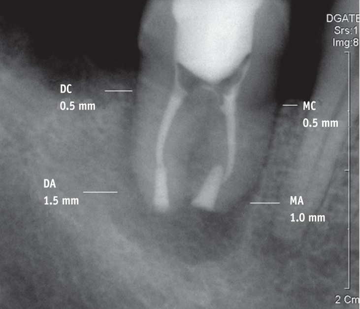
Results The average extra-oral time was 7 min 25 sec with the range of immediate to 25 min in cases which extra-oral root canal treatments were not performed while it was 9 min 15 sec when extra-oral root canal treatments were performed. The average radiographic distance between the root surface and the alveolar bone was 1.17 mm and 1.35 mm at mesial cervix and apex; they were 0.98 mm and 1.26 mm at the distal cervix and apex. When the dimensional errors between real tooth, 3D CT image model and CARP model were measured in cadavers, the average of absolute error was 0.291 mm between real teeth and CARP model.
-
Conclusions These data indicate that CARP may be of value in minimizing the extra-oral time and the gap between the donor tooth and the recipient alveolar bone in tooth transplantation.
Introduction
Materials and Methods
Case collection
Surgical Methods
1. Pre-surgical procedures
b. Fabrication of the donor tooth model
c. Practice on the recipient jaw model
2. Surgical procedures
3. Radiographic measurements of the average distance between the transplanted root surface and the alveolar bone
4. Average extra-oral time
Results
1. Measurements of the average distance between the transplanted root surface and the alveolar bone
2. Extra-oral time
Discussion
Conclusions
-
This research was supported by Basic Science Research Program through the National Research Foundation of Korea (NRF) funded by the Ministry of Education, Science and Technology (2010-0021281).
-
No potential conflict of interest relevant to this article was reported.
- 1. Lee SJ, Jung IY, Lee CY, Choi SY, Kum KY. Clinical application of computer-aided rapid prototyping for tooth transplantation. Dent Traumatol 2001;17:114-119.ArticlePubMedPDF
- 2. Andreasen JO. Interrelation between alveolar bon and periodontal ligament repair after replantation of mature permanent incisors in monkeys. J Periodontal Res 1981;16:228-235.PubMed
- 3. Hupp JG, Mesaros SV, Aukhil I, Trope M. Periodontal ligament vitality and histologic healing of teeth stored for extended periods before transplantation. Endod Dent Traumatol 1998;14:79-83.ArticlePubMed
- 4. Andreasen JO. Periodontal healing after replantation and autotransplantation of incisors in monkeys. Int J Oral Surg 1981;10:54-61.ArticlePubMed
- 5. Choi JY, Choi JH, Kim NK, Kim Y, Lee JK, Kim MK, Lee JH, Kim MJ. Analysis of errors in medical rapid prototyping models. Int J Oral Maxillofac Surg 2002;31:23-32.ArticlePubMed
- 6. Lill W, Solar P, Ulm C, Watzek G, Blahout R, Matejka M. Reproducibility of three-dimensional CT-assisted model production in the maxillofacial area. Br J Oral Maxillofac Surg 1992;30:233-236.ArticlePubMed
- 7. Lee SJ, Kim E, Kim KD, Lee SJ. In vitro study for Accuracy of computer aided rapid prototyping model(CARP model) compared with real donor tooth in autogenous tooth transplantation. J Korean Dent Assoc 2006;44:115-122.
- 8. Kim E, Jung JY, Cha IH, Kum KY, Lee SJ. Evaluation of the prognosis and causes of failure in 182 cases of autogenous tooth transplantation. Oral Surg Oral Med Oral Pathol Oral Radiol Endod 2005;100:112-119.PubMed
- 9. Sobhi MB, Rana MJ, Manzoor MA, Ibrahim M, Tasleemul-Hudda . Autotransplantation of endodontically treated third molars. J Coll Physicians Surg Pak 2003;13:372-374.PubMed
- 10. Mejàre B, Wannfors K, Jansson L. A prospective study on transplantation of third molars with complete root formation. Oral Surg Oral Med Oral Pathol Oral Radiol Endod 2004;97:231-238.ArticlePubMed
- 11. Cohen AS, Shen TC, Pogrel MA. Transplanting teeth successfully: autografts and allografts that work. J Am Dent Assoc 1995;126:481-485.ArticlePubMed
- 12. Schwartz O, Bergmann P, Klausen B. Autotransplantation of human teeth. A life-table analysis of prognostic factors. Int J Oral Surg 1985;14:245-258.PubMed
- 13. Nethander G, Andersson JE, Hirsch JM. Autogenous free tooth transplantation in man by a 2-stage operation technique. A longitudinal intra-individual radiographic assessment. Int J Oral Maxillofac Surg 1988;17:330-336.ArticlePubMed
- 14. Andreasen JO, Paulsen HU, Yu Z, Schwartz O. A long-term study of 370 autotransplanted premolars. Part III. Periodontal healing subsequent to transplantation. Eur J Orthod 1990;12:25-37.ArticlePubMed
- 15. Van Hassel HJ, Oswald RJ, Harrington GW. Replantation 2. The role of the periodontal ligament. J Endod 1980;6:506-508.ArticlePubMed
- 16. Andreasen JO. In: Gutmann JL, Harrison JW, editors. Root fractures, luxation and avulsion injuries-diagnosis and management. Proceedings of the International Conference on Oral Trauma. 1986;Chicago: American Association of Endodontics; p. 79-92.
- 17. Hammarström L, Blomlöf L, Lindskog S. Dynamics of dentoalveolar ankylosis and associated root resorption. Endod Dent Traumatol 1989;5:163-175.ArticlePubMed
- 18. Andreasen JO, Kristerson L. The effect of extra-alveolar root filling with calcium hydroxide on periodontal healing after replantation of permanent incisors in monkeys. J Endod 1981;7:349-354.ArticlePubMed
- 19. Andreasen JO, Andreasen FM. Textbook and color atlas of traumatic injuries to the teeth. 1994. 3rd ed. St. Louis: Mosby Co; p. 18.
- 20. Goerig AC, Nagy WW. Successful intentional reimplantation of mandibular molars. Quintessence Int 1988;19:585-588.PubMed
- 21. Nethander G. Periodontal conditions of teeth autogenously transplanted by a two-stage technique. J Periodontal Res 1994;29:250-258.ArticlePubMed
REFERENCES


Tables & Figures
REFERENCES
Citations

- American Dental Association and American Academy of Oral and Maxillofacial Radiology patient selection for dental radiography and cone-beam computed tomography
Erika Benavides, Joseph R. Krecioch, Trishul Allareddy, Allison Buchanan, Martha Ann Keels, Ana Karina Mascarenhas, Mai-Ly Duong, Kelly K. O’Brien, Kathleen M. Ziegler, Ruth D. Lipman, Roger T. Connolly, Lucia Cevidanes, Kitrina Cordell, Satheesh Elangova
The Journal of the American Dental Association.2026; 157(1): 20. CrossRef - American Dental Association and American Academy of Oral and Maxillofacial Radiology patient selection for dental radiography and cone-beam computed tomography
Erika Benavides, Josep R. Krecioch, Trishul Allareddy, Allison Buchanan, Martha Ann Keels, Ana Karina Mascarenhas, Mai-Ly Duong, Kelly K. O'Brien, Kathleen M. Ziegler, Ruth D. Lipman, Roger T. Connolly, Lucia Cevidanes, Kitrina Cordell, Satheesh Elangovan
Oral Surgery, Oral Medicine, Oral Pathology and Oral Radiology.2026;[Epub] CrossRef - 13-year follow-up of autotransplantation using an immature third molar: a case report
Hojin Moon
Journal of Dental Rehabilitation and Applied Science.2025; 41(1): 72. CrossRef - Three-Dimensional Printing in Dentistry: A Scoping Review of Clinical Applications, Advantages, and Current Limitations
Mi-Kyoung Jun, Jong-Woo Kim, Hye-Min Ku
Oral.2025; 5(2): 24. CrossRef - Template-Guided Autogenous Tooth Transplantation Using a CAD/CAM Dental Replica in a Complex Anatomical Scenario: A Case Report
Michael Alfertshofer, Florian Gebhart, Dirk Nolte
Dentistry Journal.2025; 13(7): 281. CrossRef - Autogenous Transplantation of Teeth Across Clinical Indications: A Systematic Review and Meta-Analysis
Martin Baxmann, Karin Christine Huth, Krisztina Kárpáti, Zoltán Baráth
Journal of Clinical Medicine.2025; 14(14): 5126. CrossRef - Effect of 3D printed replicas on the duration of third molar autotransplantation surgery: A controlled clinical trial
Miks Lejnieks, Ilze Akota, Gundega Jākobsone, Laura Neimane, Oskars Radzins, Sergio E. Uribe
Dental Traumatology.2024; 40(2): 221. CrossRef - Use of 3D printing models for donor tooth extraction in autotransplantation cases
Rui Hou, Xiaoyong Hui, Guangjie Xu, Yongqing Li, Xia Yang, Jie Xu, Yanli Liu, Minghui Zhu, Qinglin Zhu, Yu Sun
BMC Oral Health.2024;[Epub] CrossRef - Autologous Transplantation Tooth Guide Design Based on Deep Learning
Lifen Wei, Shuyang Wu, Zelun Huang, Yaxin Chen, Haoran Zheng, Liping Wang
Journal of Oral and Maxillofacial Surgery.2024; 82(3): 314. CrossRef - Anterior tooth autotransplantation: a case series
DC‐V Ong, P Goh, G Dance
Australian Dental Journal.2023; 68(3): 202. CrossRef - Dental Auto Transplantation Success Rate Increases by Utilizing 3D Replicas
Peter Kizek, Marcel Riznic, Branislav Borza, Lubos Chromy, Karolina Kamila Glinska, Zuzana Kotulicova, Jozef Jendruch, Radovan Hudak, Marek Schnitzer
Bioengineering.2023; 10(9): 1058. CrossRef - Planificación digital y guía de fresado para autotrasplante de tercer molar
Silvio Llanos, Henry García, Carlos Manresa, Carolina Bonilla, Alessandra Baasch
Reporte Imagenológico Dentomaxilofacial.2023;[Epub] CrossRef - Una alternativa a los implantes dentarios: manejo quirúrgico y endodóntico con planificación digital y guía de fresado de autotrasplantes de terceros molares. Reporte de un caso
Silvio Llanos, Henry García, Carlos Manresa, Carolina Bonilla, Julio Tebres, Stefanía Requejo, Alessandra Baasch
Latin American Journal of Oral and Maxillofacial Surgery.2023; 3(2): 80. CrossRef - Extraoral Root-End Resection May Promote Pulpal Revascularization in Autotransplanted Mature Teeth—A Retrospective Study
Petra Rugani, Barbara Kirnbauer, Irene Mischak, Kurt Ebeleseder, Norbert Jakse
Journal of Clinical Medicine.2022; 11(23): 7199. CrossRef - Three-Dimensional (3D) Stereolithographic Tooth Replicas Accuracy Evaluation: In Vitro Pilot Study for Dental Auto-Transplant Surgical Procedures
Filiberto Mastrangelo, Rossella Battaglia, Dario Natale, Raimondo Quaresima
Materials.2022; 15(7): 2378. CrossRef - Surgical Management of Impacted Lower Second Molars: A Comprehensive Review
Diane Isabel Selvido, Nattharin Wongsirichat, Pratanporn Arirachakaran, Dinesh Rokaya, Natthamet Wongsirichat
European Journal of Dentistry.2022; 16(03): 465. CrossRef - Application effect of computer-aided design combined with three-dimensional printing technology in autologous tooth transplantation: a retrospective cohort study
Shuang Han, Hui Wang, Jue Chen, Jihong Zhao, Haoyan Zhong
BMC Oral Health.2022;[Epub] CrossRef - Combined Application of Virtual Simulation Technology and 3-Dimensional-Printed Computer-Aided Rapid Prototyping in Autotransplantation of a Mature Third Molar
Hui Zhang, Min Cai, Zhiguo Liu, He Liu, Ya Shen, Xiangya Huang
Medicina.2022; 58(7): 953. CrossRef - Present status and future directions: Surgical extrusion, intentional replantation and tooth autotransplantation
Gianluca Plotino, Francesc Abella Sans, Monty S. Duggal, Nicola M. Grande, Gabriel Krastl, Venkateshbabu Nagendrababu, Gianluca Gambarini
International Endodontic Journal.2022; 55(S3): 827. CrossRef - Review on 3D printing in dentistry: conventional to personalized dental care
Shadaan Ahmad, Nazeer Hasan, Fauziya, Akash Gupta, Arif Nadaf, Lubna Ahmad, Mohd. Aqil, Prashant Kesharwani
Journal of Biomaterials Science, Polymer Edition.2022; 33(17): 2292. CrossRef - Three-dimensional printing in endodontics: A review of literature
Jyoti Chauhan, Ida de Noronha de Ataide, Marina Fernandes
IP Indian Journal of Conservative and Endodontics.2021; 6(4): 198. CrossRef - Pre- and peri-operative factors influence autogenous tooth transplantation healing in insufficient bone sites
Thanapon Suwanapong, Aurasa Waikakul, Kiatanant Boonsiriseth, Nisarat Ruangsawasdi
BMC Oral Health.2021;[Epub] CrossRef - European Society of Endodontology position statement: Surgical extrusion, intentional replantation and tooth autotransplantation
G. Plotino, F. Abella Sans, M. S. Duggal, N. M. Grande, G. Krastl, V. Nagendrababu, G. Gambarini
International Endodontic Journal.2021; 54(5): 655. CrossRef - Successful pulp revascularization of an autotransplantated mature premolar with fragile fracture apicoectomy and plasma rich in growth factors: a 3‐year follow‐up
J. F. Gaviño Orduña, M. García García, P. Dominguez, J. Caviedes Bucheli, B. Martin Biedma, F. Abella Sans, M. C. Manzanares Céspedes
International Endodontic Journal.2020; 53(3): 421. CrossRef - Clinical procedures and outcome of surgical extrusion, intentional replantation and tooth autotransplantation – a narrative review
G. Plotino, F. Abella Sans, M. S. Duggal, N. M. Grande, G. Krastl, V. Nagendrababu, G. Gambarini
International Endodontic Journal.2020; 53(12): 1636. CrossRef - 3D printing in dentistry – Exploring the new horizons
Praveen Vasamsetty, Tejaswini Pss, Divya Kukkala, Madhavi Singamshetty, Shashivardhan Gajula
Materials Today: Proceedings.2020; 26: 838. CrossRef - The use of 3D additive manufacturing technology in autogenous dental transplantation
Pau Cahuana-Bartra, Abel Cahuana-Cárdenas, Lluís Brunet-Llobet, Marta Ayats-Soler, Jaume Miranda-Rius, Alejandro Rivera-Baró
3D Printing in Medicine.2020;[Epub] CrossRef - Autotransplantation of mature impacted tooth to a fresh molar socket using a 3D replica and guided bone regeneration: two years retrospective case series
Ye Wu, Jiaming Chen, Fuping Xie, Huanhuan Liu, Gang Niu, Lin Zhou
BMC Oral Health.2019;[Epub] CrossRef - Transplantations, réimplantations
M.-A. Fauroux, E. Malthiéry, C. Favre de Thierrens, M. Zanini, J.-H. Torres
EMC - Chirurgie orale et maxillo-faciale.2019; 32(2): 1. CrossRef - Endodontic applications of 3D printing
J. Anderson, J. Wealleans, J. Ray
International Endodontic Journal.2018; 51(9): 1005. CrossRef - Applications of additive manufacturing in dentistry: A review
Aishwarya Bhargav, Vijayavenkatraman Sanjairaj, Vinicius Rosa, Lu Wen Feng, Jerry Fuh YH
Journal of Biomedical Materials Research Part B: Applied Biomaterials.2018; 106(5): 2058. CrossRef - Virtual Simulation of Autotransplantation Using 3-dimensional Printing Prototyping Model and Computer-assisted Design Program
Soram Oh, Sehoon Kim, Ha Seon Lo, Joo-Young Choi, Hyun-Jung Kim, Gil-Joo Ryu, Sun-Young Kim, Kyoung-Kyu Choi, Duck-Su Kim, Ji-Hyun Jang
Journal of Endodontics.2018; 44(12): 1883. CrossRef - Computer-aided autotransplantation of teeth with 3D printed surgical guides and arch bar: a preliminary experience
Wei He, Kaiyue Tian, Xiaoyan Xie, Enbo Wang, Nianhui Cui
PeerJ.2018; 6: e5939. CrossRef - Autotransplantation of teeth using computer-aided rapid prototyping of a three-dimensional replica of the donor tooth: a systematic literature review
J.P. Verweij, F.A. Jongkees, D. Anssari Moin, D. Wismeijer, J.P.R. van Merkesteyn
International Journal of Oral and Maxillofacial Surgery.2017; 46(11): 1466. CrossRef - Contemporary Approach to Autotransplantation of Teeth with Complete Roots Using 3D-printing Technology
Jungha Park, Sangho Lee, Nanyoung Lee, Myoungkwan Jih, Hyeran Cheong
THE JOURNAL OF THE KOREAN ACADEMY OF PEDTATRIC DENTISTRY.2017; 44(4): 461. CrossRef - 3D-printing techniques in a medical setting: a systematic literature review
Philip Tack, Jan Victor, Paul Gemmel, Lieven Annemans
BioMedical Engineering OnLine.2016;[Epub] CrossRef - Prognostic Factors for Clinical Outcomes in Autotransplantation of Teeth with Complete Root Formation: Survival Analysis for up to 12 Years
Youngjune Jang, Yoon Jeong Choi, Seung-Jong Lee, Byoung-Duck Roh, Sang Hyuk Park, Euiseong Kim
Journal of Endodontics.2016; 42(2): 198. CrossRef - Autotransplantation of an Impacted Premolar Using Collagen Sponge after Cyst Enucleation
Jae-Hyung Lim, Jong-Ki Huh, Kwang-Ho Park, Su-Jung Shin
Journal of Endodontics.2015; 41(3): 417. CrossRef - Vertical Bone Growth after Autotransplantation of Mature Third Molars: 2 Case Reports with Long-term Follow-up
Sunil Kim, Seung-Jong Lee, Yooseok Shin, Euiseong Kim
Journal of Endodontics.2015; 41(8): 1371. CrossRef - Autotransplantation of mesiodens for missing maxillary lateral incisor with cone‐beam CT‐fabricated model and orthodontics
Y. Lee, S. W. Chang, H. Perinpanayagam, Y. J. Yoo, S. M. Lim, S. R. Oh, Y. Gu, S. J. Ahn, K.‐Y. Kum
International Endodontic Journal.2014; 47(9): 896. CrossRef - Optimizing Third Molar Autotransplantation: Applications of Reverse-Engineered Surgical Templates and Rapid Prototyping of Three-Dimensional Teeth
Ji-Man Park, Jacquiline Czar I. Tatad, Maria Erika A. Landayan, Seong-Joo Heo, Sun-Jong Kim
Journal of Oral and Maxillofacial Surgery.2014; 72(9): 1653. CrossRef - Immediate autotransplantation of third molars: an experience of 57 cases
Shakil Ahmed Nagori, Ongkila Bhutia, Ajoy Roychoudhury, Ravinder Mohan Pandey
Oral Surgery, Oral Medicine, Oral Pathology and Oral Radiology.2014; 118(4): 400. CrossRef


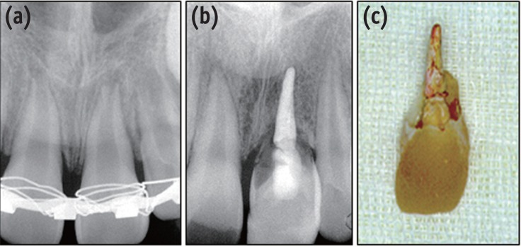
Figure 1
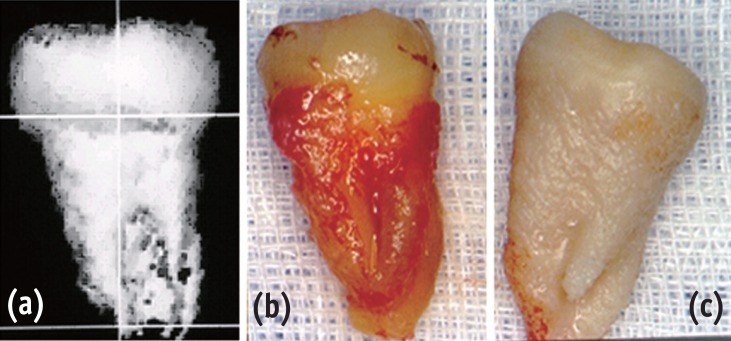
Figure 2
Figure 3
Figure 4
Absolute difference (average ± SD) between real tooth, 3D CT image model, and CARP model (Unit; micrometer; n = 12)

RT, real tooth; CTIM, computed tomography image model; CARP, computer-aided rapid prototyping.
RT, real tooth; CTIM, computed tomography image model; CARP, computer-aided rapid prototyping.

 KACD
KACD


 ePub Link
 Cite
Cite

