
Articles
- Page Path
- HOME > Restor Dent Endod > Volume 34(6); 2009 > Article
- Original Article The effects of the fluoride concentration of acidulated buffer solutions on dentine remineralization
- Won-Sub Han, Chan-Young Lee
-
2009;34(6):-536.
DOI: https://doi.org/10.5395/JKACD.2009.34.6.526
Published online: November 30, 2009
Department of Conservative Dentistry, Yonsei University, Seoul, Korea.
- Corresponding Author: Chan-Young Lee. Department of Conservative Dentistry, College of Dentistry, Yonsei University, 134, Sinchon-Dong, Seodaemun-Ku, Seoul 120-752, Korea. Tel: 82-2-2228-8700, chanyoungl@yuhs.ac
• Received: September 8, 2009 • Revised: October 23, 2009 • Accepted: October 27, 2009
Copyright © 2009 The Korean Academy of Conservative Dentistry
- 1,833 Views
- 1 Download
- 1 Crossref


Tables & Figures
REFERENCES
Citations
Citations to this article as recorded by 
- Infant Oral Health Care Concerning Education of Mothers – Part 2
Lehya Mounica Kadali, Viddyasagar Mopagar, Shilpa Shetty, Shridhar Shetty, Venkatesh Kodgi, Shantanu Chaudhari
Journal of Evolution of Medical and Dental Sciences.2021; 10(31): 2538. CrossRef
 ePub Link
 Cite
CiteThe effects of the fluoride concentration of acidulated buffer solutions on dentine remineralization


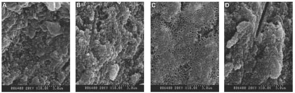

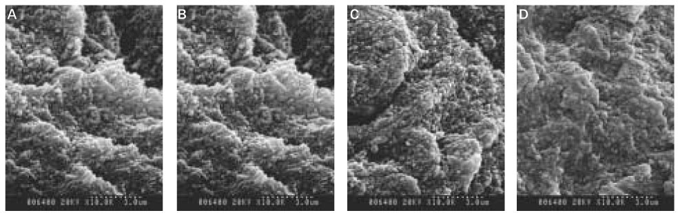


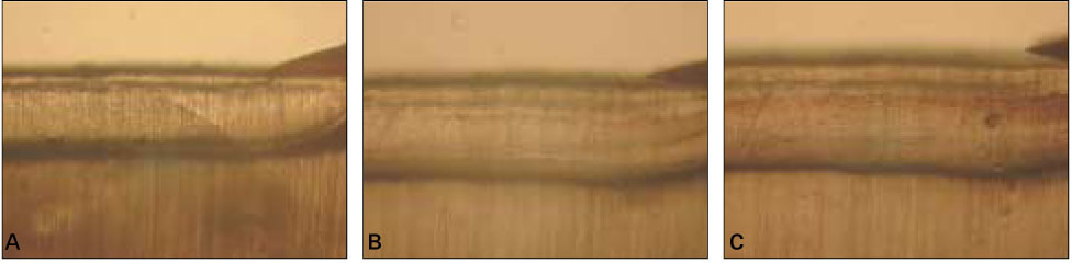
Figure 1
Representative polarizing light microscopic images of 1 ppm specimen (×100).
(A-remineralization 1day, B-remineralization 5day, C-remineralization 7day)
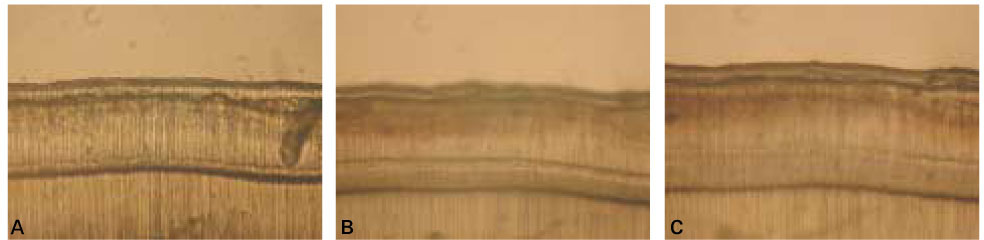
Figure 2
Representative polarizing light microscopic images of 2 ppm specimen (×100).
(A-remineralization 1day, B-remineralization 5day, C-remineralization 7day)
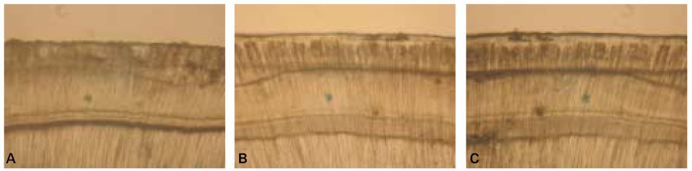
Figure 3
Representative polarizing light microscopic images of 4 ppm specimen (×100).
(A-remineralization 1day, B-remineralization 5day, C-remineralization 7day)
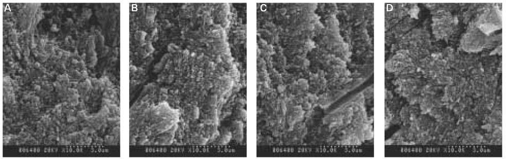
Figure 4
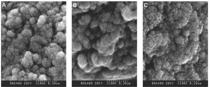
Representative scanning electron microscopic (SEM) images of 1 ppm specimen (×10,000).
(A-10 µm, B-150 µm, C-200 µm, D-250 µm each from surface)
Figure 5
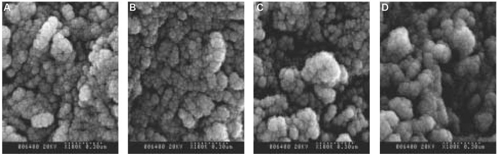
Representative SEM images of 2 ppm specimen ×10,000).
(A-50 µm, B-150 µm, C-200 µm, D-250 µm each from surface)
Figure 6
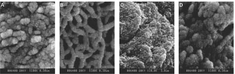
Representative SEM images of 4 ppm specimen (×10,000).
(A-50 µm, B-150 µm, C-200 µm, D-300 µm each from surface)
Figure 7
SEM images of superficial layer (×100,000).
(A-Normal dentine, B-Decalcified dentine, C-Dentine treated with 1 ppm re-mineralized solution for 7 days)
Figure 8
SEM images of upper part of lesion body (×100,000).
(A-Normal dentine, B,C and D-Dentine treated with 1,2 and 4 ppm re-mineralized solution each for 7 days)
Figure 9
SEM images of lower part of lesion body (×100,000).
(A-Normal dentine, B,C and D-Dentine treated with 1,2 and 4 ppm re-mineralized solution each for 7 days)
Figure 1
Figure 2
Figure 3
Figure 4
Figure 5
Figure 6
Figure 7
Figure 8
Figure 9
The effects of the fluoride concentration of acidulated buffer solutions on dentine remineralization
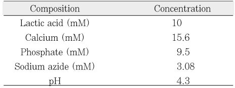
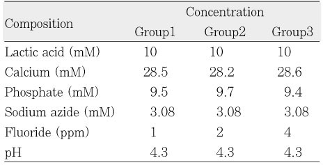
Composition of decalcification solution.
Composition of remineralinzation solution.
Table 1
Composition of decalcification solution.
Table 2
Composition of remineralinzation solution.

