
Articles
- Page Path
- HOME > Restor Dent Endod > Volume 34(4); 2009 > Article
- Original Article Management of fibrous hyperplasia in oral mucosa
- Sun-Young Ham, Chang-Kyu Song, Se-Hee Park, Jin-Woo Kim, Kyung-Mo Cho
-
2009;34(4):-345.
DOI: https://doi.org/10.5395/JKACD.2009.34.4.340
Published online: July 31, 2009
Department of Conservative Dentistry, College of Dentistry, Kangnung National University, Korea.
- Corresponding author: Kyung-Mo Cho. Department of Conservative Dentistry, College of Dentistry, Kangnung National University, Jibyun-Dong, Kangnung City, Kangwon-Do, Korea, 210-702. Tel: 82-33-640-3155, Fax: 82-33-640-3103, drbozon@kangnung.ac.kr
• Received: May 8, 2009 • Revised: May 31, 2009 • Accepted: June 5, 2009
Copyright © 2009 The Korean Academy of Conservative Dentistry
- 1,651 Views
- 5 Download
Abstract
-
There are a number of situations where the oral mucosa can be sucked or pressed to produce relatively banal but clinical distinctive changes. The labial and buccal mucosa and tongue may develop protuberances in areas where a tooth is missing or extra space is present. The mucosa is pressed and sucked into these spaces, thus leading to the development of a fibrous hyperplasia.This case report describes the management of fibrous hyperplasia in oral mucosa.Fibrous hyperplasia can be formed by habitual pressure or suction in oral mucosa. Treatment of fibrous hyperplasia consists of simple excision and, if feasible, elimination of the cause. And habit control is a important factor for preventing recurrence.
- 1. Bork , Hoede , Burgdorf , Yung . Diseases of the oral mucosa and the lip. 1996;W.B. Saunders company.
- 2. Walinski CJ. Irritation fibroma removal: A comparison of two laser wavelengths. Gen Dent. 2004;52(3):236-238.PubMed
- 3. KARAÇAY Ş, et al. Treatment of habitual lip biting. Turk J Med Sci. 2006;36(3):187-189.
REFERENCES


Tables & Figures
REFERENCES
Citations
Citations to this article as recorded by 
 ePub Link
 Cite
CiteManagement of fibrous hyperplasia in oral mucosa

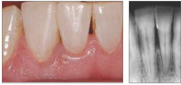
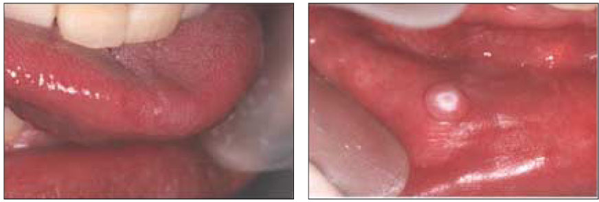
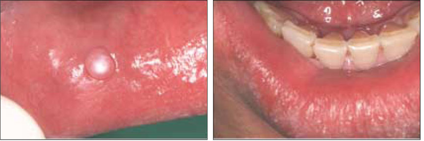
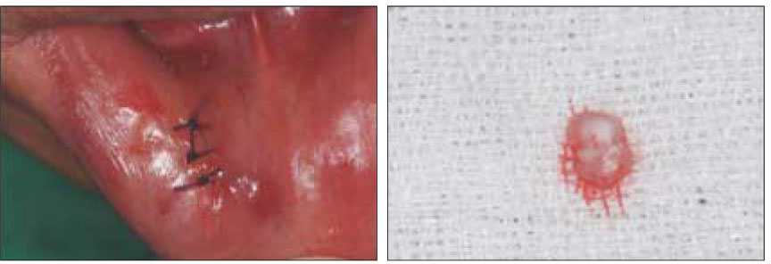


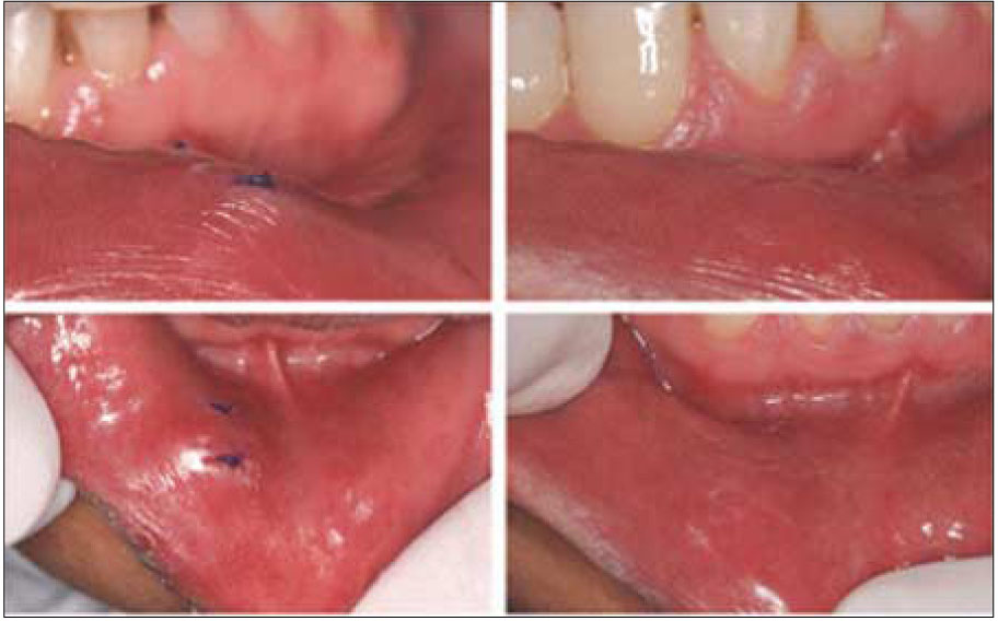
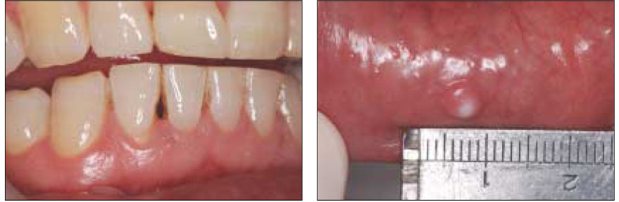
Figure 1
Interdental space & Fibrous hyperplasia in oral mucosa
Figure 2
Resin filling on #41 and 42
Figure 3
4 month follow-up
Figure 4
4 month 2 weeks follow-up
Figure 5
Surgical excision of fibrous hyperplasia
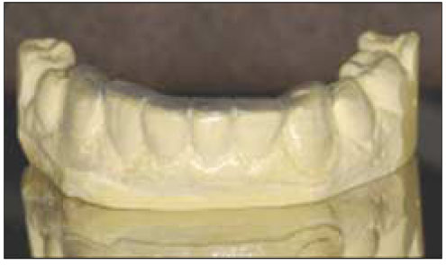
Figure 6
Mouthguard for habit control
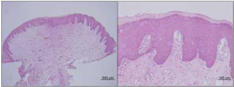
Figure 7
Histopathologic finding
Figure 8
1 and 4 weeks follow-up after surgery
Figure 1
Figure 2
Figure 3
Figure 4
Figure 5
Figure 6
Figure 7
Figure 8
Management of fibrous hyperplasia in oral mucosa

