Articles
- Page Path
- HOME > Restor Dent Endod > Volume 30(4); 2005 > Article
- Original Article Effect of anticurvature filing method on preparation of the curved root canal using ProFile
- Hyun-Ji Song, Juhea Chang1, Kyung-Mo Cho, Jin-Woo Kim
-
2005;30(4):-334.
DOI: https://doi.org/10.5395/JKACD.2005.30.4.327
Published online: July 30, 2005
Department of Conservative Dentistry, College of Dentistry, Kangnung National University, Korea.
1Department of Operative Dentistry, Indiana University, School of Dentistry, USA.
- Corresponding author: Jin-Woo Kim. Department of Conservative Dentistry, College of Dentistry, Kangnung National University, Jibyun-Dong, Kangnung City, Kangwon-Do, Korea, 210-702. Tel: 82-33-640-3189, Fax: 82-33-640-3113, mendo7@kangnung.ac.kr
Copyright © 2005 Korean Academy of Conservative Dentistry
- 1,246 Views
- 9 Download
Abstract
-
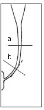
This study investigated the effect of anticurvature filing method on preparation of the curved root canal using ProFile.Thirty six resin blocks were divided equally into three groups by instrumentation motions: anticurvature filing motion, circumferential filing motion and straight up-and-down motion. Each resin block was sectioned at 8 mm level from the apex and at the greatest curvature of the canal and reassembled in metal mold by a modified Bramante technique. All groups were instrumented with the ProFile system. At each levels, image of sectioned surface were taken using CCD camera under a stereomicroscope at ×40 magnification and stored. Distances of transportation at the inner and outer area of curvature and the centering ratio were determined and compared by statistical analysis, along with the assessment of the increase of root canal cross-sectional area.The results were as follows;1. In all groups, there was no statistical difference in the mean increase of root canal cross-sectional area, the centering ratio, and the mean distances of transportation at the inner area of curvature at each level.2. At 8 mm level from the apex, the mean distances of transportation at the outer area of curvature decreases in following order anticurvature filing motion, circumferential filing motion, straight up-and-down motion but, no significant difference at the greatest curvature of the canal among three groups.Effect of anticurvature filing motion using ProFile does not seem to be different from other instrumentation motions at the inner area of curvature in curved root canal.
- 1. Schilder H. Cleaning and shaping the root canal. Dent Clin North Am. 1974;18(2):269-296.ArticlePubMed
- 2. Skidmore AE, Bjorndal AM. Root canal morphology of the human mandibular first molar. Oral Surg Oral Med Oral Pathol. 1971;32(5):778-784.ArticlePubMed
- 3. Weine F, Kelly RF, Lio PJ. The effect of preparation procedures on original canal shape and on apical foramen shape. J Endod. 1975;(8):255-262.ArticlePubMed
- 4. Meister F, Lommel TJ, Gerstein H, Davies FE. Endodontic perforation which resulted in alveolar bone loss. Oral Surg Oral Med Oral Pathol. 1979;47(5):463-470.PubMed
- 5. Abou-Rass M, Frank AL, Glick DH. The anticurvature filing method to prepare the curved root canal. J Am Dent Assoc. 1980;101(5):792-794.ArticlePubMed
- 6. Tidmarsh BG. Preparation of the root canal. Int Endod J. 1982;15(2):53-61.ArticlePubMed
- 7. Goerig AC, Michelich RJ, Schultz HH. Instrumentation of root canals in molar using the step-down technique. J Endod. 1982;8(12):550-554.ArticlePubMed
- 8. Kessler JR, Peters DD, Lorton L. Comparison of the relative risk of molar root perforations using various endodontic instrumentation techniques. J Endod. 1983;9(10):439-447.ArticlePubMed
- 9. Glossen CR, Haller RH, Dove SB, del Rio CE. A comparison of root canal preparations using Ni-Ti hand, Ni-Ti engine-driven, and K-Flex endodontic instruments. J Endod. 1995;21(3):146-151.ArticlePubMed
- 10. Calhoun G, Montgomery S. The effects of four instrumentation techniques on root canal shape. J Endod. 1988;14(6):273-277.ArticlePubMed
- 11. Thompson SA, Dummer PM. Shaping ability of Hero 642 rotary nickel-titanium instruments in simulated root canals Part 2. Int Endod J. 2000;33(3):255-261.ArticlePubMed
- 12. Schafer E. Shaping ability of Hero 642 rotary nickel-titanium instruments and stainless steel hand K-Flexofiles in simulated curved root canals. Oral Surg Oral Med Oral Pathol Oral Radiol Endod. 2001;92(2):215-220.ArticlePubMed
- 13. Thompson SA, Dummer PM. Shaping ability of Mity Roto 360 degrees and Naviflex rotary nickel-titanium instruments in simulated root canals. Part 1. J Endod. 1998;24(2):128-134.PubMed
- 14. Luiten DJ, Morgan LA, Baugartner JC, Marshall JG. A comparison of four instrumentation techniques on apical canal transportation. J Endod. 1995;21(1):26-32.ArticlePubMed
- 15. Bramante CM, Berbert A, Borges RP. A methodology for evaluation of root canal instrumentation. J Endod. 1987;13(5):243-245.ArticlePubMed
- 16. Weine FS. Endodontic Therapy. 1996;5th ed. St. Louis: Mosby Inc; 330-331.
- 17. Lim KC, Webber J. The validity of simulated root canals for the investigation of the prepared root canal shape. Int Endod J. 1985;18(4):240-246.ArticlePubMed
- 18. Dummer PMH, Alodeh MHA. A method for the construction of simulated root canals in clear resin blocks. Int Endod J. 1991;24(2):63-66.ArticlePubMed
- 19. Thompson SA, Dummer PM. Shaping ability of ProFile .04 taper series 29 rotary nickel-titanium instruments in simulated root canals Part 1. Int Endod J. 1997;30(1):1-7.ArticlePubMed
- 20. Thompson SA, Dummer PM. Shaping ability of Hero 642 rotary nickel-titanium instruments in simulated root canals Part 1. Int Endod J. 2000;33(3):248-254.ArticlePubMed
- 21. Peters OA, Barbakow F. Dynamic torque and apical forces of ProFile .04 rotary instruments during preparation of curved canals. Int Endod J. 2002;35(4):379-389.ArticlePubMed
- 22. Bonetti Filho I, Miranda Esberard R, de Toledo Leonardo R, del Rio CE. Microscopic evaluation of three endodontic files pre- and post-instrumentation. J Endod. 1998;24(7):461-464.ArticlePubMed
- 23. Lim SS, Stock CJ. The risk of perforation in the curved canal: anticurvature filing compared with the step-back technique. Int Endod J. 1987;20(1):33-39.ArticlePubMed
- 24. Song YL, Bian Z, Fan B, Fan MW, Gutmann JL, Peng B. A comparison of instrument-centering ability within the root canal for three contemporary instrumentation techniques. Int Endod J. 2004;37(4):265-271.ArticlePubMed
- 25. Lee CH, Cho KM, Hong CU. Effect of various canal preparation techniques using rotary nickel-titanium files on the maintenance of canal curvature. J Korean Acad Conserv Dent. 2003;28(1):41-49.Article
- 26. Park SH, Cho KM, Kim JW. The efficiency of the NiTi rotary files in curved simulated canals shaped by novice operators. J Korean Acad Conserv Dent. 2003;28(2):146-155.Article
- 27. Choi SD, Jin MU, Kim KO, Kim SK. Shaping ability of four rotary nickel-titanium instruments to prepare root canal at danger zone. J Korean Acad Conserv Dent. 2004;29(5):446-453.Article
REFERENCES


Tables & Figures
REFERENCES
Citations


Figure 1
Mean increase of root canal cross-sectional areas at each level in three groups (Unit: mm2, Mean ± S.D.)
Mean distances of transportation at the inner area of curvature (Unit: mm, Mean ± S.D.)
Mean distances of transportation at the outer area of curvature (Unit: mm, Mean ± S.D.)
*: Statistically significant difference, p < 0.05 by Scheffe test
Mean centering ratio for each level in three group (Mean ± S.D.)
*: Statistically significant difference, p < 0.05 by Scheffe test
*: Statistically significant difference,
*: Statistically significant difference,

 KACD
KACD


 ePub Link
 Cite
Cite

