Abstract
-
Objectives
This study assessed the shape and anatomical relationship of the mental foramen (MF) to mandibular posterior teeth in an Indian sub-population.
-
Materials and Methods
In total, 475 existing cone-beam computed tomography records exhibiting 950 MFs and including the bilateral presence of mandibular premolars and first molars were assessed. Images were evaluated 3-dimensionally to ascertain the position, shape, and anatomical proximity of MFs to mandibular teeth. The position and shape of MFs were measured and calculated. The Pythagorean theorem was used to calculate the distance between the root apex of the mandibular teeth and the MF.
-
Results
MFs exhibited a predominantly round shape (left: 67% and right: 65%) followed by oval (left: 30% and right: 31%) in both males and females and in different age groups. The root apices of mandibular second premolars (left: 71% and right: 62%) were closest to the MF, followed by distal to the first premolars and mesial to the second premolars. The mean vertical distance between the MF and the nearest tooth apex calculated on sagittal sections was 2.20 mm on the right side and 2.32 mm on the left side; no significant difference was found according to sex or age. The distance between the apices of the teeth and the MF was ≥ 4 mm (left; 4.09 ± 1.27 mm and right; 4.01 ± 1.15 mm).
-
Conclusions
These findings highlight the need for clinicians to be aware of the location of the MF in treatment planning and while performing non-surgical and surgical endodontic procedures.
-
Keywords: Anatomy; Endodontic procedures; Mandibular premolar; Mental nerve; Mental foramen; Nerve injury
INTRODUCTION
The mandibular branch of the trigeminal nerve has many divisions, one of which is the inferior alveolar nerve (IAN). The mental nerve (MN) is a branch of the IAN that exits the mandible through the mental foramen (MF). It supplies sensation to the anterior aspects of the chin and the lower lip, along with the buccal gingivae of the mandibular anterior and premolar teeth [
1]. The location of the MF ranges from the distal end of the mandibular canine in the anterior part to the posteriorly positioned first molars [
2].
Because of the position of the MF and MN relative to the root apices of mandibular posterior teeth, damage to this nerve may occur during both non-surgical and surgical endodontic procedures, presenting as paresthesia [
3]. In dentistry, paresthesia can be caused by factors such as local anesthetic injections, third molar surgery, orthognathic surgery, ablative surgery, implants, and a wide range of endodontic procedures [
4,
5].
The risk of injury to the MN increases if the MF is not properly identified and protected from insult [
6]. The likelihood of an injury further increases due to the variability in the anatomy of the mandible, the course of neurovascular bundles passing through the mandibular canal, and the type of emergence through the MF [
7]. Variations in the location of the MF, along with its accessory foramen, sometimes lead to the misdiagnosis of some pathoses. The structures that pass through the MF include the MN and accompanying vasculature [
8,
9].
The anatomic complexity of the MF region is not limited to variations in its location and dimension. The presence of 3 other elements—the anterior loop of the inferior alveolar canal, accessory mental foramina (AMFs), and the lateral lingual foramen and its associated canal—can further complicate the MF region [
10]. A comprehensive understanding of this region is important for both surgical and non-surgical endodontic treatment. AMFs contain accessory mental arteries and/or accessory MNs. The existence of an AMF affects the dimensions of the MF. The prevalence of AMFs varies across populations, with a range between 2% and 14.3% [
11,
12,
13,
14,
15,
16,
17,
18,
19,
20].
Locating the MF may be difficult at times, as there are no absolute anatomical landmarks for reference. Furthermore, in most patients, the foramen cannot be clinically visualized or palpated. Moreover, the reported anatomical position of the MF is highly variable [
21], and its location has been classified as being at the apex of the first premolar, between the apices of the first and second premolars, at the apex of the second premolar, between the apices of the second premolar and first molar, and on the mesial aspect of the first molar [
22].
Unfortunately, the non-dental causes of MN paresthesia are not in the clinicians' domain of control, whereas dental causes can be prevented through appropriate investigations and cautious work [
6]. In accordance with the principle that “prevention is better than cure,” cone-beam computed tomography (CBCT) facilitates the assessment and management of MN injuries [
23,
24].
Numerous techniques are used to identify the location of the MF, including dissection of human mandibles or the use of radiographs [
25]. However, there is a high likelihood of misidentification given the radiographic appearance of radiolucent MFs. Hence, it is necessary to obtain a 3-dimensional (3D) visualization of the adjacent teeth and the MF, which cannot be accurately assessed on standard radiographs [
26]. CBCT imaging overcomes the limitations of radiographs by improving accuracy, reducing distortion, and providing a 3D image of teeth and surrounding structures. CBCT scans were found to have an error of less than 0.6% while measuring mandibular anatomy [
27] and have been used to evaluate the anatomical relationship between the roots of the mandibular second molars and the IAN, which is related to the risk of nerve injury during endodontic treatment [
28].
It is important to consider the position of the MF for clinical and diagnostic purposes. A plethora of literature, often with inconsistent results, is available with data on the position and anatomical relationship of MF with the mandibular posterior teeth in various populations [
1,
2,
4,
26,
27]. Previous studies [
21,
22] suggested that the MF is frequently found below the apex of the second premolar. However, the sample size was insufficient to generalize the findings; to date, although some studies have been conducted to assess the MF location in the Indian population, none have had a large enough sample size to provide definitive data [
21,
22,
29,
30].
Although practicing dentists are well aware of the anatomical landmarks, it is necessary for dentists to be able to correlate the anatomical landmarks with the specific steps taken during treatment and be aware of the several complications that may arise due to even the slightest variations in the landmarks. Furthermore, the reference texts/articles to which general practitioners might refer cannot exactly predict the differences that are relevant in the Indian sub-population.
Therefore, the current study was designed to evaluate the shape and anatomical relationship between the MF and mandibular posterior teeth in a sample of the Indian population using CBCT imaging. The null hypothesis tested was that the position of the MF would be similar in both sexes and different age groups in the same population.
MATERIALS AND METHODS
The study design was approved by the Institutional Ethics Committee (EC-98/CONS12ND/2018). The study used pre-existing CBCT scans to assess the MF in the mandibular arch and adjacent teeth from previous databases that were obtained from Nair Hospital Dental College, Mumbai, India. A sample size of 950 was used based on calculations made using OpenEpi software (v.3.04). The records of patients that represented the Indian sub-population were obtained from the radiology department. The CBCT examinations were originally done for various reasons, such as assessment of bone volume for dental implant planning, diagnosis of dentoalveolar trauma, management of impacted teeth before orthodontic treatment, treatment planning before non-surgical and surgical endodontic treatment, and preoperative assessments for orthognathic surgery. Convenience sampling was used to screen the scans for eligibility. The following inclusion criteria were considered; a) patients 16–60 years of age, b) scans showing a complete mandibular arch, c) the presence of mandibular premolars and first molars on both right and left sides, and d) completely erupted mandibular premolars.
The exclusion criteria were as follows: a) presence of lesions involving the mandibular posterior teeth, b) a fracture line involving the mandibular premolar region, and c) previous surgery involving the mandibular premolar region.
A CBCT scanner (Carestream 9000 3D Unit) operating at 80 kV and 5 mA with an exposure time of 10.8 seconds was used. The voxel size of the images was 0.2 mm, and the slice thickness was 0.1 mm. A radiologist experienced in operating and acquiring CBCT scans performed the acquisition process according to the manufacturer’s recommended protocol with the minimum exposure necessary for adequate image quality. The software (CSI Imaging Software) was calibrated, and CBCT scans were interpreted in a reproducible manner. Inter-examiner (2 endodontists and 1 maxillofacial radiologist) reliability was calculated using the Cohen kappa coefficient under the supervision of experts in dental maxillofacial radiology. A kappa value of 0.91 was derived, showing almost perfect agreement.
A pilot study was carried out with 50 scans after conducting calibration to check the feasibility and validity of the methodology. Additionally, 425 CBCT scans were selected after a stringent assessment via the inclusion and exclusion criteria.
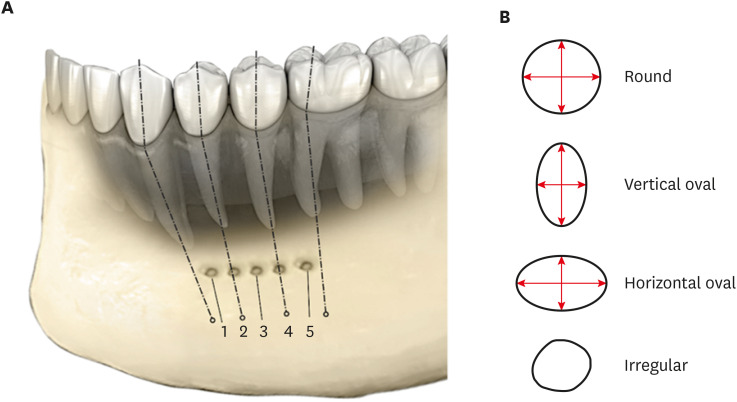
The method of evaluating the relationship between the MF and the roots of mandibular posterior teeth was based on the method proposed by Chong
et al. [
1]. Measurements in millimeters (mm) were obtained. The location of the MF corresponding to the long axis of mandibular teeth was as follows: 1) mesial to the long axis of the first premolar (
mFP); 2) along the long axis of the first premolar (FP); 3) between the long axis of the first and second premolars (FP
d-
mSP); 4) along the long axis of the second premolar (SP); or 5) distal to the long axis of the second premolar (SP
d) (
Figure 1A). The shapes of the MF were defined as follows: round (both diameters being the same,
i.e., vertical and horizontal), oval vertical/horizontal (one of the diameters being ≥ 2 times the other), and irregular (
Figure 1B) [
31,
32]. The MFs with any one diameter which was greater (>) than the other or < 2 times was classified as irregular.
Figure 1 Location and shapes of the mental foramen (MF). (A) Different locations of the MF found in the current study. 1) mFP: mesial to the first premolar, 2) FP: apex of the first premolar, 3) FPd-mSP: distal to the first premolar and mesial to the second premolar, 4) SP: apex of the second premolar, and 5) SPd-mFM: distal to the second premolar and mesial to the first molar. (B) Different shapes of the MF: round - both the diameters are the same; oval - long:short diameter ≥ 2; and irregular.

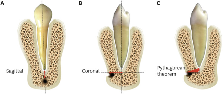
The vertical distance was measured in millimeters from the superior border of the MF to the apex of the closest root (
Figure 2A). The horizontal distance was measured from the opening of the MF in the coronal plane to the long axis of the closest tooth (
Figure 2B) [
33]. The shape of the MF in 3D was confirmed by measuring any 2 diameters, and the shape was classified as circular, oval, or irregular. The actual 3D distance of the proximate tooth and MF was measured using the Pythagorean theorem in millimeters (mm) [
1] (
Figure 2C), where a = vertical distance, b = horizontal distance, and c = the actual distance of the MF from the apex of the premolar tooth. The formula used was a
2 + b
2 = c
2. The effects of age and sex on the location of the MF were also evaluated.
Figure 2 Measuring the distance of the mental foramen. (A) Vertical distance in a sagittal section; (B) horizontal distance in a coronal section; and (C) application of the Pythagorean theorem.

The obtained data were statistically analyzed using SPSS version 21 (IBM Corp., Armonk, NY, USA). Frequency statistics were applied to evaluate the prevalence of different positions and shapes of the MF on the left and right sides of the mandible. The normality of the data distribution was checked using the Shapiro-Wilk test. The mean distances from the root apex of the mandibular teeth to the MF on both sides of the mandibular arch were assessed, and the unpaired t-test was used to evaluate the significance of differences. All statistical analyses were done at a 95% confidence level.
RESULTS
According to the inclusion and exclusion criteria, 475 scans were evaluated from 231 male and 244 female patients. The distribution according to age and sex is presented in
Table 1. The positions of the MF on the left and right sides related to the mandibular teeth are presented in
Table 2. Out of the evaluated 475 scans with 950 MFs, the MFs were most often located at the apex of the mandibular second premolar (632/950). On the left side, 71% (337/475) of cases had the root apex of the second premolar closest to the MF, whereas on the right side, 62% (295/475) of cases had the root apex of the second premolar closest to the MF, which is similar to the pattern found on the contralateral side. The next most frequent location of the MF was the FP
d -
mSP position, which was found in 14% (67/475) of cases on the left side and in 26% (124/475) of cases on the right side. All of the above findings were statistically significant. In our study, the association between the location of the MF on both sides and sex was evaluated. On both the left and right sides, both males and females predominantly showed the MF in the second premolar region, followed by distal to the first premolar and medial to the second premolar. This difference was not statistically significant. (
p = 0.112 and
p = 0.224) (
Table 3). The association between the location of the MF on both sides and age was also investigated. On both sides, the 16–30, 31–45, and 46–60 age groups predominantly showed the MF in the second premolar region, followed by distal to the first premolar and medial to the second premolar. This difference was statistically significant for the age group of 16–30 years as compared to other age groups on both sides (
p = 0.012 for the left side and
p = 0.021 for the right side) (
Table 4). The most common shape of the MF was round on both sides (
Table 5). The shape of the MF was oval (horizontal or vertical) in 30% of scans (143/475) and round in 67% (318/475) of scans on the left side, while 65% (308/475) were round and 31% (147/475) were oval on the right side. The association between the shape of the MF on the left and right sides and sex was determined. A round shape was most common, followed by an oval horizontal shape, in both sexes and on both sides, without a statistically significant difference (
p = 0.112 and
p = 0.224) (
Table 6).
Table 1 Age and sex distributions of the sample in this study
|
Age of the patients |
Sex |
Total |
|
Male |
Female |
|
16–30 |
101 (44) |
126 (52) |
227 (47) |
|
31–45 |
74 (32) |
57 (23) |
131 (28) |
|
46–60 |
56 (24) |
61 (25) |
117 (25) |
|
Total |
231 (100) |
244 (100) |
475 (100) |
Table 2 Prevalence of the location of the mental foramen (MF) on the left and right sides
|
Location of MF |
No. of scans and percentage |
|
Left |
Right |
|
FP |
47 (10) |
28 (6) |
|
FPd-mSP |
67 (14) |
124 (26) |
|
SP |
337 (71) |
295 (62) |
|
SPd
|
24 (5) |
28 (6) |
|
Total |
475 (100) |
475 (100) |
Table 3 Association between the location of the mental foramen on the left and right sides and sex
|
Sex\Location |
Left |
Right |
|
FP |
FPd-mSP |
SP |
SPd
|
FP |
FPd-mSP |
SP |
SPd
|
|
Male |
10 |
61 |
150 |
10 |
15 |
41 |
170 |
5 |
|
Female |
15 |
69 |
145 |
15 |
32 |
26 |
167 |
19 |
|
p value |
0.224 |
0.112 |
Table 4 Association between the location of the mental foramen on the left and right sides and age
|
Age\Location |
Left |
Right |
|
FP |
FPd-mSP |
SP |
SPd
|
FP |
FPd-mSP |
SP |
SPd
|
|
16–30 |
15 |
40 |
167 |
5 |
12 |
80 |
120 |
15 |
|
31–45 |
11 |
20 |
90 |
10 |
0 |
10 |
120 |
1 |
|
46–60 |
21 |
7 |
80 |
9 |
16 |
34 |
55 |
12 |
|
p value |
0.012*
|
0.021*
|
Table 5 Prevalence of shapes of the mental foramen (MF) on the left and right sides
|
Shape of MF |
No. of scans and percentage |
|
Left |
Right |
|
Round |
318 (67) |
308 (65) |
|
Oval horizontal |
81 (17) |
76 (16) |
|
Oval vertical |
62 (13) |
71 (15) |
|
Irregular |
14 (3) |
20 (4) |
|
Total |
475 (100) |
475 (100) |
Table 6 Association between the shape of the mental foramen on the left and right sides and sex
|
Sex\Shape |
Left |
Right |
|
Round |
Oval horizontal |
Oval vertical |
Irregular |
Round |
Oval horizontal |
Oval vertical |
Irregular |
|
Male |
150 |
50 |
20 |
11 |
160 |
41 |
20 |
10 |
|
Female |
168 |
31 |
42 |
3 |
148 |
35 |
51 |
0 |
|
p value |
0.324 |
0.185 |
The association between the shape of the MF on both sides and age was investigated. On both sides, the round shape predominated in all 3 age groups (16–30, 31–45, and 46–60 years), followed by the oral horizontal shape, with the exception of the 31–45 age group, wherein the oval vertical shape was the second most common. The differences were not statistically significant (
p = 0.201 and
p = 0.732) (
Table 7).
Table 7 Association between the shape of the mental foramen on the left and right sides and age
|
Age\Shape |
Left |
Right |
|
Round |
Oval horizontal |
Oval vertical |
Irregular |
Round |
Oval horizontal |
Oval vertical |
Irregular |
|
16–30 |
120 |
57 |
46 |
4 |
115 |
50 |
50 |
12 |
|
31–45 |
101 |
10 |
10 |
10 |
99 |
9 |
15 |
8 |
|
46–60 |
97 |
14 |
6 |
0 |
94 |
17 |
6 |
0 |
|
p value |
0.201 |
0.732 |
The mean distance calculated using the Pythagorean theorem between the MF and root apex of mandibular teeth on the right and the left side was 4.01 ± 1.15 mm and 4.09 ± 1.27 mm, respectively (
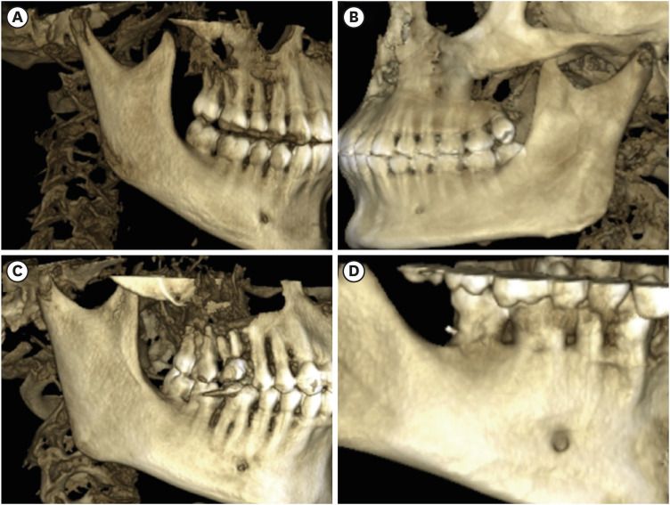
Table 8). The positions of the MFs relative to the mandibular teeth ranged from the first premolar to the distal position in the second premolars (
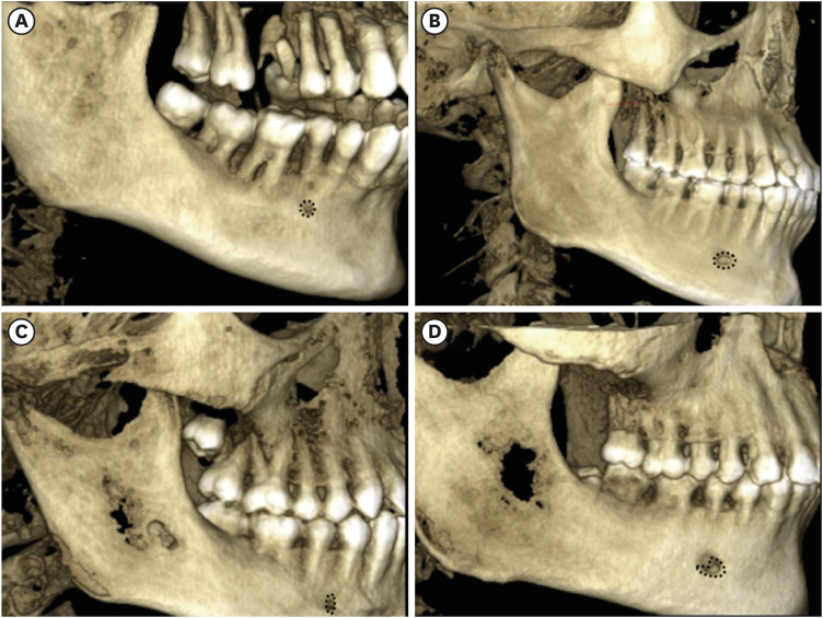
Figure 3). The MFs also exhibited different shapes (
Figure 4).
Table 8 Distance in millimeters (mm) from mental foramen to the root
|
Distance*
|
Minimum |
Maximum |
Mean |
Standard deviation |
|
Right |
2.33 |
8.92 |
4.01 |
1.15 |
|
Left |
1.60 |
9.20 |
4.09 |
1.27 |
Figure 3 Position of the mental foramen. (A) FP; (B) FPd-mSP; (C) SP; and (D) SPd-mFM. FP: first premolar, SP: second premolar, FPd-mSP: between the distal FP and medial SP, SPd: distal to the SP.

Figure 4 Different shapes of the mental foramen. (A) Round; (B) oval (horizontal); (C) oval (vertical); and (D) irregular.

The mean vertical distance between the MF and the nearest tooth apex calculated on sagittal sections was 2.20 mm on the right side and 2.32 mm on the left side, and no significant difference was found according to sex or age (p = 0.253).
DISCUSSION
In this study, the shape and position of MFs were evaluated in different age groups and both sexes in an Indian sub-population using CBCT. Based on the results, the null hypothesis was rejected.
Endodontic-related paresthesia requires a thorough assessment to determine the causative factors. To prevent paresthesia or to minimize its occurrence, the endodontist must be aware of the proximity of the apices of the teeth to the nerve structures prior to any non-surgical or surgical procedures [
34]. The inconsistent position of the MF should always be considered while investigating radiographic periapical areas and while performing periodontal or endodontic surgery in the area between the canine and the mesial root of the first molar. The MF is a significant anatomical landmark in the orofacial region. Furthermore, the position of the MF varies across ethnic groups [
21], which should alert the clinician to consider variability in their patient populations.
Based on the findings presented by Santini and Alayan [
22], in Indian and Chinese populations, the MF was in a similar position, although the mandible was smaller in Indians. The MF showed a consistent size in the mandibles of European and Chinese populations. However, the MF was further forward relative to the mandible in the European cohort [
35].
When paresthesia occurs in the mandibular area, it is usually assumed that the nerve tissue is directly damaged. This could be due to the operative procedure itself (for example, over-extension of endodontic files), chemical damage from the materials used (by their neurotoxicity), or compression as a causative factor of nerve degeneration (for example, over-extended material being in contact with the nerve with resultant pressure) [
36,
37,
38]. The risk of nerve injury during endodontic procedures is always of concern, and to overcome this issue, diagnostic and treatment approaches are suggested. To diagnose a nerve injury, it is important to consider a combination of a thorough anamnesis, a proper clinical evaluation, and an adjunct radiographic evaluation whenever indicated [
25]. Precise and accurate determination of the working length is of the utmost importance when performing non-surgical root canal procedures. If an intimate relationship between the MF and the root apex is detected, then the working length should be cautiously verified at every stage of canal preparation.
CBCT images were used due to their advantages over 2-dimensional intraoral periapical radiography. These scans encompass a greater 3D area of hard and soft tissues in a continuous view, while allowing more accurate localization of the MF in both horizontal and vertical dimensions [
39,
40]. CBCT scans were found to have an error of less than 0.6% while measuring mandibular anatomy [
27] and have been used to evaluate the anatomical relationship between the roots of the mandibular second molars and the IAN, which is related to the risk of nerve injury during endodontic treatment [
28].
Al-Mahalawy
et al. [
15] reported that the mean distance from the nearest tooth apex to the MF was 3.1 mm. In contrast, Kalender
et al. [
16] examined 386 sites in 193 Turkish patients and found that the mean distance from the nearest root apex to the MF was 4.2 mm. This disparity could possibly be attributed to racial/ethnic differences.
This retrospective study assessed the anatomical relationship and proximity of the MF with mandibular premolars using CBCT and estimated the risk of MN paresthesia while performing endodontic procedures. The root apex of the second premolar on both sides of the mandibular arch was closest to the MF in the sagittal plane, which is in agreement with previous studies [
1,
41,
42]. MN paresthesia arising after the start of endodontic procedures has been documented in previous studies [
3,
38,
39].
The risk of MN injury during a surgical endodontic procedure is higher if the MF is situated closer to the distal side of the mandibular premolars [
43,
44]. Based upon the CBCT scans evaluated in the sagittal plane, the mean distance from the distal side of the root apex of the mandibular first premolars to the MF on both sides of the arch was ≥ 4 mm. By marking the distance of 4 mm from the MF, the risk of a potential injury can be avoided. Therefore, preoperative radiographs can provide information on the appropriate distance and relevant anatomical structures in the vicinity of the MN during endodontic procedures.
Significant variations are found in the literature regarding the normal location of the MF in different ethnic groups over the globe. In the Malay population, Brazilian population, and Malawian population, the MF was most often located in line with the second premolar, which is in agreement with our findings [
42,
45,
46]. Kqiku
et al.'s study [
47] conducted in the Kosovarian population found that the MF was precisely located between the first and second mandibular premolars. The results of the present study corroborated the data from Indian populations reported by Bhagat
et al. [
22], Alok
et al. [
21], Swamy
et al. [
30], and Srivastava
et al. [
29], which conclusively stated that the most common position of MF in the Indian population was below the mandibular second premolar, followed by the position between the 2 premolars.
The incidence of altered sensation after periapical surgery appears to be relatively high (14%), with a higher incidence found in premolars than in molars. Therefore, thorough knowledge of the anatomical structures in the MF region is crucial, and CBCT imaging enables a clear identification of the anatomical relationship of the root apices to important neighboring anatomical structures in any plane the clinician wishes to view.
Usually, when the MN is damaged, the patient might experience anesthesia or paresthesia of the lower lip. The affected tissues may regain normal sensation over varying periods, depending on the type and extent of damage to the nerve bundle. Some patients report a return to the normal sensation after several days, while others may take several weeks or even months to recover [
48]. Meticulous preoperative planning of endodontic treatment through CBCT scans will minimize the risk of injury to the MN [
49]. Furthermore, CBCT is currently the best available imaging technique to determine the location of the MF accurately. However, CBCT is not considered as a standard assessment method, and its indication should be justified on an individual basis; in particular, it is necessary to consider whether the potential benefits outweigh the potential risks to a patient. A recently updated joint position statement on the use of CBCT in endodontics by the American Association of Endodontists and the American Academy of Oral and Maxillofacial Radiology states that, in general, CBCT should be cautiously used to assess and treat complex endodontic conditions, which involve pre-surgical case planning to determine the exact location of the root apex/apices and the proximity of adjacent anatomical structures [
49].
Historically, researchers have shown keen interest and efforts in identifying anatomical variation in the position and shape of the MF, while presently, greater significance is placed on this structure and its characteristics due to the advanced dental rehabilitation procedures that benefit from more accurate 3D diagnostic images. Moreover, this study assessed, to some extent, various anatomical variations that could be associated with ethnic origins. A few limitations of the present study include the analysis of a smaller sub-population, the limited number of examiners who assessed the anatomical landmarks, and the limitation of the study to individuals of a specific ethnic origin.
Future research should expand upon this study by evaluating and comparing differences among anatomical landmarks of the mandible in populations of different ethnic and racial origins. Furthermore, clinical parameters can be correlated with this anatomical landmark to achieve successful clinical outcomes of treatment by considering the crestal bone/MF distance or other relevant factors. Moreover, the microsurgical aspects of these findings should also be taken into consideration in future studies.
CONCLUSIONS
The results obtained through this study provide insights into the type and position of MF in an Indian sub-population. The MF in the studied population exhibited proximity to the mandibular second premolar in both males and females and in different age groups. The most common shape of the MF was found to be round, followed by oval horizontal, in the studied population. The findings of this study may be used as a guideline for dental professionals in locating the MF while performing the dental procedures in that region.
-
Conflict of Interest: No potential conflict of interest relevant to this article was reported.
-
Author Contributions:
Conceptualization: Pawar AM, Sheth K.
Data curation: Sheth K.
Formal analysis: Pawar AM, Sheth K, Banga KS.
Investigation: Sheth K.
Methodology: Sheth K, Pawar AM, Banga KS.
Project administration: Sheth K.
Software: Sheth K.
Supervision: Gutmann JL, Kim HC.
Validation: Pawar AM, Kim HC.
Visualization: Sheth K.
Writing - original draft: Pawar AM, Sheth K.
Writing - review & editing: Gutmann JL, Kim HC.
REFERENCES
- 1. Chong BS, Gohil K, Pawar R, Makdissi J. Anatomical relationship between mental foramen, mandibular teeth and risk of nerve injury with endodontic treatment. Clin Oral Investig 2017;21:381-387.ArticlePubMedPDF
- 2. Greenstein G, Tarnow D. The mental foramen and nerve: clinical and anatomical factors related to dental implant placement: a literature review. J Periodontol 2006;77:1933-1943.ArticlePubMed
- 3. Ahonen M, Tjäderhane L. Endodontic-related paresthesia: a case report and literature review. J Endod 2011;37:1460-1464.ArticlePubMed
- 4. Renton T. Prevention of iatrogenic inferior alveolar nerve injuries in relation to dental procedures. Dent Update 2010;37:350-352. 354-356. 358-360.ArticlePubMed
- 5. Moon S, Lee SJ, Kim E, Lee CY. Hypoesthesia after IAN block anesthesia with lidocaine: management of mild to moderate nerve injury. Restor Dent Endod 2012;37:232-235.ArticlePubMedPMC
- 6. Juodzbalys G, Wang HL, Sabalys G. Anatomy of mandibular vital structures. Part II: mandibular incisive canal, mental foramen and associated neurovascular bundles in relation with dental implantology. J Oral Maxillofac Res 2010;1:e3.Article
- 7. von Arx T, Friedli M, Sendi P, Lozanoff S, Bornstein MM. Location and dimensions of the mental foramen: a radiographic analysis by using cone-beam computed tomography. J Endod 2013;39:1522-1528.ArticlePubMed
- 8. Montagu MF. The direction and position of the mental foramen in the great apes and man. Am J Phys Anthropol 1954;12:503-518.ArticlePubMed
- 9. Tebo HG, Telford IR. An analysis of the variations in position of the mental foramen. Anat Rec 1950;107:61-66.ArticlePubMed
- 10. von Arx T. The mental foramen or “the crossroads of the mandible.” An anatomic and clinical observation. Schweiz Monatsschr Zahnmed 2013;123:205-225.PubMed
- 11. Naitoh M, Hiraiwa Y, Aimiya H, Gotoh K, Ariji E. Accessory mental foramen assessment using cone-beam computed tomography. Oral Surg Oral Med Oral Pathol Oral Radiol Endod 2009;107:289-294.ArticlePubMed
- 12. Iwanaga J, Watanabe K, Saga T, Tabira Y, Kitashima S, Kusukawa J, Yamaki K. Accessory mental foramina and nerves: Application to periodontal, periapical, and implant surgery. Clin Anat 2016;29:493-501.ArticlePubMed
- 13. Katakami K, Mishima A, Shiozaki K, Shimoda S, Hamada Y, Kobayashi K. Characteristics of accessory mental foramina observed on limited cone-beam computed tomography images. J Endod 2008;34:1441-1445.ArticlePubMed
- 14. Sawyer DR, Kiely ML, Pyle MA. The frequency of accessory mental foramina in four ethnic groups. Arch Oral Biol 1998;43:417-420.ArticlePubMed
- 15. Al-Mahalawy H, Al-Aithan H, Al-Kari B, Al-Jandan B, Shujaat S. Determination of the position of mental foramen and frequency of anterior loop in Saudi population. A retrospective CBCT study. Saudi Dent J 2017;29:29-35.ArticlePubMedPMC
- 16. Kalender A, Orhan K, Aksoy U. Evaluation of the mental foramen and accessory mental foramen in Turkish patients using cone-beam computed tomography images reconstructed from a volumetric rendering program. Clin Anat 2012;25:584-592.ArticlePubMed
- 17. Orhan AI, Orhan K, Aksoy S, Ozgül O, Horasan S, Arslan A, Kocyigit D. Evaluation of perimandibular neurovascularization with accessory mental foramina using cone-beam computed tomography in children. J Craniofac Surg 2013;24:e365-e369.ArticlePubMed
- 18. Alam MK, Alhabib S, Alzarea BK, Irshad M, Faruqi S, Sghaireen MG, Patil S, Basri R. 3D CBCT morphometric assessment of mental foramen in Arabic population and global comparison: imperative for invasive and non-invasive procedures in mandible. Acta Odontol Scand 2018;76:98-104.ArticlePubMed
- 19. Gümüşok M, Akarslan ZZ, Başman A, Üçok Ö. Evaluation of accessory mental foramina morphology with cone-beam computed tomography. Niger J Clin Pract 2017;20:1550-1554.ArticlePubMed
- 20. Wei X, Gu P, Hao Y, Wang J. Detection and characterization of anterior loop, accessory mental foramen, and lateral lingual foramen by using cone beam computed tomography. J Prosthet Dent 2020;124:365-371.ArticlePubMed
- 21. Alok A, Singh ID, Panat SR, Singh S, Kishore M, Jha A. Position and symmetry of mental foramen: A radiographic study in bareilly population. J Indian Acad Oral Med Radiol 2017;29:16-19.Article
- 22. Bhagat J, Shah D, Fernandes G. Prevalence of the mental foramen location in an Indian subpopulation: a retrospective orthopantomogram study. Int J Oral Care Res 2018;6:89-92.
- 23. Rowe AH. Damage to the inferior dental nerve during or following endodontic treatment. Br Dent J 1983;155:306-307.ArticlePubMedPDF
- 24. Tsompanides G, Konstantinos I, Christos A, Lambrianidis T. The contribution of cone beam CT in the assessment and management of endodontic-related mental nerve paraesthesia: a report of two cases. ENDO 2014;8:63-70.
- 25. Aminoshariae A, Su A, Kulild JC. Determination of the location of the mental foramen: a critical review. J Endod 2014;40:471-475.ArticlePubMed
- 26. Currie CC, Meechan JG, Whitworth JM, Carr A, Corbett IP. Determination of the mental foramen position in dental radiographs in 18–30 year olds. Dentomaxillofac Radiol 2016;45:20150195.ArticlePubMed
- 27. Ludlow JB, Laster WS, See M, Bailey LJ, Hershey HG. Accuracy of measurements of mandibular anatomy in cone beam computed tomography images. Oral Surg Oral Med Oral Pathol Oral Radiol Endod 2007;103:534-542.ArticlePubMed
- 28. Chong BS, Quinn A, Pawar RR, Makdissi J, Sidhu SK. The anatomical relationship between the roots of mandibular second molars and the inferior alveolar nerve. Int Endod J 2015;48:549-555.ArticlePubMed
- 29. Srivastava S, Patil RK, Tripathi A, Khanna V, Sharna P. Evaluation of Mental Foramen in U.P. population – A CBCT study. J Otolaryngol ENT Res 2017;8:00253.
- 30. Swamy NN, Nagaraj T, Ghouse N, Jagadish CD, Sreelakshmi N, Goswami RD. Radiographic study of mental foramen type and position in Bangalore population. J Med Radiol Pathol Surg 2015;1:5-8.Article
- 31. Oliveira Junior EM, Araújo AL, da Silva CM, de Sousa-Rodrigues CF, de Lima FJ. Morphological and morphometric study of the mental foramen on the M-CP-18 jiachenjiang point. Int J Morphol 2009;27:231-238.
- 32. Zhang L, Zheng Q, Zhou X, Lu Y, Huang D. Anatomic relationship between mental foramen and peripheral structures observed by cone-beam computed tomography. Anat Physiol 2015;5:182.Article
- 33. dos Santos Oliveira R, Rodrigues Coutinho M, Kühl Panzarella F. Morphometric analysis of the mental foramen using cone-beam computed tomography. Int J Dent 2018;2018:4571895.PubMedPMC
- 34. Alves FR, Coutinho MS, Gonçalves LS. Endodontic-related facial paresthesia: systematic review. J Can Dent Assoc 2014;80:e13.PubMed
- 35. Santini A, Alayan I. A comparative anthropometric study of the position of the mental foramen in three populations. Br Dent J 2012;212:E7.ArticlePubMedPDF
- 36. Pogrel MA. The results of microneurosurgery of the inferior alveolar and lingual nerve. J Oral Maxillofac Surg 2002;60:485-489.ArticlePubMed
- 37. Ørstavik D, Brodin P, Aas E. Paraesthesia following endodontic treatment: survey of the literature and report of a case. Int Endod J 1983;16:167-172.PubMed
- 38. Pogrel MA, Bryan J, Regezi J. Nerve damage associated with inferior alveolar nerve blocks. J Am Dent Assoc 1995;126:1150-1155.ArticlePubMed
- 39. Afkhami F, Haraji A, Boostani HR. Radiographic localization of the mental foramen and mandibular canal. J Dent (Tehran) 2013;10:436-442.PubMedPMC
- 40. Sakdajeyont W, Chaiyasamut T, Arayasantiparb R, Wongsirichat N. The mental foramen in panoramic versus cone beam computed tomogram. Mo Dent J 2018;38:159-167.
- 41. Phillips JL, Weller RN, Kulild JC. The mental foramen: 1. Size, orientation, and positional relationship to the mandibular second premolar. J Endod 1990;16:221-223.PubMed
- 42. Ngeow WC, Yuzawati Y. The location of the mental foramen in a selected Malay population. J Oral Sci 2003;45:171-175.ArticlePubMed
- 43. Concepcion M, Rankow HJ. Accessory branch of the mental nerve. J Endod 2000;26:619-620.ArticlePubMed
- 44. Zahedi S, Mostafavi M, Lotfirikan N. Anatomic study of mandibular posterior teeth using cone-beam computed tomography for endodontic surgery. J Endod 2018;44:738-743.ArticlePubMed
- 45. Amorim MM, Prado FB, Borini CB, Bittar TO, Volpato MC, Groppo FC, Caria PH. The mental foramen position in dentate and edentulous Brazilian's mandible. Int J Morphol 2008;26:981-987.
- 46. Igbigbi PS, Lebona S. The position and dimensions of the mental foramen in adult Malawian mandibles. West Afr J Med 2005;24:184-189.ArticlePubMed
- 47. Kqiku L, Weiglein A, Kamberi B, Hoxha V, Meqa K, Städtler P. Position of the mental foramen in Kosovarian population. Coll Antropol 2013;37:545-549.PubMed
- 48. Abbott PV. Lower lip paraesthesia following restoration of a second premolar tooth. Case report. Aust Dent J 1997;42:297-301.ArticlePubMed
- 49. Wang X, Chen K, Wang S, Tiwari SK, Ye L, Peng L. Relationship between the mental foramen, mandibular canal, and the surgical access line of the mandibular posterior teeth: a cone beam computed tomographic analysis. J Endod 2017;43:1262-1266.ArticlePubMed
 , Kulvinder Singh Banga1
 , Ajinkya M. Pawar1
 , James L. Gutmann2
 , Hyeon-Cheol Kim3


 KACD
KACD


 ePub Link
 Cite
Cite

