
Search
- Page Path
- HOME > Search
Research Articles
- Anatomical analysis of the resected roots of mandibular first molars after failed non-surgical retreatment
- Jiyoung Yoon, Byeong-Hoon Cho, Jihyun Bae, Yonghoon Choi
- Restor Dent Endod 2018;43(2):e16. Published online March 5, 2018
- DOI: https://doi.org/10.5395/rde.2018.43.e16
-
 Abstract
 PDF
PDF PubReader
 ePub
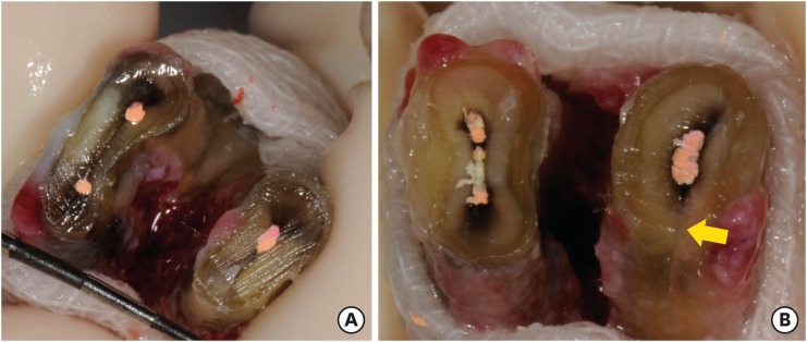
ePub Objectives Understanding the reason for an unsuccessful non-surgical endodontic treatment outcome, as well as the complex anatomy of the root canal system, is very important. This study examined the cross-sectional root canal structure of mandibular first molars confirmed to have failed non-surgical root canal treatment using digital images obtained during intentional replantation surgery, as well as the causative factors of the failed conventional endodontic treatments.
Materials and Methods This study evaluated 115 mandibular first molars. Digital photographic images of the resected surface were taken at the apical 3 mm level and examined. The discolored dentin area around the root canal was investigated by measuring the total surface area, the treated areas as determined by the endodontic filling material, and the discolored dentin area.
Results Forty 2-rooted teeth showed discolored root dentin in both the mesial and distal roots. Compared to the original filled area, significant expansion of root dentin discoloration was observed. Moreover, the mesial roots were significantly more discolored than the distal roots. Of the 115 molars, 92 had 2 roots. Among the mesial roots of the 2-rooted teeth, 95.7% of the roots had 2 canals and 79.4% had partial/complete isthmuses and/or accessory canals.
Conclusions Dentin discoloration that was not visible on periapical radiographs and cone-beam computed tomography was frequently found in mandibular first molars that failed endodontic treatment. The complex anatomy of the mesial roots of the mandibular first molars is another reason for the failure of conventional endodontic treatment.
-
Citations
Citations to this article as recorded by
- In vitro evaluation of the sealing ability of combined use of iRoot BP Plus and iRoot SP for root-end filling
Xu Dong, Qian Xie, Xin Xu
Clinical Oral Investigations.2023; 27(6): 2969. CrossRef - The Impact of the Preferred Reporting Items for Case Reports in Endodontics (PRICE) 2020 Guidelines on the Reporting of Endodontic Case Reports
Sofian Youssef, Phillip Tomson, Amir Reza Akbari, Natalie Archer, Fayjel Shah, Jasmeet Heran, Sunmeet Kandhari, Sandeep Pai, Shivakar Mehrotra, Joanna M Batt
Cureus.2023;[Epub] CrossRef - Clinical diagnostic approach in the treatment of chronic periodontitis in mandibular molars: Clinical cases
M. A. Postnikov, A. M. Golovachev, S. E. Chigarina, D. N. Kudryashov, I. A. Zakharova, S. A. Burakshaev
Kuban Scientific Medical Bulletin.2023; 30(5): 100. CrossRef - Evaluation of interorifice distance in permanent mandibular first molar with middle mesial canal in Bengaluru city, Karnataka: A cone-beam computed tomography study
Shruthika Mahajan, N. Meena, Anithakumari Rangappa, Ali Mohammed Mashood, Chethana Murthy, M. Lokapriya
Endodontology.2023; 35(2): 100. CrossRef - A comparative study of the effects of gutta‐percha solvents on human osteoblasts and murine fibroblasts
Gul Ipek Gundogan, Sare Durmus, Gulgun Cansu Ozturk, Nazmi Kucukyesil, Yasin Talat Acar, Rumeysa Balaban, Cenk Kig
Australian Endodontic Journal.2021; 47(3): 569. CrossRef - Endodontic retreatment of curved root canals using the dual wavelength erbium, chromium:yttrium, scandium, gallium, garnet, and diode 940-nm lasers and the XP-Endoshaper/finisher technique
Riman Nasher, Ralf-Dieter Hilgers, Norbert Gutknecht
Lasers in Dental Science.2020; 4(4): 211. CrossRef - Evaluation of gutta-percha removal from the dentinal tubules using different instrumentation techniques with or without solvent: An In vitro study
MukeshKumar Hasija, Babita Meena, Deepti Wadhwa, KulvinderKaur Wadhwani, Virender Yadav
Journal of the International Clinical Dental Research Organization.2020; 12(1): 27. CrossRef
- In vitro evaluation of the sealing ability of combined use of iRoot BP Plus and iRoot SP for root-end filling
- 2,385 View
- 10 Download
- 7 Crossref

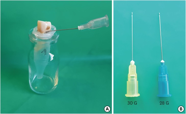
- Influence of size and insertion depth of irrigation needle on debris extrusion and sealer penetration
- Emel Uzunoglu-Özyürek, Hakan Karaaslan, Sevinç Aktemur Türker, Bahar Özçelik
- Restor Dent Endod 2018;43(1):e2. Published online December 22, 2017
- DOI: https://doi.org/10.5395/rde.2018.43.e2
-
Abstract
PDFPubReaderePub
Objectives To determine the effect of size and insertion depth of irrigation needle on the amount of apical extruded debris and the amount of penetration depth of sealer using a confocal laser scanning microscope (CLSM).
Materials and Methods Twenty maxillary premolars were assigned to 2 groups (
n = 10), according to the size of needle tip, 28 G or 30 G. Buccal roots of samples were irrigated with respective needle type inserted 1 mm short of the working length (WL), while palatal roots were irrigated with respective needle type inserted 3 mm short of the WL. Prepared teeth were removed from the pre-weighed Eppendorf tubes. Canals were filled with F3 gutta-percha cone and rhodamine B dye-labeled AH 26 sealer. Teeth were transversally sectioned at 1 and 3 mm levels from the apex and observed under a CLSM. Eppendorf tubes were incubated to evaporate the irrigant and were weighed again. The difference between pre- and post-weights was calculated, and statistical evaluation was performed.Results Inserting needles closer to the apex and using needles with wider diameters were associated with significantly more debris extrusion (
p < 0.05). The position of needles and level of sections had statistically significant effects on sealer penetration depth (p < 0.05 for both).Conclusions Following preparation, inserting narrower needles compatible with the final apical diameter of the prepared root canal at 3 mm short of WL during final irrigation might prevent debris extrusion and improve sealer penetration in the apical third.
-
Citations
Citations to this article as recorded by- Effect of laser-induced pulpal anesthesia of single-rooted teeth with irreversible pulpitis treated by single-visit root canal therapy - A randomized clinical trial
Geeta Asthana, Dhwani Morakhia, Ravina Parmar, Rajashree Tamuli
Endodontology.2025; 37(3): 244. CrossRef - Efficacy of different irrigation needles used in endodontics: an in silico and an in vitro investigation
Maulee Sheth, Ankit Arora, Sonali Kapoor, Balraj Shukla
Biomaterial Investigations in Dentistry.2025; 12: 264. CrossRef - Preliminary insights: exploring irrigation practices during endodontic treatment among general dental practitioners in Malaysia
Kai Qi Chiew, Xin Ni Lim, Shekhar Bhatia, Naveen Chhabra
British Dental Journal.2024;[Epub] CrossRef - Efficiency of diode laser in control of post-endodontic pain: a randomized controlled trial
Hend H. Ismail, Maram Obeid, Ehab Hassanien
Clinical Oral Investigations.2023; 27(6): 2797. CrossRef - Endodontic management of an aberrant germinated composite odontome: A case report
Ankit Arora, Kavina Desai, Sonali Kapoor, Seema Gajera
Australian Endodontic Journal.2023; 49(3): 684. CrossRef - Potentials of 3D-Modeling in the Preclinical Stage of Root Needle Research
Aleksandr V. Kuligin, Larisa N. Kazakova, Oksana S. Tereshchuk, Vadim V. Bokov
I.P. Pavlov Russian Medical Biological Herald.2022; 30(1): 95. CrossRef - Effect of root canal geometry and needle type on apical extrusion of irrigant: an ex vivo study
Büşra SERÇE FİKİRLİ, Bülent ALTUNKAYNAK, Güven KAYAOĞLU
Acta Odontologica Turcica.2022; 39(3): 58. CrossRef - An in vitro radiological evaluation of irrigant penetration in the root canals using three different irrigation systems: Waterpik WP-100 device, passive irrigation, and manual dynamic irrigation systems
Suragani Hemalatha, Archana Srinivasan, A Srirekha, Lekha Santhosh, C Champa, Ashwija Shetty
Journal of Conservative Dentistry.2022; 25(4): 403. CrossRef - Preparation Ability of ProTaper Next and XP-endo Shaper Instruments in Isthmus-containing Root Canal System
Mustafa Sarıkahya, Tayfun Alaçam
Conservative Dentistry and Endodontic Journal.2021; 5(2): 28. CrossRef - Penetration depth of irrigants into root dentine after sonic, ultrasonic and photoacoustic activation
K. M. Galler, V. Grubmüller, R. Schlichting, M. Widbiller, A. Eidt, C. Schuller, M. Wölflick, K.‐A. Hiller, W. Buchalla
International Endodontic Journal.2019; 52(8): 1210. CrossRef
- Effect of laser-induced pulpal anesthesia of single-rooted teeth with irreversible pulpitis treated by single-visit root canal therapy - A randomized clinical trial
- 2,572 View
- 25 Download
- 10 Crossref
 First
First Prev
Prev

