
Search
- Page Path
- HOME > Search
Research Article
-
The influence of sodium hypochlorite concentration on the fibrin structure of human blood clots and transforming growth factor-beta 1 release: an
ex vivo study - Anisha Mishra, Velmurugan Natanasabapathy, Nandini Suresh
- Restor Dent Endod 2022;47(4):e42. Published online October 31, 2022
- DOI: https://doi.org/10.5395/rde.2022.47.e42
-
 Abstract
 PDF
PDF Supplementary Material
 PubReader
 ePub
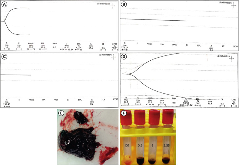
ePub Objective This study investigated the effects of various concentrations of sodium hypochlorite (NaOCl) on human whole-blood clotting kinetics, the structure of the blood clots formed, and transforming growth factor (TGF)-β1 release.
Materials and Methods Human whole blood was collected from 5 healthy volunteers and divided into 4 groups: CG (control, 0.5 mL of blood), BN0.5 (0.5 mL of blood with 0.5 mL of 0.5% NaOCl), BN3 (0.5 mL of blood with 0.5 mL of 3% NaOCl), and BN5.25 (0.5 mL of blood with 0.5 mL of 5.25% NaOCl). The effects of NaOCl on clotting kinetics, structure of fibrin and cells, and release of TGF-β1 were assessed using thromboelastography (TEG), scanning electron microscopy (SEM), and enzyme-linked immunosobent assay, respectively. Statistical analysis was conducted using the Kruskal Wallis and Mann-Whitney
U tests, followed by thepost hoc Dunn test. Ap value < 0.05 indicated statistical significance.Results The blood samples in BN0.5 and BN3 did not clot, whereas the TEG of BN5.25 showed altered clot formation. Samples from the CG and BN3 groups could only be processed with SEM, which showed that the latter lacked fibrin formation and branching of fibers, as well as clumping of red blood cells with surface roughening and distortion. TGF-β1 release was significantly highest in BN3 when all groups were compared to CG (
p < 0.05).Conclusions Each concentration of NaOCl affected the release of TGF-β1 from blood clots and altered the clotting mechanism of blood by affecting clotting kinetics and cell structure.
-
Citations
Citations to this article as recorded by
- Evaluation of the cytotoxicity of a broad-spectrum antiseptic using a model of erythrocyte hemolysis in an in vitro experiment
S. P. Rubnikovich, O. E. Bekjanova, L. E. Khasanova, Sh. F. Shamsieva, S. X. Alimova, M. M. Astanakulova, N. T. Babadjanova, X. Sh. Mirzaev
Proceedings of the National Academy of Sciences of Belarus, Medical series.2026; 23(2): 95. CrossRef - Effect of Adjunctive Ozone Application Protocols on Dentin-Derived Growth Factor Release: An In Vitro Study
Sude Göbüt, Melis Oya Ateş, Ali Keleş, Fatma Avcıoğlu
Journal of Clinical Medicine.2026; 15(11): 4277. CrossRef - Cytotoxic Effects of Synthetic and Herbal Endodontic Irrigants on Human Red Blood Cells: An In Vitro Study
Panna Mangat, Bhaviya Chandel, Mampi Biswas, Sara Trivedy, Akshata Gupta, Nayan Shree, Seema Gupta
Cureus.2025;[Epub] CrossRef
- Evaluation of the cytotoxicity of a broad-spectrum antiseptic using a model of erythrocyte hemolysis in an in vitro experiment
- 3,058 View
- 39 Download
- 1 Web of Science
- 3 Crossref

Original Articles
- The effect of mineral trioxide aggregate on the production of growth factors and cytokine by human periodontal ligament fibroblasts
- Ji-Yoon Kwon, Sung-Sam Lim, Seung-Ho Baek, Kwang-Shik Bae, Myung-Hoe Kang, Woocheol Lee
- J Korean Acad Conserv Dent 2007;32(3):191-197. Published online May 31, 2007
- DOI: https://doi.org/10.5395/JKACD.2007.32.3.191
-
Abstract
PDFPubReaderePub
Mineral trioxide aggregate (MTA) would influence healing of periapical tissues by modulating the production of growth factors and cytokines from PDL fibroblasts, however, the studies are insufficient. Therefore, the purpose of this study was to monitor the expression of transforming growth factor-beta1 (TGF-β1), fibroblast growth factor-2 (FGF-2), and interleukin-6 (IL-6) from PDL fibroblasts in the presence of MTA. The human PDL fibroblasts were seeded onto the set MTA or IRM at a level of 1 × 10⁵ cells per unit well, and further incubated for 6, 12, 24, and 48 hours. The levels of TGF-β1, FGF-2, and IL-6 from the supernatant were measured by enzyme-linked immunosorbent assay (ELISA). The data were analyzed using one-way ANOVA. The level of TGF-β1 was down-regulated when the cells were grown in the presence of MTA except at 6 hours. The levels of FGF-2 release were significantly suppressed when PDL fibroblasts were grown in the presence of MTA or IRM at all time intervals (p < 0.05). The expressions of IL-6 from MTA treated cells were comparable to those of untreated control cells throughout the observation periods. We presume that this material inhibits the stimulatory function of growth factors on granulation tissue formation and in turn, it promotes the healing process modulated by other bone-remodeling cells.
-
Citations
Citations to this article as recorded by- Osteo/odontogenic Differentiation of Human Mesenchymal Stem Cells with Platelet-rich Plasma and Mineral Trioxide Aggregate
Shanthi Vanka, Amit Vanka, Sandeep Kumar Vishwakarma, Manohar K Bhat, Othman Wali, Aleem A Khan
The Journal of Contemporary Dental Practice.2019; 20(10): 1171. CrossRef - The effect of several root-end filling materials on MG63 osteoblast-like cells
Jeong-Ho Lee, Won-Jun Shon, WooCheol Lee, Seung-Ho Baek
Journal of Korean Academy of Conservative Dentistry.2010; 35(3): 222. CrossRef - Biocompatibility of experimental mixture of mineral trioxide aggregate and glass ionomer cement
Min-Jae Oh, Yu-Na Jeong, In-Ho Bae, So-Young Yang, Bum-Jun Park, Jeong-Tae Koh, Yun-Chan Hwang, In-Nam Hwang, Won-Mann Oh
Journal of Korean Academy of Conservative Dentistry.2010; 35(5): 359. CrossRef - Biocompatibility of bioaggregate cement on human pulp and periodontal ligament (PDL) derived cells
Choo-Ryung Chung, Euiseong Kim, Su-Jung Shin
Journal of Korean Academy of Conservative Dentistry.2010; 35(6): 473. CrossRef - Effects of condensation techniques and canal sizes on the microleakage of orthograde MTA apical plug in simulated canals
Deuk-Lim Nam, Jeong-Kil Park, Bock Hur, Hyeon-Cheol Kim
Journal of Korean Academy of Conservative Dentistry.2009; 34(3): 208. CrossRef
- Osteo/odontogenic Differentiation of Human Mesenchymal Stem Cells with Platelet-rich Plasma and Mineral Trioxide Aggregate
- 2,157 View
- 5 Download
- 5 Crossref
-
Effects of
Enterococcus faecalis sonicated extracts on IL-2, IL-4 and TGF-β1 production from human lymphocytes - Hyeon-Sik Kim, Seok-Woo Jang, Wan-Jun Shon, Song-Takg Lee, Cheol-Ho Kim, Woo-Cheol Lee, Sung-Sam Lim
- J Korean Acad Conserv Dent 2005;30(1):1-6. Published online January 31, 2005
- DOI: https://doi.org/10.5395/JKACD.2005.30.1.001
-
Abstract
PDFPubReaderePub
In order to examine the immunoresponse of host cells to
Enterococcus faecalis , this in vitro study monitored the production of Interleukin-2 (IL-2), Interleukin-4 (IL-4) and Transforming growth factor-β1 (TGF-β1) in human lymphocytes. Lymphocytes were activated with PHA in the presence or abscence of sonicated extracts ofE. Faecalis (SEF) and further incubated for 72 hours. The level of each cytokine was measured by ELISA. Data were analyzed with Kruskal-Wallis test and Mann-Whitney U test (P < 0.05). PHA-activated group did exhibit higher level of IL-2 and IL-4 than untreated control group. The levels of expression of both cytokines were significantly decreased following the treatment of high (25µg/ml ) and medium concentration (12.5µg/ml ) of SEF (P <0 .05) than those of PHA activated group. But low concentration (5µg/ml ) of SEF showed the similar level of IL-2 and IL-4 production as those of PHA activated group. TGF-β1 was unaffected by SEF treatment. These results suggested thatE. faecalis may suppress IL-2 and IL-4 production by lymphocytes and this could be one of possible factors whyE. faecalis are found frequently in the teeth with failed endodontic treatment.-
Citations
Citations to this article as recorded by- Immune System of Dental Pulp in Inflamed and Normal Tissue
Sepideh Sarfi, Ehsaneh Azaryan, Mohsen Naseri
DNA and Cell Biology.2024; 43(8): 369. CrossRef - Role(s) of cytokines in pulpitis: Latest evidence and therapeutic approaches
Mohammad M.Y. Khorasani, Gholamhossein Hassanshahi, Aniela Brodzikowska, Hossein Khorramdelazad
Cytokine.2020; 126: 154896. CrossRef - Educational needs of an integrated health and oral health project for community dental hygienists
Su-kyung Park, Yang-Keum Han, Young-Kyung Kim, Hyun-Ju Lim, Yang-Ok Kown, Han-Mi Kim, Mag-Yup Oh, Nam-Hee Kim
Journal of Korean Academy of Oral Health.2015; 39(2): 127. CrossRef - Microorganism penetration in dentinal tubules of instrumented and retreated root canal walls.In vitroSEM study
Saad Al-Nazhan, Alaa Al-Sulaiman, Fellwa Al-Rasheed, Fatimah Alnajjar, Bander Al-Abdulwahab, Abdulhakeem Al-Badah
Restorative Dentistry & Endodontics.2014; 39(4): 258. CrossRef
- Immune System of Dental Pulp in Inflamed and Normal Tissue
- 2,297 View
- 2 Download
- 4 Crossref
 First
First Prev
Prev

