Search
- Page Path
- HOME > Search
- Bioblock technique to treat severe internal resorption with subsequent periapical pathology: a case report
- Márk Fráter, Tekla Sáry, Sufyan Garoushi
- Restor Dent Endod 2020;45(4):e43. Published online August 18, 2020
- DOI: https://doi.org/10.5395/rde.2020.45.e43

-
 Abstract
 PDF
PDF PubReader
 ePub
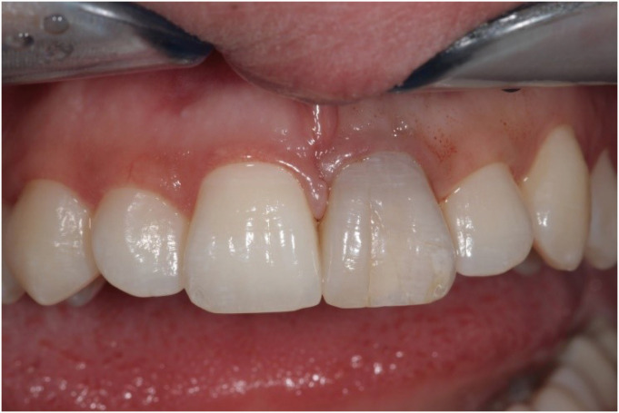
ePub A variety of therapeutic modalities can be used for the endodontic treatment of a traumatized tooth with internal root resorption (IRR). The authors present a case report of the successful restoration of a traumatized upper central incisor that was weakened due to severe IRR and subsequent periapical lesion formation. A 20-year-old female patient was referred to our clinic with severe internal resorption and subsequent periapical pathosis destroying the buccal bone wall. Root canal treatment had been initiated previously at another dental practice, but at that time, the patient's condition could not be managed even with several treatments. After cone-beam computed tomography imaging and proper chemomechanical cleaning, the tooth was managed with a mineral trioxide aggregate plug followed by root canal filling using short fiber-reinforced composite, known as the Bioblock technique. This report is the first documentation of the use of the Bioblock technique in the restoration of a traumatized tooth. The Bioblock technique appears to be ideal for restoring wide irregular root canals, as in cases of severe internal resorption, because it can uniquely fill out the hollow irregularities of the canal. However, further long-term clinical investigations are required to provide additional information about this new technique.
-
Citations
Citations to this article as recorded by- Üvegszálas fogászati kompozit tömőanyag keménysége a gyökércsatornában: nanoindentációs vizsgálat
András Jakab, Kata Lilla Vánkay, Tamás Tarjányi, Gábor Gulyás, Krisztián Bali, Pál Patrik Dézsi, Márton Sámi, Márk Fráter
Fogorvosi Szemle.2024; 117(2): 47. CrossRef - Evaluation of microhardness of short fiber-reinforced composites inside the root canal after different light curing methods – An in vitro study
Márk Fráter, János Grosz, András Jakab, Gábor Braunitzer, Tamás Tarjányi, Gábor Gulyás, Krisztián Bali, Paula Andrea Villa-Machado, Sufyan Garoushi, András Forster
Journal of the Mechanical Behavior of Biomedical Materials.2024; 150: 106324. CrossRef - Imaging techniques and various treatment modalities used in the management of internal root resorption: A systematic review
R. S Digholkar, S D Aggarwal, P S Kurtarkar, P. B Dhatavkar, V L Neil, D N Agarwal
Endodontology.2023; 35(2): 85. CrossRef - The Impact of the Preferred Reporting Items for Case Reports in Endodontics (PRICE) 2020 Guidelines on the Reporting of Endodontic Case Reports
Sofian Youssef, Phillip Tomson, Amir Reza Akbari, Natalie Archer, Fayjel Shah, Jasmeet Heran, Sunmeet Kandhari, Sandeep Pai, Shivakar Mehrotra, Joanna M Batt
Cureus.2023;[Epub] CrossRef - Fatigue performance of endodontically treated premolars restored with direct and indirect cuspal coverage restorations utilizing fiber-reinforced cores
Márk Fráter, Tekla Sáry, Janka Molnár, Gábor Braunitzer, Lippo Lassila, Pekka K. Vallittu, Sufyan Garoushi
Clinical Oral Investigations.2022; 26(4): 3501. CrossRef
- Üvegszálas fogászati kompozit tömőanyag keménysége a gyökércsatornában: nanoindentációs vizsgálat
- 3,217 View
- 93 Download
- 5 Crossref

- Fibre reinforcement in a structurally compromised endodontically treated molar: a case report
- Renita Soares, Ida de Noronha de Ataide, Marina Fernandes, Rajan Lambor
- Restor Dent Endod 2016;41(2):143-147. Published online February 22, 2016
- DOI: https://doi.org/10.5395/rde.2016.41.2.143
-
 Abstract
 PDF
PDF PubReader
 ePub
ePub The reconstruction of structurally compromised posterior teeth is a rather challenging procedure. The tendency of endodontically treated teeth (ETT) to fracture is considerably higher than vital teeth. Although posts and core build-ups followed by conventional crowns have been generally employed for the purpose of reconstruction, this procedure entails sacrificing a considerable amount of residual sound enamel and dentin. This has drawn the attention of researchers to fibre reinforcement. Fibre-reinforced composite (FRC), designed to replace dentin, enables the biomimetic restoration of teeth. Besides improving the strength of the restoration, the incorporation of glass fibres into composite resins leads to favorable fracture patterns because the fibre layer acts as a stress breaker and stops crack propagation. The following case report presents a technique for reinforcing a badly broken-down ETT with biomimetic materials and FRC. The proper utilization of FRC in structurally compromised teeth can be considered to be an economical and practical measure that may obviate the use of extensive prosthetic treatment.
-
Citations
Citations to this article as recorded by- Performance of direct and indirect onlay restorations for structurally compromised teeth
Khaled Abid Althaqafi
The Journal of Prosthetic Dentistry.2025; 133(6): 1513. CrossRef - Endodontically Treated Teeth with Fiber-Reinforced Composite Resins
Ridhima Gupta, Ashwini B. Prasad, Deepak Raisingani, Deeksha Khurana, Prachi Mital, Vaishali Moryani
Journal of Dental Research and Review.2022; 9(4): 310. CrossRef - Survival and success of endocrowns: A systematic review and meta-analysis
Raghad A. Al-Dabbagh
The Journal of Prosthetic Dentistry.2021; 125(3): 415.e1. CrossRef - Short fiber‐reinforced composite restorations: A review of the current literature
Sufyan Garoushi, Ausama Gargoum, Pekka K. Vallittu, Lippo Lassila
Journal of Investigative and Clinical Dentistry.2018;[Epub] CrossRef
- Performance of direct and indirect onlay restorations for structurally compromised teeth
- 1,852 View
- 42 Download
- 4 Crossref

- The effect of reinforcing methods on fracture strength of composite inlay bridge
- Chang-Won Byun, Sang-Hyuk Park, Sang-Jin Park, Kyoung-Kyu Choi
- J Korean Acad Conserv Dent 2007;32(2):111-120. Published online March 31, 2007
- DOI: https://doi.org/10.5395/JKACD.2007.32.2.111
-
 Abstract
 PDF
PDF PubReader
 ePub
ePub The purpose of this study is to evaluate the effects of surface treatment and composition of reinforcement material on fracture strength of fiber reinforced composite inlay bridges.
The materials used for this study were I-beam, U-beam TESCERA ATL system and ONE STEP(Bisco, IL, USA). Two kinds of surface treatments were used; the silane and the sandblast. The specimens were divided into 11 groups through the composition of reinforcing materials and the surface treatments.
On the dentiform, supposing the missing of Maxillary second pre-molar and indirect composite inlay bridge cavities on adjacent first pre-molar disto-occlusal cavity, first molar mesio-occlusal cavity was prepared with conventional high-speed inlay bur.The reinforcing materials were placed on the proximal box space and build up the composite inlay bridge consequently. After the curing, specimen was set on the testing die with ZPC. Flexural force was applied with universal testing machine (EZ-tester; Shimadzu, Japan). at a cross-head speed of 1 mm/min until initial crack occurred. The data wasanalyzed using one-way ANOVA/Scheffes' post-hoc test at 95% significance level.
Groups using I-beam showed the highest fracture strengths (p < 0.05) and there were no significant differences between each surface treatment (p > 0.05). Most of the specimens in groups that used reinforcing material showed delamination.
The use of I-beam represented highest fracture strengths (p < 0.05).
In groups only using silane as a surface treatment showed highest fracture strength, but there were no significant differences between other surface treatments (p > 0.05).
The reinforcing materials affect the fracture strength and pattern of composites inlay bridge.
The holes at the U-beam did not increase the fracture strength of composites inlay bridge.
-
Citations
Citations to this article as recorded by- Esthetic rehabilitation of single anterior edentulous space using fiber-reinforced composite
Hyeon Kim, Min-Ju Song, Su-Jung Shin, Yoon Lee, Jeong-Won Park
Restorative Dentistry & Endodontics.2014; 39(3): 220. CrossRef
- Esthetic rehabilitation of single anterior edentulous space using fiber-reinforced composite
- 1,031 View
- 3 Download
- 1 Crossref


 KACD
KACD

 First
First Prev
Prev


