Search
- Page Path
- HOME > Search
- Endodontic micro-resurgery and guided tissue regeneration of a periapical cyst associated to recurrent root perforation: a case report
- Fernando Córdova-Malca, Hernán Coaguila-Llerena, Lucía Garré-Arnillas, Jorge Rayo-Iparraguirre, Gisele Faria
- Restor Dent Endod 2022;47(4):e35. Published online September 3, 2022
- DOI: https://doi.org/10.5395/rde.2022.47.e35

-
 Abstract
 PDF
PDF PubReader
 ePub
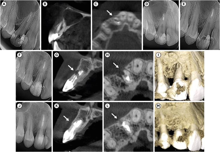
ePub Although the success rates of microsurgery and micro-resurgery are very high, the influence of a recurrent perforation combined with radicular cyst remains unclear. A 21-year-old white female patient had a history of root perforation in a previously treated right maxillary lateral incisor. Analysis using cone-beam computed tomography (CBCT) revealed an extensive and well-defined periapical radiolucency, involving the buccal and palatal bone plate. The perforation was sealed with bioceramic material (Biodentine) in the pre-surgical phase. In the surgical phase, guided tissue regeneration (GTR) was performed by combining xenograft (lyophilized bovine bone) and autologous platelet-rich fibrin applied to the bone defect. The root-end preparation was done using an ultrasonic tip. The retrograde filling was performed using a bioceramic material (Biodentine). Histopathological analysis confirmed a radicular cyst. The patient returned to her referring practitioner to continue the restorative procedures. CBCT analysis after 1-year recall revealed another perforation in the same place as the first intervention, ultimately treated by micro-resurgery using the same protocol with GTR, and a bioceramic material (MTA Angelus). The 2-year recall showed healing and bone neoformation. In conclusion, endodontic micro-resurgery with GTR showed long-term favorable results when a radicular cyst and a recurrent perforation compromised the success.
-
Citations
Citations to this article as recorded by- Outcome of endodontic micro-resurgery: A systematic review
Faisal Alnassar, Riyadh Alroomy, Qamar Hashem, Abdullah Alqedairi, Nabeel Almotairy
Saudi Endodontic Journal.2025; 15(2): 112. CrossRef - Platelet-Rich Plasma and Platelet-Rich Fibrin in Endodontics: A Scoping Review
Simão Rebimbas Guerreiro, Carlos Miguel Marto, Anabela Paula, Joana Rita de Azevedo Pereira, Eunice Carrilho, Manuel Marques-Ferreira, Siri Vicente Paulo
International Journal of Molecular Sciences.2025; 26(12): 5479. CrossRef - Non-surgical Approach to a Maxillary Cyst-Like Lesion: Orthograde Endodontic Treatment With Neodymium-Doped Yttrium Aluminum Garnet (Nd:YAG) Decontamination of the Canal System
Beatrice Spaggiari, Paolo Vescovi, Silvia Pizzi, Roberta Iaria, Ilaria Giovannacci
Cureus.2025;[Epub] CrossRef - Persistent Periradicular Lesion Associated With Concurrent Root Fracture and Odontogenic Keratocyst: A Case Report
Mehdi Vatanpour, Fatemeh Rezaei
Clinical Case Reports.2025;[Epub] CrossRef - Management of Apico-marginal Defects With Endodontic Microsurgery and Guided Tissue Regeneration: A Report of Thirteen Cases
Abayomi O. Baruwa, Jorge N.R. Martins, Mariana D. Pires, Beatriz Pereira, Pedro May Cruz, António Ginjeira
Journal of Endodontics.2023; 49(9): 1207. CrossRef
- Outcome of endodontic micro-resurgery: A systematic review
- 2,736 View
- 53 Download
- 4 Web of Science
- 5 Crossref

- Progression of periapical cystic lesion after incomplete endodontic treatment
- Jong-Ki Huh, Dong-Kyu Yang, Kug-Jin Jeon, Su-Jung Shin
- Restor Dent Endod 2016;41(2):137-142. Published online February 22, 2016
- DOI: https://doi.org/10.5395/rde.2016.41.2.137
-
 Abstract
 PDF
PDF PubReader
 ePub
ePub We report a case of large radicular cyst progression related to endodontic origin to emphasize proper intervention and follow-up for endodontic pathosis. A 25 yr old man presented with an endodontically treated molar with radiolucency. He denied any intervention because of a lack of discomfort. Five years later, the patient returned. The previous periapical lesion had drastically enlarged and involved two adjacent teeth. Cystic lesion removal and apicoectomy were performed on the tooth. Histopathological analysis revealed that the lesion was an inflammatory radicular cyst. The patient did not report any discomfort except for moderate swelling 3 days after the surgical procedure. Although the patient had been asymptomatic, close follow-ups are critical to determine if any periapical lesions persist after root canal treatment.
-
Citations
Citations to this article as recorded by- Prognosis of Vital Teeth Involved in Large Cystic Lesions After a Surgical Intervention: A Longitudinal Ambidirectional Cohort Study
Khalid A. Merdad, Maha Shawky, Khalid A. Aljohani, Rawia Alghamdi, Saja Alzahrani, Omar R. Alkhattab, Abdulaziz Bakhsh
Dentistry Journal.2025; 13(2): 83. CrossRef - Assessing the efficacy of apicoectomy without retrograde filling in treating periapical inflammatory cysts
Jeong-Kui Ku, Woo-Young Jeon, Seung-O Ko, Ji-Young Yoon
Journal of the Korean Association of Oral and Maxillofacial Surgeons.2024; 50(3): 140. CrossRef - Cystic lesion between a deciduous tooth and the succeeding permanent tooth: a retrospective analysis of 87 cases
Changmo Sohn, Jihye Ryu, Inhye Nam, Sang-Hun Shin, Jae-Yeol Lee
Journal of the Korean Association of Oral and Maxillofacial Surgeons.2022; 48(6): 342. CrossRef - The effectiveness of antibacterial treatment of the root canal in chronic apical periodontitis using an erbium-chromium laser
M. A. Postnikov, A. Yu. Rozenbaum, S. E. Chigarina, D. N. Kudryashov, M. B. Khaikin, I. V. Khramova, G. N. Belanov
Endodontics Today.2022; 20(2): 115. CrossRef - Factors Associated with Incomplete Endodontic Care
Carla Y. Falcon, Anthony R. Arena, Rebecca Hublall, Craig S. Hirschberg, Paul A. Falcon
Journal of Endodontics.2021; 47(9): 1398. CrossRef - Tratamento cirúrgico e conservador de cisto periapical de grande proporção: relato de caso
Maraísa Aparecida Pinto Resende, Neuza Maria Souza Picorelli Assis, Augusto César Sette-Dias, Evandro Guimarães de Aguiar, Bruno Salles Sotto-Maior
HU Revista.2018; 43(2): 191. CrossRef
- Prognosis of Vital Teeth Involved in Large Cystic Lesions After a Surgical Intervention: A Longitudinal Ambidirectional Cohort Study
- 2,601 View
- 14 Download
- 6 Crossref


 KACD
KACD

 First
First Prev
Prev


