
Search
- Page Path
- HOME > Search
Case Reports
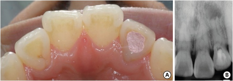
- A case report of multiple bilateral dens invaginatus in maxillary anteriors
- Shin Hye Chung, You-Jeong Hwang, Sung-Yeop You, Young-Hye Hwang, Soram Oh
- Restor Dent Endod 2019;44(4):e39. Published online October 21, 2019
- DOI: https://doi.org/10.5395/rde.2019.44.e39
-
 Abstract
 PDF
PDF PubReader
 ePub
ePub The present report presents a case of dens invaginatus (DI) in a patient with 4 maxillary incisors. A 24-year-old female complained of swelling of the maxillary left anterior region and discoloration of the maxillary left anterior tooth. The maxillary left lateral incisor (tooth #22) showed pulp necrosis and a chronic apical abscess, and a periapical X-ray demonstrated DI on bilateral maxillary central and lateral incisors. All teeth responded to a vitality test, except tooth #22. The anatomic form of tooth #22 was similar to that of tooth #12, and both teeth had lingual pits. In addition, panoramic and periapical X-rays demonstrated root canal calcification, such as pulp stones, in the maxillary canines, first and second premolars, and the mandibular incisors, canines, and first premolars bilaterally. The patient underwent root canal treatment of tooth #22 and non-vital tooth bleaching. After a temporary filling material was removed, the invaginated mass was removed using ultrasonic tips under an operating microscope. The working length was established, and the root canal was enlarged up to #50 apical size and obturated with gutta-percha and AH 26 sealer using the continuous wave of condensation technique. Finally, non-vital bleaching was performed, and the access cavity was filled with composite resin.
-
Citations
Citations to this article as recorded by
- Non-surgical endodontic management of dens invaginatus type II in an immature maxillary lateral incisor using a bioceramic apical plug: A 3-year follow-up case report
Yahya Raja Alharbi, Qayed Saad Alharbi, Shaul Hameed Kolarkodi
Saudi Endodontic Journal.2026; 16(1): 115. CrossRef - The use of three-dimensional-printed guides, static navigation, and bioactive materials to treat bilateral and double dens invaginatus
Parth Patel, Nidhi Bharti, Ankit Arora, C. Nimisha Shah
Saudi Endodontic Journal.2025; 15(2): 207. CrossRef - Endodontic Management of Dens in Dente – A Systematic Review of Case Reports and Case Series
Sanket Dilip Aras, Anamika Chetan Borkar, Sonal Kale, Sayali Maral, Prakriti Jaggi, Shailendra Sonawane
Journal of the International Clinical Dental Research Organization.2024; 16(1): 17. CrossRef - Dens invaginatus of fourteen teeth in a pediatric patient
Momoko Usuda, Tatsuya Akitomo, Mariko Kametani, Satoru Kusaka, Chieko Mitsuhata, Ryota Nomura
Pediatric Dental Journal.2023; 33(3): 240. CrossRef - The Impact of the Preferred Reporting Items for Case Reports in Endodontics (PRICE) 2020 Guidelines on the Reporting of Endodontic Case Reports
Sofian Youssef, Phillip Tomson, Amir Reza Akbari, Natalie Archer, Fayjel Shah, Jasmeet Heran, Sunmeet Kandhari, Sandeep Pai, Shivakar Mehrotra, Joanna M Batt
Cureus.2023;[Epub] CrossRef - Root Maturation of an Immature Dens Invaginatus Despite Unsuccessful Revitalization Procedure: A Case Report and Recommendations for Educational Purposes
Julia Ludwig, Marcel Reymus, Alexander Winkler, Sebastian Soliman, Ralf Krug, Gabriel Krastl
Dentistry Journal.2023; 11(2): 47. CrossRef - Conservative Management of Infraorbital Space Infection Secondary to Type III B Dens Invaginatus: A Case Report
Ashima Goyal, Aditi Kapur, Manoj A Jaiswal, Gauba Krishan, Raja Raghu, Sanjeev K Singh
Journal of Postgraduate Medicine, Education and Research.2022; 56(4): 192. CrossRef
- Non-surgical endodontic management of dens invaginatus type II in an immature maxillary lateral incisor using a bioceramic apical plug: A 3-year follow-up case report
- 3,597 View
- 35 Download
- 7 Crossref

- Endodontic management of a maxillary first molar with three roots and seven root canals with the aid of cone-beam computed tomography
- Gurudutt Nayak, Kamal Krishan Singh, Rhitu Shekhar
- Restor Dent Endod 2015;40(3):241-248. Published online June 3, 2015
- DOI: https://doi.org/10.5395/rde.2015.40.3.241
-
Abstract
PDFPubReaderePub
Variation in root canal morphology, especially in maxillary first molar presents a constant challenge for a clinician in their detection and management. This case report describes the successful root canal treatment of a three rooted right maxillary first molar presenting with three canals each in the mesiobuccal and distobuccal roots and one canal in the palatal root. The clinical detection of this morphologic aberration was made using a dental operating microscope, and the canal configuration was established after correlating and computing the clinical, radiographic and cone-beam computed tomography (CBCT) scan findings. CBCT images confirmed the configuration of the canals in the mesiobuccal and distobuccal roots to be Al-Qudah and Awawdeh type (3-2) and type (3-2-1), respectively, whereas the palatal root had a Vertucci type I canal pattern. This report reaffirms the importance of careful examination of the floor of the pulp chamber with a dental operating microscope and the use of multiangled preoperative radiographs along with advanced diagnostic aids such as CBCT in identification and successful management of aberrant canal morphologies.
-
Citations
Citations to this article as recorded by- Inhibition potential of rhamnolipid biosurfactant against Corynespora cassiicola – a phytopathogen of king chilli
Nilam Sarma, Suresh Deka, Hemen Deka
Studia Biologica.2025; 19(3): 153. CrossRef - Endodontic Management of Maxillary First Molar with Seven Root Canals Diagnosed Using Cone-beam Computed Tomography: A Case Report
Ravindranath Megha, Venkatachalam Prakash
World Journal of Dentistry.2021; 12(1): 89. CrossRef - The MB3 canal in maxillary molars: a micro-CT study
Ronald Ordinola-Zapata, Jorge N. R. Martins, Hugo Plascencia, Marco A. Versiani, Clovis M. Bramante
Clinical Oral Investigations.2020; 24(11): 4109. CrossRef - Maxillary first molar with 7 root canals diagnosed using cone-beam computed tomography
Evaldo Rodrigues, Antônio Henrique Braitt, Bruno Ferraz Galvão, Emmanuel João Nogueira Leal da Silva
Restorative Dentistry & Endodontics.2017; 42(1): 60. CrossRef - Endodontic management of a maxillary first molar with seven root canal systems evaluated using cone-beam computed tomography scanning
VijayReddy Venumuddala, Sridhar Moturi, SV Satish, BKalyan Chakravarthy, Sudhakar Malapati
Journal of International Society of Preventive and Community Dentistry.2017; 7(5): 297. CrossRef
- Inhibition potential of rhamnolipid biosurfactant against Corynespora cassiicola – a phytopathogen of king chilli
- 3,222 View
- 15 Download
- 5 Crossref
 First
First Prev
Prev

