Search
- Page Path
- HOME > Search
- Guided endodontics, precision and predictability: a case series of mineralized anterior teeth with follow-up cone-beam computed tomography
- Rafael Fernández-Grisales, Wilder Javier Rojas-Gutierrez, Pamela Mejía, Carolina Berruecos-Orozco, Néstor Ríos-Osorio
- Restor Dent Endod 2025;50(1):e4. Published online January 6, 2025
- DOI: https://doi.org/10.5395/rde.2025.50.e4

-
 Abstract
 PDF
PDF PubReader
 ePub
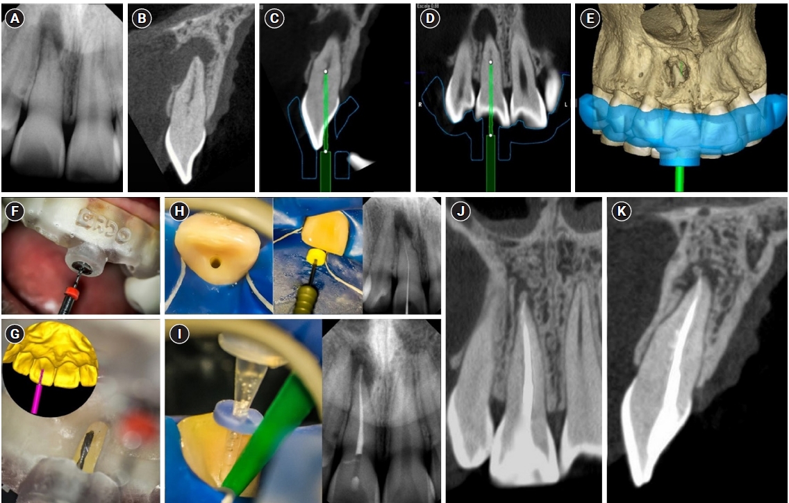
ePub - Pulp chamber and root canal obliteration (PCO/RCO) presents a challenge for clinicians when nonsurgical endodontic treatment is indicated. Guided endodontics (GE) aims to precisely locate the root canal (RC) system while preserving as much pericervical dentin as possible. GE involves integrating cone-beam computed tomography (CBCT) of the affected tooth with a digital impression of the maxillary/mandibular arch, allowing for careful planning of the drilling path to the RC system through a three-dimensional (3D) static guide. This article reports four cases of teeth with PCO/RCO, accompanied by additional diagnoses of internal and external root resorption and horizontal tooth fracture, all successfully treated with GE. These cases highlight the clinical and radiographic success of GE treatments using CBCT, establishing this technique as a predictable approach for managing mineralized teeth.
- 3,805 View
- 309 Download

- Fiber-reinforced composite post removal using guided endodontics: a case report
- Changgi Cho, Hyo Jin Jo, Jung-Hong Ha
- Restor Dent Endod 2021;46(4):e50. Published online September 23, 2021
- DOI: https://doi.org/10.5395/rde.2021.46.e50

-
 Abstract
 PDF
PDF PubReader
 ePub
ePub Although several techniques have been proposed to remove fiber-reinforced composite (FRC) post, no safe and efficient technique has been established. Recently, a guided endodontics technique has been introduced in cases of pulp canal obliteration. This study describes 2 cases of FRC post removal from maxillary anterior teeth using this guided endodontics technique with a dental operating microscope. Optically scanned data set from plaster cast model was superimposed with the data set of cone-beam computed tomography. By implant planning software, the path of a guide drill was selected. Based on them, a customized stent was fabricated and utilized to remove the FRC post. Employing guided endodontics, the FRC post was removed quickly and safely with minimizing the loss of the remaining tooth structure. The guided endodontics was a useful option for FRC post removal.
-
Citations
Citations to this article as recorded by- Application of 3D-printed resin guides for the removal of molar fiber posts
Yumin Wu, Lumei Huang, Bing Ge, Yuhang Zhang, Juan Zhang, Haifeng Xie, Ye Zhu, Chen Chen
Journal of Dentistry.2025; 153: 105462. CrossRef - Guided Removal of Long and Short Fiber Posts Using Endodontic Static Guides: A Case Report
Sahar Shafagh, Mamak Adel, Atiyeh Sabzpai
Clinical Case Reports.2025;[Epub] CrossRef - Guided versus non-guided fiber post removal: A systematic review and meta-analysis of the accuracy, efficiency, and dentin preservation of static navigation techniques in the removal of fiber posts
Mohamad Elabdalla, Farshad Khosraviani, Shahryar Irannejadrankouhi, Niloofar Ghadimi, Turgut Yağmur Yalçın, Shaheen Wathiq Tawfeeq Al Hajaj, Mahmood Dashti
The Journal of Prosthetic Dentistry.2025; 134(3): 630.e1. CrossRef - Top 100 Most-cited Scientific Articles in Guided Endodontic 2018–2024: A Bibliometric Analysis
Gustavo Adrián Morales Valladares, Raquel Esmeralda Guillén Guillén, Martha Elena Gallegos Intriago, Mary Yussely Burgos Barreiro, Claudia Jhelissa Campos Vélez, Andrés Alexander Castillo Chacón, Silvana Beatriz Terán Ayala
The Open Dentistry Journal.2025;[Epub] CrossRef - Nonsurgical Management of a Tooth With Intracanal Fiber Post and Periapical Lesion Using Guided Endodontic Technique
Mamak Adel, Zohreh Asgari
Clinical Case Reports.2025;[Epub] CrossRef - Comparing the Effectiveness of a Robotic and Dynamic Navigation System in Fiber Post removal: An In Vitro Study
Duo Zhou, Fulu Xu, Jiayun Dai, Xingyang Wang, Yifan Ping, Juan Wang
Journal of Endodontics.2025;[Epub] CrossRef - Impact of Guided Endodontics on the Success of Endodontic Treatment: An Umbrella Review of Systematic Reviews and Meta-Analyses
Aakansha Puri, Dax Abraham, Alpa Gupta
Cureus.2024;[Epub] CrossRef - Endodontia guiada por tomografia computadorizada de feixe cônico
Maysa Gaudereto Laurindo, Celso Neiva Campos, Anamaria Pessoa Pereira Leite, Paola Cantamissa Rodrigues Ferreira
Cadernos UniFOA.2024; 19(54): 1. CrossRef - Removal of fiber posts using conventional versus guided endodontics: a comparative study of dentin loss and complications
R. Krug, F. Schwarz, C. Dullin, W. Leontiev, T. Connert, G. Krastl, F. Haupt
Clinical Oral Investigations.2024;[Epub] CrossRef - Accuracy and Efficiency of the Surgical-Guide-Assisted Fiber Post Removal Technique for Anterior Teeth: An Ex Vivo Study
Ryota Ito, Satoshi Watanabe, Kazuhisa Satake, Ryuma Saito, Takashi Okiji
Dentistry Journal.2024; 12(10): 333. CrossRef - Endodontic management of severely calcified mandibular anterior teeth using guided endodontics: A report of a case and a review of the literature
Mina Davaji, Sahar Karimpour
Saudi Endodontic Journal.2024; 14(2): 245. CrossRef - A laboratory study comparing the static navigation technique using a bur with a conventional freehand technique using ultrasonic tips for the removal of fibre posts
Francesc Abella Sans, Zeena Tariq Alatiya, Gonzalo Gómez Val, Venkateshbabu Nagendrababu, Paul Michael Howell Dummer, Fernando Durán‐Sindreu Terol, Juan Gonzalo Olivieri
International Endodontic Journal.2024; 57(3): 355. CrossRef - A three‐dimensional printed assembled sleeveless guide system for fiber‐post removal
Yang Xue, Lei Zhang, Ye Cao, Yongsheng Zhou, Qiufei Xie, Xiaoxiang Xu
Journal of Prosthodontics.2023; 32(2): 178. CrossRef - Accuracy of a 3D printed sleeveless guide system used for fiber post removal: An in vitro study
Siyi Mo, Yongwei Xu, Lei Zhang, Ye Cao, Yongsheng Zhou, Xiaoxiang Xu
Journal of Dentistry.2023; 128: 104367. CrossRef - Expert consensus on digital guided therapy for endodontic diseases
Xi Wei, Yu Du, Xuedong Zhou, Lin Yue, Qing Yu, Benxiang Hou, Zhi Chen, Jingping Liang, Wenxia Chen, Lihong Qiu, Xiangya Huang, Liuyan Meng, Dingming Huang, Xiaoyan Wang, Yu Tian, Zisheng Tang, Qi Zhang, Leiying Miao, Jin Zhao, Deqin Yang, Jian Yang, Junqi
International Journal of Oral Science.2023;[Epub] CrossRef - Knowledge, attitude, practice and perception survey on post and core restorations
Aruna Kumari Veronica, Shamini Sai, Anand V Susila
Endodontology.2023; 35(3): 228. CrossRef
- Application of 3D-printed resin guides for the removal of molar fiber posts
- 3,960 View
- 84 Download
- 10 Web of Science
- 16 Crossref

- Traditional and minimally invasive access cavities in endodontics: a literature review
- Ioanna Kapetanaki, Fotis Dimopoulos, Christos Gogos
- Restor Dent Endod 2021;46(3):e46. Published online August 13, 2021
- DOI: https://doi.org/10.5395/rde.2021.46.e46
-
 Abstract
 PDF
PDF PubReader
 ePub
ePub The aim of this review was to evaluate the effects of different access cavity designs on endodontic treatment and tooth prognosis. Two independent reviewers conducted an unrestricted search of the relevant literature contained in the following electronic databases: PubMed, Science Direct, Scopus, Web of Science, and OpenGrey. The electronic search was supplemented by a manual search during the same time period. The reference lists of the articles that advanced to second-round screening were hand-searched to identify additional potential articles. Experts were also contacted in an effort to learn about possible unpublished or ongoing studies. The benefits of minimally invasive access (MIA) cavities are not yet fully supported by research data. There is no evidence that this approach can replace the traditional approach of straight-line access cavities. Guided endodontics is a new method for teeth with pulp canal calcification and apical infection, but there have been no cost-benefit investigations or time studies to verify these personal opinions. Although the purpose of MIA cavities is to reflect clinicians' interest in retaining a greater amount of the dental substance, traditional cavities are the safer method for effective instrument operation and the prevention of iatrogenic complications.
-
Citations
Citations to this article as recorded by- Benefits of Using Magnification in Access Cavity Preparation by Undergraduate Dental Students: A Micro‐Computed Tomography Study
Manal Almaslamani, Okba Mahmoud, Aya Ali, Mawada Abdelmagied
European Journal of Dental Education.2025;[Epub] CrossRef - Effect of access cavity design on canal instrumentation efficiency and fracture resistance in mandibular molars: A cone-beam computed tomography study
Dalia Al-Harith, Rawan Meshal AlOtaibi
Saudi Journal of Oral Sciences.2025; 12(1): 72. CrossRef - Full-coverage Porcelain-fused-to-metal Crown with Guided Access for Future Endodontic Treatment: A Comparative Pilot In Vitro Study
Mohammed Mashyakhy, Hemant Chourasia, Hafiz Adawi, Abdulaziz Abu-Melha, Elham Khudhayr, Rafif Bakri, Taif Kameli, Khalid Moashy, Hitesh Chohan
The Journal of Contemporary Dental Practice.2025; 26(3): 234. CrossRef - Comparative evaluation of root canal morphology in mandibular first premolars with deep radicular grooves using direct vision, dental operating microscope, 2D radiographic visualisation and micro-computed tomography
Mohmed Isaqali Karobari, Hany Mohamed Aly Ahmed, Mohd Fadhli Bin Khamis, Norliza Ibrahim, Tahir Yusuf Noorani, Miriam Fatima Zaccaro Scelza
PLOS One.2025; 20(7): e0329439. CrossRef - Impact of conservative and traditional endodontic accesses on the strength of maxillary zirconia crowns
Carlos A. Jurado, Gustavo Morrice, Mark Antal, Silvia Rojas‐Rueda, Francisco X. Azpiazu‐Flores, Brian R. Morrow, Franklin Garcia‐Godoy, Damian J. Lee
Journal of Prosthodontics.2025;[Epub] CrossRef - A Finite Element Method Study of Stress Distribution in Dental Hard Tissues: Impact of Access Cavity Design and Restoration Material
Mihaela-Roxana Boțilă, Dragos Laurențiu Popa, Răzvan Mercuț, Monica Mihaela Iacov-Crăițoiu, Monica Scrieciu, Sanda Mihaela Popescu, Veronica Mercuț
Bioengineering.2024; 11(9): 878. CrossRef - Impact of Access Cavity Design on Fracture Resistance of Endodontically Treated Maxillary First Premolar: In Vitro
Anju Daniel, Abdul Rahman Saleh, Anas Al-Jadaa, Waad Kheder
Brazilian Dental Journal.2024;[Epub] CrossRef - Management of Traumatized Teeth With Severely Calcified Canals and Minimally Invasive Access Cavity Using the AReneto® System: A Case Report
Pucha Sai Manaswini, Varun Prabhuji, Champa C, Srirekha A, Veena S Pai
Cureus.2024;[Epub] CrossRef - Exploring the Impact of Access Cavity Designs on Canal Orifice Localization and Debris Presence: A Scoping Review
Mario Dioguardi, Davide La Notte, Diego Sovereto, Cristian Quarta, Andrea Ballini, Vito Crincoli, Riccardo Aiuto, Mario Alovisi, Angelo Martella, Lorenzo Lo Muzio
Clinical and Experimental Dental Research.2024;[Epub] CrossRef - The effect of computer aided navigation techniques on the precision of endodontic access cavities: A systematic review and meta-analysis
P. R. Kesharani, S. D. Aggarwal, N. K. Patel, J. A. Patel, D. A. Patil, S. H. Modi
Endodontics Today.2024; 22(3): 244. CrossRef - Minimally Invasive Access Cavity Designs: A Review
Sushmita Rane, Varsha Pandit, Ashwini Gaikwad, Shivani Chavan, Rajlaxmi Patil, Mrunal Shinde
Journal of Pharmacy and Bioallied Sciences.2024; 16(Suppl 3): S1971. CrossRef - Influence of Cavity Designs on Fracture Resistance: Analysis of the Role of Different Access Techniques to the Endodontic Cavity in the Onset of Fractures: Narrative Review
Mario Dioguardi, Davide La Notte, Diego Sovereto, Cristian Quarta, Angelo Martella, Andrea Ballini, Cornelis H. Pameijer
The Scientific World Journal.2024;[Epub] CrossRef - Digital precision meets dentin preservation: PriciGuide™ system for guided access opening
Varun Prabhuji, A. Srirekha, Veena Pai, Archana Srinivasan, S. M. Laxmikanth, Shwetha Shanbhag
Journal of Conservative Dentistry and Endodontics.2024; 27(8): 884. CrossRef - Minimal Invasive Endodontics: A Comprehensive Narrative Review
Jaydip Marvaniya, Kishan Agarwal, Dhaval N Mehta, Nirav Parmar, Ritwik Shyamal , Jenee Patel
Cureus.2022;[Epub] CrossRef
- Benefits of Using Magnification in Access Cavity Preparation by Undergraduate Dental Students: A Micro‐Computed Tomography Study
- 6,720 View
- 183 Download
- 8 Web of Science
- 14 Crossref

- Guided endodontics: a case report of maxillary lateral incisors with multiple dens invaginatus
- Afzal Ali, Hakan Arslan
- Restor Dent Endod 2019;44(4):e38. Published online October 21, 2019
- DOI: https://doi.org/10.5395/rde.2019.44.e38

-
 Abstract
 PDF
PDF PubReader
 ePub
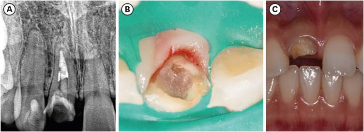
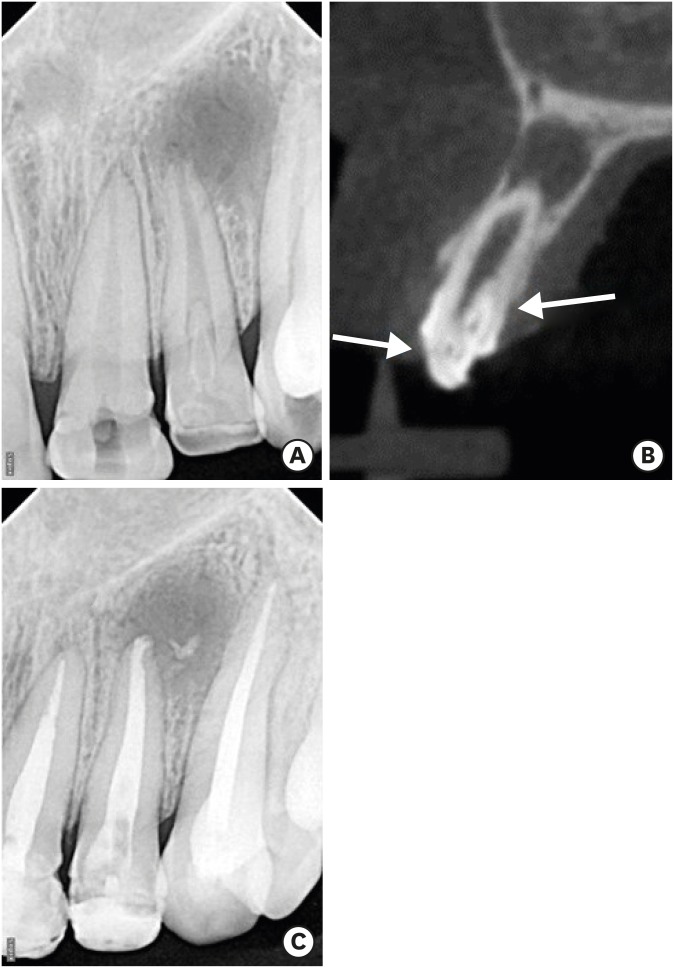
ePub Navigation of the main root canal and dealing with a dens invaginatus (DI) is a challenging task in clinical practice. Recently, the guided endodontics technique has become an alternative method for accessing root canals, surgical cavities, and calcified root canals without causing iatrogenic damage to tissue. In this case report, the use of the guided endodontics technique for two maxillary lateral incisors with multiple DIs is described. A 16-year-old female patient was referred with the chief complaint of pain and discoloured upper front teeth. Based on clinical and radiographic findings, a diagnosis of pulp necrosis and chronic periapical abscess associated with double DI (Oehler's type II) was established for the upper left lateral maxillary incisor (tooth #22). Root canal treatment and the sealing of double DI with mineral trioxide aggregate was planned for tooth #22. For tooth #12 (Oehler's type II), preventive sealing of the DI was planned. Minimally invasive access to the double DI and the main root canal of tooth #22, and to the DI of tooth #12, was achieved using the guided endodontics technique. This technique can be a valuable tool because it reduces chair-time and, more importantly, the risk of iatrogenic damage to the tooth structure.
-
Citations
Citations to this article as recorded by- Guided endodontics in the application of personalized mini-invasive treatment in clinical cases: a literature review
Shuangshuang Ren, Wanping Wang, Mingyue Cheng, Wenyue Tang, Yue Zhao, Leiying Miao
The Saudi Dental Journal.2025;[Epub] CrossRef - Navigating Calcified Challenges: Guided Endodontic Treatment of a Maxillary Central Incisor
Saide Nabavi, Sara Navabi, Iman Shiezadeh, SeyedehZahra JamaliMotlagh
Clinical Case Reports.2025;[Epub] CrossRef - Successful nonsurgical management of Oehler’s type III dens invaginatus in maxillary lateral incisor: A case report as per CARE guidelines
Anshul Sachdeva, Gurdeep Singh Gill, Adel Al Obied, Suraj Arora, Ali Y. Alsaeed, Waled Abdulmalek Alanesi, Gotam Das
Medicine.2025; 104(31): e42725. CrossRef - Guided Endodontics in Managing Root Canal Treatment for Anomalous Teeth—A Narrative Review
Pouya Sabanik, Mohammad Samiei, Shiva Tavakkoli Avval, Bruno Cavalcanti
Australian Endodontic Journal.2025;[Epub] CrossRef - Efficacy of Computer-aided Static Navigation on Accuracy of Guided Endodontic Root Canal Treatment: A Systematic Review and Meta-analysis
Ashish Jain, Rahul D Rao, Meenakshi R Verma, Rishabhkumar N Jain, Shreya Sivasailam, Anandita Sinha
World Journal of Dentistry.2024; 14(11): 1004. CrossRef - Application of personalized templates in minimally invasive management of coronal dens invaginatus: a report of two cases
Mingming Li, Guosong Wang, Fangzhi Zhu, Han Jiang, Yingming Yang, Ran Cheng, Tao Hu, Ru Zhang
BMC Oral Health.2024;[Epub] CrossRef - Endodontic management of severely calcified mandibular anterior teeth using guided endodontics: A report of a case and a review of the literature
Mina Davaji, Sahar Karimpour
Saudi Endodontic Journal.2024; 14(2): 245. CrossRef - Application of an Endodontic Static Guide in Fiber Post Removal from a Compromised Tooth
Mehran Farajollahi, Omid Dianat, Samaneh Gholami, Shima Saber Tahan, Sivakumar Nuvvula
Case Reports in Dentistry.2023;[Epub] CrossRef - The Impact of the Preferred Reporting Items for Case Reports in Endodontics (PRICE) 2020 Guidelines on the Reporting of Endodontic Case Reports
Sofian Youssef, Phillip Tomson, Amir Reza Akbari, Natalie Archer, Fayjel Shah, Jasmeet Heran, Sunmeet Kandhari, Sandeep Pai, Shivakar Mehrotra, Joanna M Batt
Cureus.2023;[Epub] CrossRef - Effectiveness of guided endodontics in locating calcified root canals: a systematic review
F. Peña-Bengoa, M. Valenzuela, M. J. Flores, N. Dufey, K. P. Pinto, E. J. N. L. Silva
Clinical Oral Investigations.2023; 27(5): 2359. CrossRef - Expert consensus on digital guided therapy for endodontic diseases
Xi Wei, Yu Du, Xuedong Zhou, Lin Yue, Qing Yu, Benxiang Hou, Zhi Chen, Jingping Liang, Wenxia Chen, Lihong Qiu, Xiangya Huang, Liuyan Meng, Dingming Huang, Xiaoyan Wang, Yu Tian, Zisheng Tang, Qi Zhang, Leiying Miao, Jin Zhao, Deqin Yang, Jian Yang, Junqi
International Journal of Oral Science.2023;[Epub] CrossRef - Prevalence and morphological analysis of dens invaginatus in anterior teeth using cone beam computed tomography: A systematic review and meta-analysis
Guilherme Nilson Alves dos Santos, Manoel Damião Sousa-Neto, Helena Cristina Assis, Fabiane Carneiro Lopes-Olhê, André L. Faria-e-Silva, Matheus L. Oliveira, Jardel Francisco Mazzi-Chaves, Amanda Pelegrin Candemil
Archives of Oral Biology.2023; 151: 105715. CrossRef - Root Maturation of an Immature Dens Invaginatus Despite Unsuccessful Revitalization Procedure: A Case Report and Recommendations for Educational Purposes
Julia Ludwig, Marcel Reymus, Alexander Winkler, Sebastian Soliman, Ralf Krug, Gabriel Krastl
Dentistry Journal.2023; 11(2): 47. CrossRef - Guided Endodontics as a Personalized Tool for Complicated Clinical Cases
Wojciech Dąbrowski, Wiesława Puchalska, Adam Ziemlewski, Iwona Ordyniec-Kwaśnica
International Journal of Environmental Research and Public Health.2022; 19(16): 9958. CrossRef - Present status and future directions – Guided endodontics
Thomas Connert, Roland Weiger, Gabriel Krastl
International Endodontic Journal.2022; 55(S4): 995. CrossRef - Treatment options for dens in dente: state-of-art literature review
Volodymyr Fedak
Ukrainian Dental Journal.2022; 1(1): 37. CrossRef - Dens Invaginatus: Clinical Implications and Antimicrobial Endodontic Treatment Considerations
José F. Siqueira, Isabela N. Rôças, Sandra R. Hernández, Karen Brisson-Suárez, Alessandra C. Baasch, Alejandro R. Pérez, Flávio R.F. Alves
Journal of Endodontics.2022; 48(2): 161. CrossRef - Guided Endodontics: Static vs. Dynamic Computer-Aided Techniques—A Literature Review
Diana Ribeiro, Eva Reis, Joana A. Marques, Rui I. Falacho, Paulo J. Palma
Journal of Personalized Medicine.2022; 12(9): 1516. CrossRef - Application of CBCT Data and Three-Dimensional Printing for Endodontic Diagnosis and Treatment
Srinidhi Vishnu Ballulaya, Neha Taufin, Nenavath Deepthi, Venu Babu Devella
Nigerian Journal of Experimental and Clinical Biosciences.2021; 9(3): 206. CrossRef - When to consider the use of CBCT in endodontic treatment planning in adults
Nisha Patel, Andrew Gemmell, David Edwards
Dental Update.2021; 48(11): 932. CrossRef
- Guided endodontics in the application of personalized mini-invasive treatment in clinical cases: a literature review
- 2,849 View
- 94 Download
- 20 Crossref


 KACD
KACD

 First
First Prev
Prev


