Search
- Page Path
- HOME > Search
- Resin infiltrant protects deproteinized dentin against erosive and abrasive wear
- Ana Theresa Queiroz de Albuquerque, Bruna Oliveira Bezerra, Isabelly de Carvalho Leal, Maria Denise Rodrigues de Moraes, Mary Anne S. Melo, Vanara Florêncio Passos
- Restor Dent Endod 2022;47(3):e29. Published online July 1, 2022
- DOI: https://doi.org/10.5395/rde.2022.47.e29

-
 Abstract
 PDF
PDF PubReader
 ePub
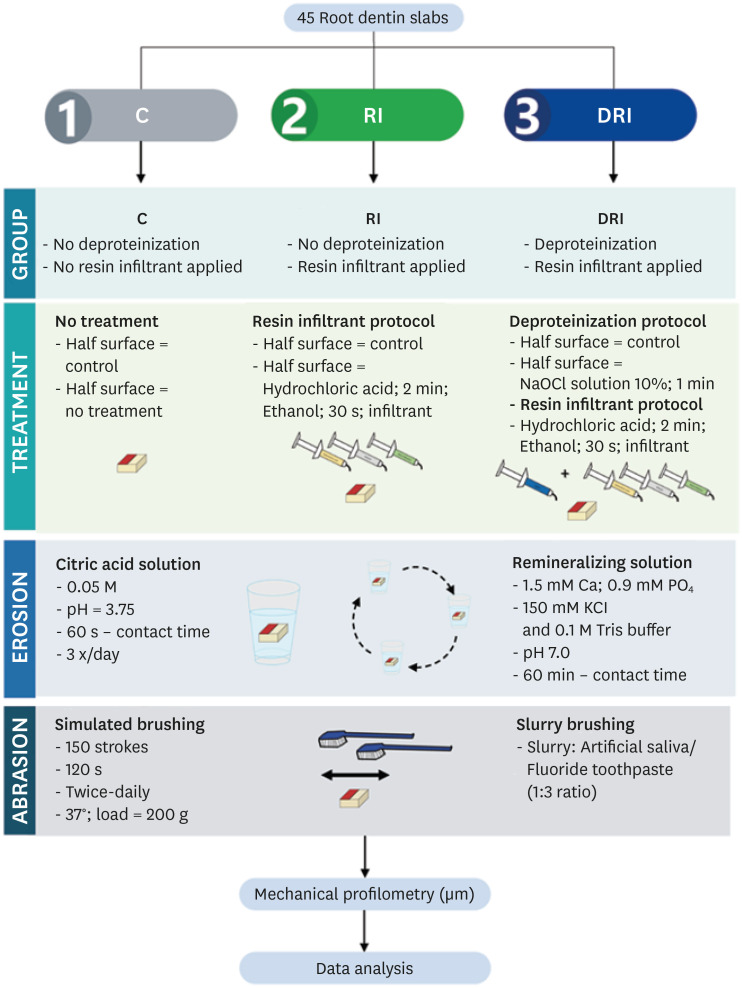
ePub Objectives This study aimed to investigate the anti-erosive/abrasive effect of resin infiltration of previous deproteinized dentin.
Materials and Methods Dentin slabs were randomly assigned to 3 groups (
n = 15): Control (no deproteinization; no resin infiltrant applied), RI (no deproteinization; resin infiltrant applied), and DRI (deproteinization; resin infiltrant applied). After undergoing the assigned treatment, all slabs were subjected to anin vitro cycling model for 5 days. The specimens were immersed in citric acid (0.05 M, pH = 3.75; 60 seconds; 3 times/day) and brushed (150 strokes). Between the challenges, the specimens were exposed to a remineralizing solution (60 minutes). The morphological alterations were analyzed by mechanical profilometry (µm) and scanning electron microscopy (SEM). Data were submitted to one-way analysis of variance (ANOVA) and Tukey tests (p < 0.05).Results Control and RI groups presented mineral wear and did not significantly differ from each other (
p = 0.063). DRI maintained a protective layer preserving the dentin (p < 0.001). After erosive/abrasive cycles, it was observed that in group RI, only 25% of the slabs partially evidenced the presence of the infiltrating, while, in the DRI group, 80% of the slabs presented the treated surface entirely covered by a resin-component layer protecting the dentin surface as observed in SEM images.Conclusions The removal of the organic content allows the resin infiltrant to efficiently protect the dentin surface against erosive/abrasive lesions.
-
Citations
Citations to this article as recorded by- Acidic/abrasive challenges on simulated non-carious cervical lesions development and morphology
Giovanna C. Denucci, Ian Towle, Cecilia P. Turssi, George J. Eckert, Anderson T. Hara
Archives of Oral Biology.2025; 169: 106120. CrossRef - Physio‐Mechanic and Microscopic Analyses of Bioactive Glass‐Based Resin Infiltrants
Syed Zubairuddin Ahmed, Abdul Samad Khan, Wejdan Waleed Nasser, Methayel Abdulrahman Alrushaid, Zahrah Mohammed Alfaraj, Moayad Mohammed Aljeshi, Asma Tufail Shah, Budi Aslinie Md Sabri, Sultan Akhtar, Mohamed Ibrahim Abu Hassan
Microscopy Research and Technique.2025; 88(2): 595. CrossRef - Resin Infiltration Treatment of Developmental Enamel Defects in a Patient With Hydrocephalus and Cerebral Palsy: A Case Report on the Impact on the Maternal Caregiver
Eduarda Martins Fontes Cantarella de Almeida, Anna Luísa Araujo Pimenta, Francisco Wanderley Garcia de Paula‐Silva, Fabricio Kitazono de Carvalho, Laurindo Borelli‐Neto, Susanne Effenberger, Fernanda de Carvalho Panzeri, Silmara Aparecida Milori Corona, K
Special Care in Dentistry.2025;[Epub] CrossRef
- Acidic/abrasive challenges on simulated non-carious cervical lesions development and morphology
- 2,133 View
- 43 Download
- 3 Web of Science
- 3 Crossref

- Management of dental erosion induced by gastro-esophageal reflux disorder with direct composite veneering aided by a flexible splint matrix
- Sherin Jose Chockattu, Byathnal Suryakant Deepak, Anubhav Sood, Nandini T. Niranjan, Arun Jayasheel, Mallikarjun K. Goud
- Restor Dent Endod 2018;43(1):e13. Published online February 6, 2018
- DOI: https://doi.org/10.5395/rde.2018.43.e13

-
 Abstract
 PDF
PDF PubReader
 ePub
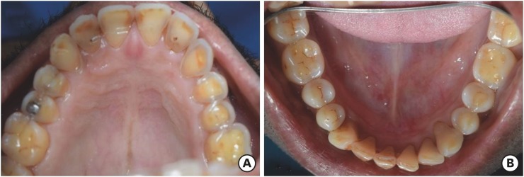
ePub Dental erosion is frequently overlooked in clinical practice. The management of erosion-induced damage to the dentition is often delayed, such that extensive occlusal rehabilitation is required. These cases can be diagnosed by a careful clinical examination and a thorough review of the patient's medical history and/or lifestyle habits. This case report presents the diagnosis, categorization, and management of a case of gastro-esophageal reflux disease-induced palatal erosion of the maxillary teeth. The early management of such cases is of utmost importance to delay or prevent the progression of damage both to the dentition and to occlusal stability. Non-invasive adhesively bonded restorations aid in achieving this goal.
-
Citations
Citations to this article as recorded by- Effect of Acidic Media on Surface Topography and Color Stability of Two Different Glass Ceramics
Fatma Makkeyah, Nesrine A. Elsahn, Mahmoud M. Bakr, Mahmoud Al Ankily
European Journal of Dentistry.2025; 19(01): 173. CrossRef - Mechanical Performance and Surface Roughness of Lithium Disilicate and Zirconia-Reinforced Lithium Silicate Ceramics Before and After Exposure to Acidic Challenge
Ahmed Elsherbini, Salma M. Fathy, Walid Al-Zordk, Mutlu Özcan, Amal A. Sakrana
Dentistry Journal.2025; 13(3): 117. CrossRef - Biomechanical reinforcement by CAD-CAM materials affects stress distributions of posterior composite bridges: 3D finite element analysis.
Alaaeldin Elraggal, Islam M. Abdelraheem, David C. Watts, Sandipan Roy, Vamsi Krishna Dommeti, Abdulrahman Alshabib, Khaled Abid Althaqafi, Rania R. Afifi
Dental Materials.2024; 40(5): 869. CrossRef - Surface Properties and Wear Resistance of Injectable and Computer-Aided Design/Computer Aided Manufacturing–Milled Resin Composite Thin Occlusal Veneers
Nesrine A. Elsahn, Hatem M. El-Damanhoury, Zainab Shirazi, Abdul Rahman M. Saleh
European Journal of Dentistry.2023; 17(03): 663. CrossRef - Effect of acidic media on flexural strength and fatigue of CAD-CAM dental materials
Alaaeldin Elraggal, Rania. R Afifi, Rasha A. Alamoush, Islam Abdel Raheem, David C. Watts
Dental Materials.2023; 39(1): 57. CrossRef - Three-year Follow-up of Conservative Direct Composite Veneers on Eroded Teeth
RQ Ramos, NF Coelho, GC Lopes
Operative Dentistry.2022; 47(2): 131. CrossRef - The effects of intrinsic and extrinsic acids on nanofilled and bulk fill resin composites: Roughness, surface hardness, and scanning electron microscopy analysis
Milena F. Alencar, Mirella T. Pereira, Maria D. R. De‐Moraes, Sérgio L. Santiago, Vanara F. Passos
Microscopy Research and Technique.2020; 83(2): 202. CrossRef
- Effect of Acidic Media on Surface Topography and Color Stability of Two Different Glass Ceramics
- 2,028 View
- 16 Download
- 7 Crossref

- Carbohydrate-electrolyte drinks exhibit risks for human enamel surface loss
- Mary Anne Sampaio de Melo, Vanara Florêncio Passos, Juliana Paiva Marques Lima, Sérgio Lima Santiago, Lidiany Karla Azevedo Rodrigues
- Restor Dent Endod 2016;41(4):246-254. Published online August 16, 2016
- DOI: https://doi.org/10.5395/rde.2016.41.4.246
-
 Abstract
 PDF
PDF PubReader
 ePub
ePub Objectives The aim of this investigation was to give insights into the impact of carbohydrate-electrolyte drinks on the likely capacity of enamel surface dissolution and the influence of human saliva exposure as a biological protective factor.
Materials and Methods The pH, titratable acidity (TA) to pH 7.0, and buffer capacity (β) of common beverages ingested by patients under physical activity were analyzed. Then, we randomly distributed 50 specimens of human enamel into 5 groups. Processed and natural coconut water served as controls for testing three carbohydrate-electrolyte drinks. In all specimens, we measured surface microhardness (Knoop hardness numbers) and enamel loss (profilometry, µm) for baseline and after simulated intake cycling exposure model. We also prepared areas of specimens to be exposed to human saliva overnight prior to the simulated intake cycling exposure. The cycles were performed by alternated immersions in beverages and artificial saliva. ANOVA two-way and Tukey HDS tests were used.
Results The range of pH, TA, and β were 2.85 - 4.81, 8.33 - 46.66 mM/L and 3.48 - 10.25 mM/L × pH, respectively. The highest capacity of enamel surface dissolution was found for commercially available sports drinks for all variables. Single time human saliva exposure failed to significantly promote protective effect for the acidic attack of beverages.
Conclusions In this study, carbohydrate-electrolyte drinks usually consumed during endurance training may have a greater capacity of dissolution of enamel surface depending on their physicochemical proprieties associated with pH and titratable acidity.
-
Citations
Citations to this article as recorded by- Evaluation of developmentally hypomineralised enamel after surface pretreatment with Papacarie Duo gel and different etching modes: an in vitro SEM and AFM study
Y.-L. Lee, K. C. Li, C. K. Y. Yiu, D. H. Boyd, M. Ekambaram
European Archives of Paediatric Dentistry.2022; 23(1): 117. CrossRef - Is the consumption of beverages and food associated to dental erosion? A cross-sectional study in Portuguese athletes
M.-R.G. Silva, M.-A. Chetti, H. Neves, M.-C. Manso
Science & Sports.2021; 36(6): 477.e1. CrossRef - Assessment of surface roughness changes on orthodontic acrylic resin by all-in-one spray disinfectant solutions
Kuei-ling Hsu, Abdulrahman A. Balhaddad, Isadora Martini Garcia, Fabricio Mezzomo Collares, Louis DePaola, Mary Anne Melo
Journal of Dental Research, Dental Clinics, Dental Prospects.2020; 14(2): 77. CrossRef - Nitrate-rich beetroot juice offsets salivary acidity following carbohydrate ingestion before and after endurance exercise in healthy male runners
Mia C. Burleigh, Nicholas Sculthorpe, Fiona L. Henriquez, Chris Easton, Yi-Hung Liao
PLOS ONE.2020; 15(12): e0243755. CrossRef - Dental erosion’ prevalence and its relation to isotonic drinks in athletes: a systematic review and meta-analysis
Pedro Henrique Pereira de Queiroz Gonçalves, Ludmila Silva Guimarães, Fellipe Navarro Azevedo de Azeredo, Letícia Maira Wambier, Lívia Azeredo A. Antunes, Leonardo Santos Antunes
Sport Sciences for Health.2020; 16(2): 207. CrossRef - Atomic force microscopy analysis of enamel nanotopography after interproximal reduction
Shadi Mohebi, Nazila Ameli
American Journal of Orthodontics and Dentofacial Orthopedics.2017; 152(3): 295. CrossRef
- Evaluation of developmentally hypomineralised enamel after surface pretreatment with Papacarie Duo gel and different etching modes: an in vitro SEM and AFM study
- 1,875 View
- 14 Download
- 6 Crossref

-
How to design
in situ studies: an evaluation of experimental protocols - Young-Hye Sung, Hae-Young Kim, Ho-Hyun Son, Juhea Chang
- Restor Dent Endod 2014;39(3):164-171. Published online May 13, 2014
- DOI: https://doi.org/10.5395/rde.2014.39.3.164
-
 Abstract
 PDF
PDF PubReader
 ePub
ePub Objectives Designing
in situ models for caries research is a demanding procedure, as both clinical and laboratory parameters need to be incorporated in a single study. This study aimed to construct an informative guideline for planningin situ models relevant to preexisting caries studies.Materials and Methods An electronic literature search of the PubMed database was performed. A total 191 of full articles written in English were included and data were extracted from materials and methods. Multiple variables were analyzed in relation to the publication types, participant characteristics, specimen and appliance factors, and other conditions. Frequencies and percentages were displayed to summarize the data and the Pearson's chi-square test was used to assess a statistical significance (
p < 0.05).Results There were many parameters commonly included in the majority of
in situ models such as inclusion criteria, sample sizes, sample allocation methods, tooth types, intraoral appliance types, sterilization methods, study periods, outcome measures, experimental interventions, etc. Interrelationships existed between the main research topics and some parameters (outcome measures and sample allocation methods) among the evaluated articles.Conclusions It will be possible to establish standardized
in situ protocols according to the research topics. Furthermore, data collaboration from comparable studies would be enhanced by homogeneous study designs.-
Citations
Citations to this article as recorded by- What is the effectiveness of titanium tetrafluoride to prevent or treat dental caries and tooth erosion? A systematic review
Ana Beatriz Chevitarese, Karla Lorene de França Leite, Guido A. Marañón-Vásquez, Danielle Masterson, Matheus Pithon, Lucianne Cople Maia
Acta Odontologica Scandinavica.2022; 80(6): 441. CrossRef - Effect of fluoride group on dental erosion associated or not with abrasion in human enamel: A systematic review with network metanalysis
Bruna Machado da Silva, Daniela Rios, Gerson Aparecido Foratori-Junior, Ana Carolina Magalhães, Marília Afonso Rabelo Buzalaf, Silvia De Carvalho Sales Peres, Heitor Marques Honório
Archives of Oral Biology.2022; 144: 105568. CrossRef - Multimodal Human and Environmental Sensing for Longitudinal Behavioral Studies in Naturalistic Settings: Framework for Sensor Selection, Deployment, and Management
Brandon M Booth, Karel Mundnich, Tiantian Feng, Amrutha Nadarajan, Tiago H Falk, Jennifer L Villatte, Emilio Ferrara, Shrikanth Narayanan
Journal of Medical Internet Research.2019; 21(8): e12832. CrossRef - Evaluation of an antibacterial orthodontic adhesive incorporated with niobium-based bioglass: an in situ study
Felipe Weidenbach DEGRAZIA, Aline Segatto Pires ALTMANN, Carolina Jung FERREIRA, Rodrigo Alex ARTHUR, Vicente Castelo Branco LEITUNE, Susana Maria Werner SAMUEL, Fabrício Mezzomo COLLARES
Brazilian Oral Research.2019;[Epub] CrossRef - A Review of the Common Models Used in Mechanistic Studies on Demineralization-Remineralization for Cariology Research
Ollie Yiru Yu, Irene Shuping Zhao, May Lei Mei, Edward Chin-Man Lo, Chun-Hung Chu
Dentistry Journal.2017; 5(2): 20. CrossRef - Effects of rinsing with arginine bicarbonate and urea solutions on initial enamel lesions in situ
Y Yu, X Wang, C Ge, B Wang, C Cheng, Y‐H Gan
Oral Diseases.2017; 23(3): 353. CrossRef - The cariogenicity of commercial infant formulas: a systematic review
S. F. Tan, H. J. Tong, X. Y. Lin, B. Mok, C. H. Hong
European Archives of Paediatric Dentistry.2016; 17(3): 145. CrossRef - In situ antibiofilm effect of glass-ionomer cement containing dimethylaminododecyl methacrylate
Jin Feng, Lei Cheng, Xuedong Zhou, Hockin H.K. Xu, Michael D. Weir, Markus Meyer, Hans Maurer, Qian Li, Matthias Hannig, Stefan Rupf
Dental Materials.2015; 31(8): 992. CrossRef
- What is the effectiveness of titanium tetrafluoride to prevent or treat dental caries and tooth erosion? A systematic review
- 1,321 View
- 4 Download
- 8 Crossref


 KACD
KACD

 First
First Prev
Prev


