
 KACD
KACD
Search
- Page Path
- HOME > Search
Research Articles
- Classification proposal of obliterated canals based on the position of the target point and the caliber of the root canal
- Gustavo Adolfo Castillo Barrios, Patricia Rodríguez Sánchez, Anuar Elian Barona Triviño, Carlos Humberto Martínez Cajas
- Restor Dent Endod 2026;51(3):e32. Published online August 4, 2026
- DOI: https://doi.org/10.5395/rde.2026.51.e32
-
 Abstract
 PDF
PDF - Objectives
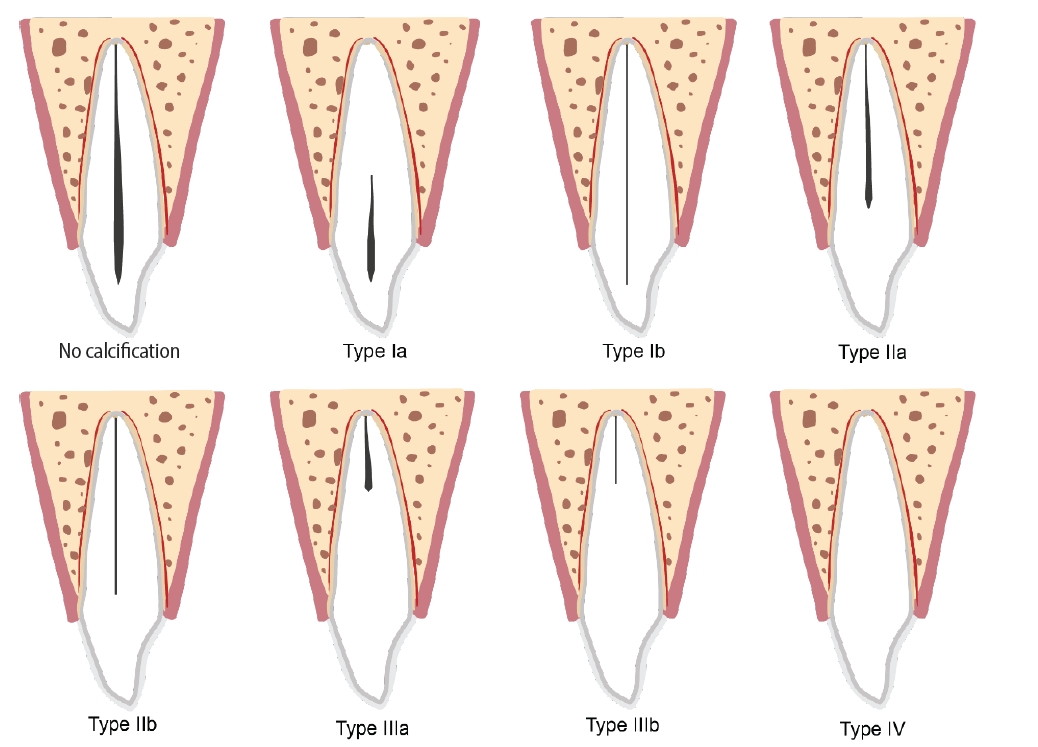
This study aimed to propose a classification system for calcified root canals based on the location of the target point or canal starting point and root canal lumen caliber to facilitate the evaluation of dynamically guided techniques in endodontic treatment. Methods: A retrospective analysis of eight cases with calcified root canals was conducted. Periapical radiographs and cone-beam computed tomography were used to classify cases based on root location and degree of obliteration. Clinical and radiographic outcomes were evaluated. Results: Eight classification types are identified, ranging from non-obliteration and partial obliteration to total obliteration. The location of the root canal lumen varies from the pulp chamber to the apical third of the root. Guided endodontics with dynamic navigation has been demonstrated to be an effective treatment modality in cases of advanced obliteration, thereby substantiating its efficacy in complex scenarios. Conclusions: The proposed classification system provides a framework to understand the complexity of calcified root canals. Guided endodontics with dynamically guided techniques can be valuable in managing these challenging cases, particularly when the root canal lumen is in the apical third. Further studies are needed to validate the classification system and evaluate the long-term outcomes of the different treatment approaches. -
Citations
Citations to this article as recorded by
- Failure of Guided Endodontic Access in a Severely Obliterated Root Canal: A Case Report of Drill Deviation
Stephanie Isabel Diaz Zamalloa, Iandara Lima Scardini, Shirley Maklane Gomes Soares, Daniel de Almeida Decurcio, Marcelo dos Santos, Celso Luiz Caldeira, Hannah Wesley
Case Reports in Dentistry.2026;[Epub] CrossRef
- Failure of Guided Endodontic Access in a Severely Obliterated Root Canal: A Case Report of Drill Deviation
- 1,328 View
- 120 Download
- 1 Web of Science
- 1 Crossref

- Determination of optimal horizontal beam angulations for canal separation in mandibular molars using cone-beam computed tomography: a retrospective image-based analysis
- Benedikt Schneider, Tamina Tepe, Daniel Rapp, Wilhelm Frank, Maria Lessani, Constantin von See, Sebastian Fitzek, Jörg Philipp Tchorz
- Restor Dent Endod 2026;51(1):e9. Published online February 26, 2026
- DOI: https://doi.org/10.5395/rde.2026.51.e9
-
Abstract
PDF
- Objectives
Two-dimensional intraoral radiographs often obscure canals due to superimposition, especially in mandibular molars with complex anatomy. This cone-beam computed tomography (CBCT) study identified the horizontal beam angles at which first and second molar canals overlap and derived clinically applicable angulations for enhanced canal separation.
Methods
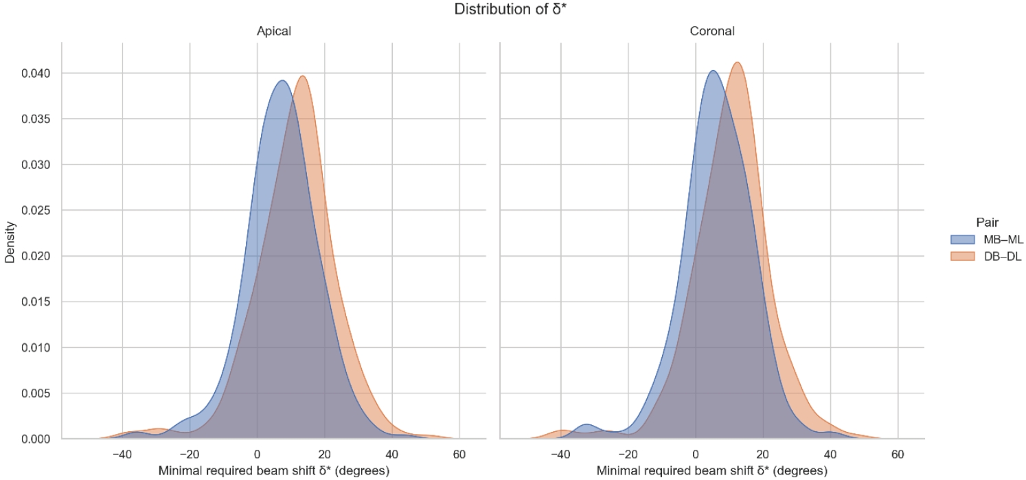
Eighty-five CBCT datasets from 100 patients met the inclusion criteria, yielding 318 mandibular molars (160 first, 158 second). Using ImageJ, absolute horizontal overlap angles (α) were measured to determine the corresponding theoretical separation angles defined as δ* = 90° – α. Separability was modeled across horizontal beam angulation increments from −45° to +45° in five steps, and Wilson’s 95% confidence intervals were computed. Group comparisons used the Mann-Whitney U and independent t-tests (p ≤ 0.05)
Results
Minimal mesial beam angulations for effective canal separability (δ* = 90° − α) ranged from approximately 7° to 15° for mesial roots and approximately 10° to 13° for distal roots. No significant mesial differences were observed between first and second molars (p > 0.30). Distal roots of second molars exhibited significantly higher angulations (p = 0.003 coronal, p < 0.001 apical). Mesial canals achieved ≥95% separability at approximately 25° and ≥99% at approximately 35°; distal canals required approximately 30° and approximately 40°.
Conclusions
A mesial beam angulation of 30° to 35° provides probable canal differentiation in mandibular molars, separating mesial canals in ≥99% and distal canals in ≥95% of cases. This range refines previous recommendations and supports the as low as reasonably achievable (ALARA) principle.
- 1,895 View
- 42 Download
 First
First Prev
Prev


