Search
- Page Path
- HOME > Search
- Influence of access cavity design on calcium hydroxide removal using different cleaning protocols: a confocal laser scanning microscopy study
- Seda Falakaloğlu, Merve Yeniçeri Özata, Betül Güneş, Emmanuel João Nogueira Leal Silva, Mustafa Gündoğar, Burcu Güçyetmez Topal
- Restor Dent Endod 2023;48(3):e25. Published online July 24, 2023
- DOI: https://doi.org/10.5395/rde.2023.48.e25

-
 Abstract
 PDF
PDF PubReader
 ePub
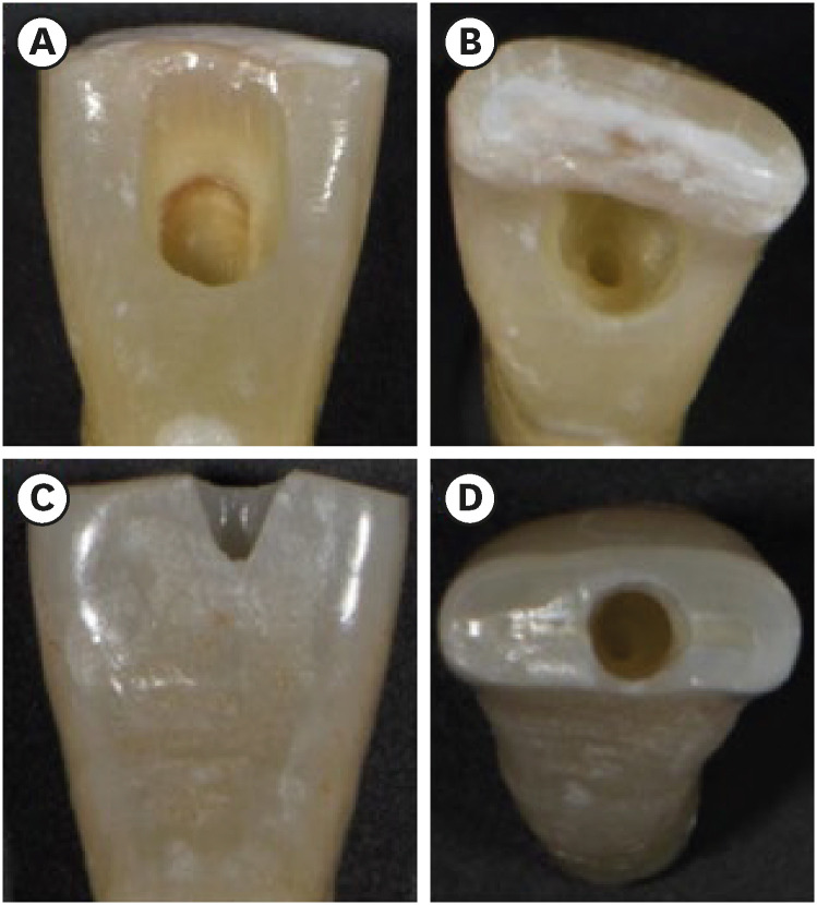
ePub Objectives The purpose of this study was to evaluate the influence of endodontic access cavities design on the removal of calcium hydroxide medication of the apical third of mandibular incisor root canal walls and dentinal tubules with different cleaning protocols: EDDY sonic activation, Er,Cr:YSGG laser-activated irrigation, or conventional irrigation with IrriFlex.
Materials and Methods Seventy-eight extracted human mandibular incisors were assigned to 6 experimental groups (
n = 13) according to the endodontic access cavity and cleaning protocol for calcium hydroxide removal: traditional access cavity (TradAC)/EDDY; ultraconservative access cavity performed in the incisal edge (UltraAC.Inc)/EDDY; TradAC/Er,Cr:YSGG; UltraAC.Inc/Er,Cr:YSGG; TradAC/IrriFlex; or UltraAC.Inc/IrriFlex. Confocal laser scanning microscopy images were used to measure the non-penetration percentage, maximum residual calcium hydroxide penetration depth, and penetration area at 2 and 4 mm from the apex. Data were statistically analyzed using Shapiro-Wilk and WRS2 package for 2-way comparison of non-normally distributed parameters (depth of penetration, area of penetration, and percentage of non-penetration) according to cavity and cleaning protocol with the significance level set at 5%.Results The effect of cavity and cleaning protocol interactions on penetration depth, penetration area and non-penetration percentage was not found statistically significant at 2 and 4 mm levels (
p > 0.05).Conclusions The present study demonstrated that TradAC or UltraAC.Inc preparations with different cleaning protocols in extracted mandibular incisors did not influence the remaining calcium hydroxide at 2 and 4 mm from the apex.
-
Citations
Citations to this article as recorded by- Effect of Apical Preparation Size and Preparation Taper on Smear Layer Removal Using Two Different Irrigation Needles: A Scanning Electron Microscopy Study
Rania Lebbos, Naji Kharouf, Deepak Mehta, Jamal Jabr, Cynthia Kamel, Roula El Hachem, Youssef Haikel, Marc Krikor Kaloustian
European Journal of Dentistry.2025; 19(03): 678. CrossRef - Combination of Chitosan Nanoparticles, EDTA, and Irrigation Activation Enhances TGF-β1 Release from Dentin: A Laboratory Study
Sıla Nur Usta, Emre Avcı, Ayşe Nur Oktay, Cangül Keskin
Journal of Endodontics.2025; 51(8): 1081. CrossRef
- Effect of Apical Preparation Size and Preparation Taper on Smear Layer Removal Using Two Different Irrigation Needles: A Scanning Electron Microscopy Study
- 2,707 View
- 68 Download
- 2 Web of Science
- 2 Crossref

- Stress analysis of maxillary premolars with composite resin restoration of notch-shaped class V cavity and access cavity; Three-dimensional finite element study
- Seon-Hwa Lee, Hyeon-Cheol Kim, Bock Hur, Kwang-Hoon Kim, Kwon Son, Jeong-Kil Park
- J Korean Acad Conserv Dent 2008;33(6):570-579. Published online November 30, 2008
- DOI: https://doi.org/10.5395/JKACD.2008.33.6.570
-
 Abstract
 PDF
PDF PubReader
 ePub
ePub The purpose of this study was to investigate the distribution of tensile stress of canal obturated maxillary second premolar with access cavity and notch-shaped class V cavity restored with composite resin using a 3D finite element analysis.
The tested groups were classified as 8 situations by only access cavity or access cavity with notch-shaped class VS cavity (S or N), loading condition (L1 or L2), and with or without glass ionomer cement base (R1 or R2). A static load of 500 N was applied at buccal and palatal cusps. Notch-shaped cavity and access cavity were filled microhybrid composite resin (Z100) with or without GIC base (Fuji II LC). The tensile stresses presented in the buccal cervical area, palatal cervical area and occlusal surface were analyzed using ANSYS.
Tensile stress distributions were similar regardless of base. When the load was applied on the buccal cusp, excessive high tensile stress was concentrated around the loading point and along the central groove of occlusal surface. The tensile stress values of the tooth with class V cavity were slightly higher than that of the tooth without class V cavity. When the load was applied the palatal cusp, excessive high tensile stress was concentrated around the loading point and along the central groove of occlusal surface. The tensile stress values of the tooth without class V cavity were slightly higher than that of the tooth with class V cavity.
- 1,009 View
- 0 Download

- The influence of different access cavity designs on the fracture strength in endodontically treated mandibular anterior teeth
- Young-Gyun Lee, Hye-Jin Shin, Se-Hee Park, Kyung-Mo Cho, Jin-Woo Kim
- J Korean Acad Conserv Dent 2004;29(6):515-519. Published online November 30, 2004
- DOI: https://doi.org/10.5395/JKACD.2004.29.6.515
-
 Abstract
 PDF
PDF PubReader
 ePub
ePub Straight access cavity design allows the operator to locate all canals, helps in proper cleaning and shaping, ultimately facilitates the obturation of the canal system. However, change in the fracture strength according to the access cavity designs was not clearly demonstrated yet. The purpose of this study was to determine the influence of different access cavity designs on the fracture strength in endodontically treated mandibular anterior teeth.
Recently extracted mandibular anterior teeth that have no caries, cervical abrasion, and fracture were divided into three groups (Group 1 : conventional lingual access cavity, Group 2 : straight access cavity, Group 3 : extended straight access cavity) according to the cavity designs. After conventional endodontic treatment, cavities were filled with resin core material. Compressive loads parallel to the long axis of the teeth were applied at a crosshead speed of 2mm/min until the fracture occurred. The fracture strength analyzed with ANOVA and the Scheffe test at the 95% confidence level.
The results of this study were as follows :
1. The mean fracture strength decrease in following sequence Group 1 (558.90 ± 77.40 N), Group 2 (494.07 ± 123.98 N) and Group 3 (267.33 ± 27.02 N).
2. There was significant difference between Group 3 and other groups (P = 0.00).
Considering advantage of direct access to apical third and results of this study, straight access cavity is recommended for access cavity form of the mandibular anterior teeth.
-
Citations
Citations to this article as recorded by- Fracture resistance of crown-root fractured teeth repaired with dual-cured composite resin and horizontal posts
Seok-Woo Chang, Yong-Keun Lee, Seung-Hyun Kyung, Hyun-Mi Yoo, Tae-Seok Oh, Dong-Sung Park
Journal of Korean Academy of Conservative Dentistry.2009; 34(5): 383. CrossRef
- Fracture resistance of crown-root fractured teeth repaired with dual-cured composite resin and horizontal posts
- 978 View
- 2 Download
- 1 Crossref

- The effect of the endodontic access cavity on the marginal leakage of crowns
- Euiseong Kim, Jinho Chung, Yongkun Kim
- J Korean Acad Conserv Dent 2002;27(4):389-393. Published online July 31, 2002
- DOI: https://doi.org/10.5395/JKACD.2002.27.4.389
-
 Abstract
 PDF
PDF PubReader
 ePub
ePub The marginal integrity of the crown can be broken during endodontic access cavity preparation due to the vibration of burs. Therefore, the purpose of this study was to evaluate the effect of endodontic access cavity preparation on the marginal leakage of full veneer gold crowns. 24 intact molars were mounted in acrylic resin blocks and prepared for crowns by a restorative dentist and crowns were cast with gold alloy. 20 Crowns were cemented with glass ionomer cement and 2 crowns were not cemented for positive control. 200 thermo-cycles from 5℃ to 50℃ with a travel time of 20s were completed. Then samples were randomly divided into 2 experimental groups of 9 each. Endodontic access preparation and zinc-oxide eugenol temporary fillings were done in Group 1. Teeth in Group 2 were not treated. Samples were coated with 2 layers of nail varnish and were immersed in 1% methylene blue dye for 20 hrs. Endodontic access was prepared in 2 samples, which were coated with nail varnish on all surfaces for negative control. After washing in running water, gold crowns were cut with a #330 bur. Four buccolingual sections, 2 mm apart, were cut from the central section of each tooth and were examined and scored under the microscope for dye leakage. Score 1: leakage to the cervical 1/3 of the axial wall, Score 2: leakage to the middle 1/3 of the axial wall, Score 3: leakage to the coronal 1/3 of the axial wall, Score 4: leakage to the occlusal surface. The median value for Group 1 is 4 and for Group 2 is 2. The result of this study showed that samples in Group 1 leaked more than those in Group 2. This finding was significant(P<0.001).
- 843 View
- 3 Download


 KACD
KACD

 First
First Prev
Prev


