
 KACD
KACD
Previous issues
- Page Path
- HOME > Browse articles > Previous issues
- Volume 51 (2); May 2026
-


Editorial
- Artificial intelligence hallucinations in endodontics: implications for scientific integrity and clinical decision-making
- Emmanuel João Nogueira Leal da Silva, Fernanda Nehme Simão Jorge Riche
- Restor Dent Endod 2026;51(2):e18. Published online April 7, 2026
- DOI: https://doi.org/10.5395/rde.2026.51.e18
-
 PDF
PDF -
Citations
Citations to this article as recorded by
- Guarding the Gate: Research Integrity in Orthodontic Publishing in the Age of Generative AI
Mridula Trehan
Journal of Indian Orthodontic Society.2026; 60(4): 269. CrossRef
- Guarding the Gate: Research Integrity in Orthodontic Publishing in the Age of Generative AI
- 2,306 View
- 180 Download
- 1 Crossref

Review Article
- Effectiveness of silver diamine fluoride in managing hypersensitivity of molar-incisor hypomineralization affected molars: a scoping review
- Vo Truong Nhu Ngoc, Do Trong Hieu, Tran Anh Tuan, Vo Nhat Minh, Trinh Khanh Linh
- Restor Dent Endod 2026;51(2):e19. Published online April 13, 2026
- DOI: https://doi.org/10.5395/rde.2026.51.e19
-
 Abstract
PDF
Abstract
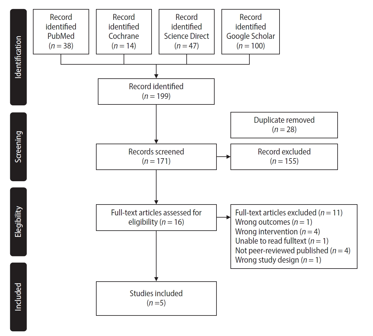
PDF - This study aimed to evaluate the efficacy of dentinal hypersensitivity treatment with silver diamine fluoride (SDF) in molar-incisor hypomineralization (MIH)-affected molars. This scoping review was designed and structured according to the guidelines of the Preferred Reporting Items for Systematic Reviews and Meta-Analyses and its extension for scoping reviews. A search strategy was conducted across PubMed, The Cochrane Library, ScienceDirect, and Google Scholar to identify articles related to the topic. Two authors screened titles, abstracts, and full texts for review. Five studies met the eligibility criteria, comprising four randomized controlled trials and one case report, with sample sizes ranging from four to 200 participants. All included studies reported improvements in clinical outcomes, including reduced hypersensitivity following SDF application, as indicated by lower Schiff cold air sensitivity scale scores. SDF is a promising treatment strategy for reducing hypersensitivity in MIH-affected molars; however, further research using SDF alone is needed to evaluate its exact effectiveness.
- 3,132 View
- 292 Download
Research Articles
- The recovery effect of dentin biomodifiers on microtensile bond strength and sealer-penetration depth of coronal and radicular dentin: an in vitro experimental study
- Mona Rizk Aboelwafa, Yasmin Tawfik Mohamed Sobh
- Restor Dent Endod 2026;51(2):e15. Published online April 7, 2026
- DOI: https://doi.org/10.5395/rde.2026.51.e15
-
Abstract
PDF
- Objectives
This study aimed to assess the outcomes of bromelain enzyme and chlorhexidine (CHX) following endodontic irrigation by evaluating coronal dentin microtensile bond strength (μTBS) and radicular dentin sealer penetration depth.
Methods
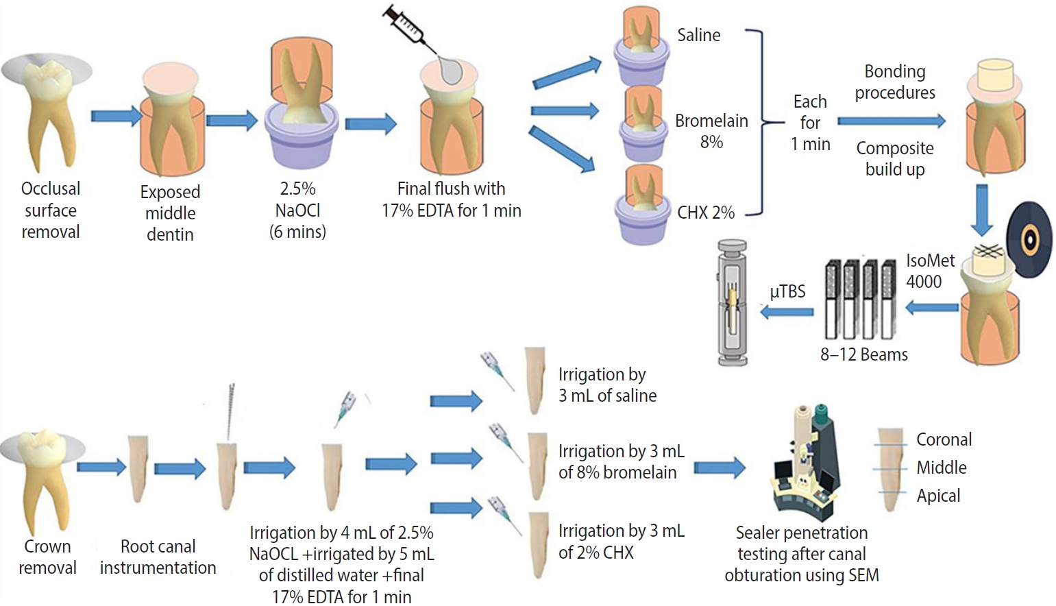
Fifty-one human molars with flat mid-dentin surfaces were soaked in sodium hypochlorite, then randomly assigned to three groups relying on the biomodification approach (n = 17): group 1, saline; group 2, 8% bromelain; and group 3, 2% CHX. After bonding and resin composite build-ups, the μTBS, failure mode, and bond interface were evaluated. Forty-two root canals of human molars were mechanically prepared and randomly distributed among three groups (n = 14), similar to the coronal-dentin biomodification protocol. The sealer-penetration depth was measured utilizing the scanning electron microscope. One- and two-way analyses of variance and the pairwise t- and chi-square tests were utilized.
Results
The bromelain group showed the highest statistically significant resin-dentin μTBS values, followed by the CHX and control groups. For sealer-penetration assessment, the bromelain group showed the highest penetration at the middle and apical root levels, whereas CHX demonstrated the highest penetration at the coronal level.
Conclusions
Bromelain biomodification positively influenced the resin-dentin bond strength and the sealer-penetration depth in apical and middle levels.
- 2,047 View
- 261 Download
- Effect of sugar and sweetener on the bleachability of coffee and tea-induced stains on composites: an in vitro experimental study
- Nilay Bayraktar, Osman Kerim Arda Karaca, Yunus Ekşılı, Mustafa Furkan Yıldırım, Osman Tolga Harorli
- Restor Dent Endod 2026;51(2):e16. Published online April 1, 2026
- DOI: https://doi.org/10.5395/rde.2026.51.e16
-
Abstract
PDF
- Objectives
This in vitro study evaluated the effects of various sugary and non-sugary beverages on the color change of a dental composite and the subsequent bleaching efficacy.
Methods
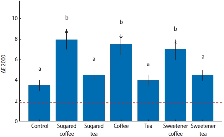
Forty-nine disc-shaped composite samples (Neo Spectra ST, Dentsply Sirona) were split into seven groups at random (n = 7). Distilled water was used to hydrate each sample for 24 hours at 37°C. After 24 hours, the first color measurements (T0) were made by using a clinical spectrophotometer (VITA Easyshade Compact; VITA Zahnfabrik). Color measurements were repeated after 7 days (T1) and 14 days (T2) of immersion in distilled water (control), tea, coffee, sugary tea, sugary coffee, tea with sweetener added, and coffee with sweetener added. After staining for 2 weeks, the specimens were bleached for 6 hours a day for a week using 16% carbamide peroxide (Opalescence Ultradent Products). Color measurements were taken again after bleaching (T3). Using CIEDE2000, color differences (ΔE) were computed. Analysis of variance (ANOVA) and repeated measures ANOVA with a Tukey post hoc test were used to evaluate the data.
Results
After 1 week, coffee-containing solutions produced significantly greater discoloration than the control (p < 0.001). By 2 weeks, tea groups exhibited similar discoloration to coffee groups (p < 0.001). The addition of sugar or sweetener had no significant effect (p > 0.05). Post-bleaching, coffee groups showed lower Whiteness Index values than the control, without statistical significance (p > 0.05).
Conclusions
Coffee and tea markedly stain resin composites, with discoloration persisting post-bleaching, while sugar or sweetener additions exert no significant effect. -
Citations
Citations to this article as recorded by- Effects of surface treatment methods on the color stability, whiteness, and surface roughness of bleach-shade composite resins
İlayda Kutlu, Muhammet Fi̇dan
BMC Oral Health.2026;[Epub] CrossRef
- Effects of surface treatment methods on the color stability, whiteness, and surface roughness of bleach-shade composite resins
- 2,085 View
- 140 Download
- 1 Web of Science
- 1 Crossref
- Clinical outcomes of tooth autotransplantation: a systematic review and meta-analysis of survival
- Jasmine Wong, Elise Hoi Wan Fok, Kar Yan Li, Chengfei Zhang, Gary Shun Pan Cheung
- Restor Dent Endod 2026;51(2):e17. Published online April 2, 2026
- DOI: https://doi.org/10.5395/rde.2026.51.e17
-
Abstract
PDF
- Objectives
Autotransplantation is a procedure that involves the extraction and transplantation of a tooth from one site to another within the same individual. This systematic review and meta-analysis aimed to investigate how clinical outcomes of autotransplanted teeth evolve over time and the principal reasons for extraction.
Methods
Studies were identified from five databases. A meta-analysis was performed to estimate the survival rates in the short-term (1 to ≤4 years), medium-term (>4 to ≤8 years), and long-term (>8 years) periods. Subgroup analysis was performed for age and root development. Risks of bias, reasons for extraction, and patient-reported outcome measures were evaluated.
Results
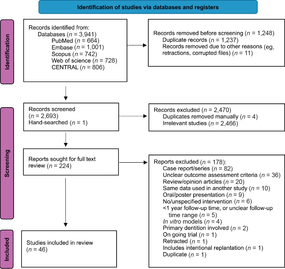
Of the 3,941 reports initially identified, 46 were included. The estimated short-, medium-, and long-term survival rates were 96.31% (95% confidence interval [CI], 94.80–97.82), 88.23% (95% CI, 85.59–90.87), and 84.80% (95% CI, 76.70– 92.91), respectively. There were no significant differences in outcomes between age and root development groups. The most common reason for tooth loss was root resorption. High patient satisfaction rates were reported.
Conclusions
Autotransplanted teeth exhibit high survival rates in the short- to medium-term. Minimizing root surface damage and excluding pulpal contaminants may promote longevity. The procedure appeared equally successful for teeth at different stages of root development and across various age groups.
- 4,002 View
- 237 Download
- Initial attachment, viability, proliferation, and migration of osteoblast-like SaOS-2 cells on two resorbable xenogeneic membranes for guided tissue regeneration: ab in vitro experimental study
- Rafael Fernández-Grisales, Giovanna García-Suárez, Ximena Guerrero-Rodríguez, Carolina Berruecos-Orozco, Marco Calle-Jaramillo, Wilder Javier Rojas, Vanessa Esmeralda Duque, Daniela Serna-Guisao, Néstor Ríos-Osorio
- Restor Dent Endod 2026;51(2):e20. Published online April 13, 2026
- DOI: https://doi.org/10.5395/rde.2026.51.e20
-
Abstract
PDF
- Objectives
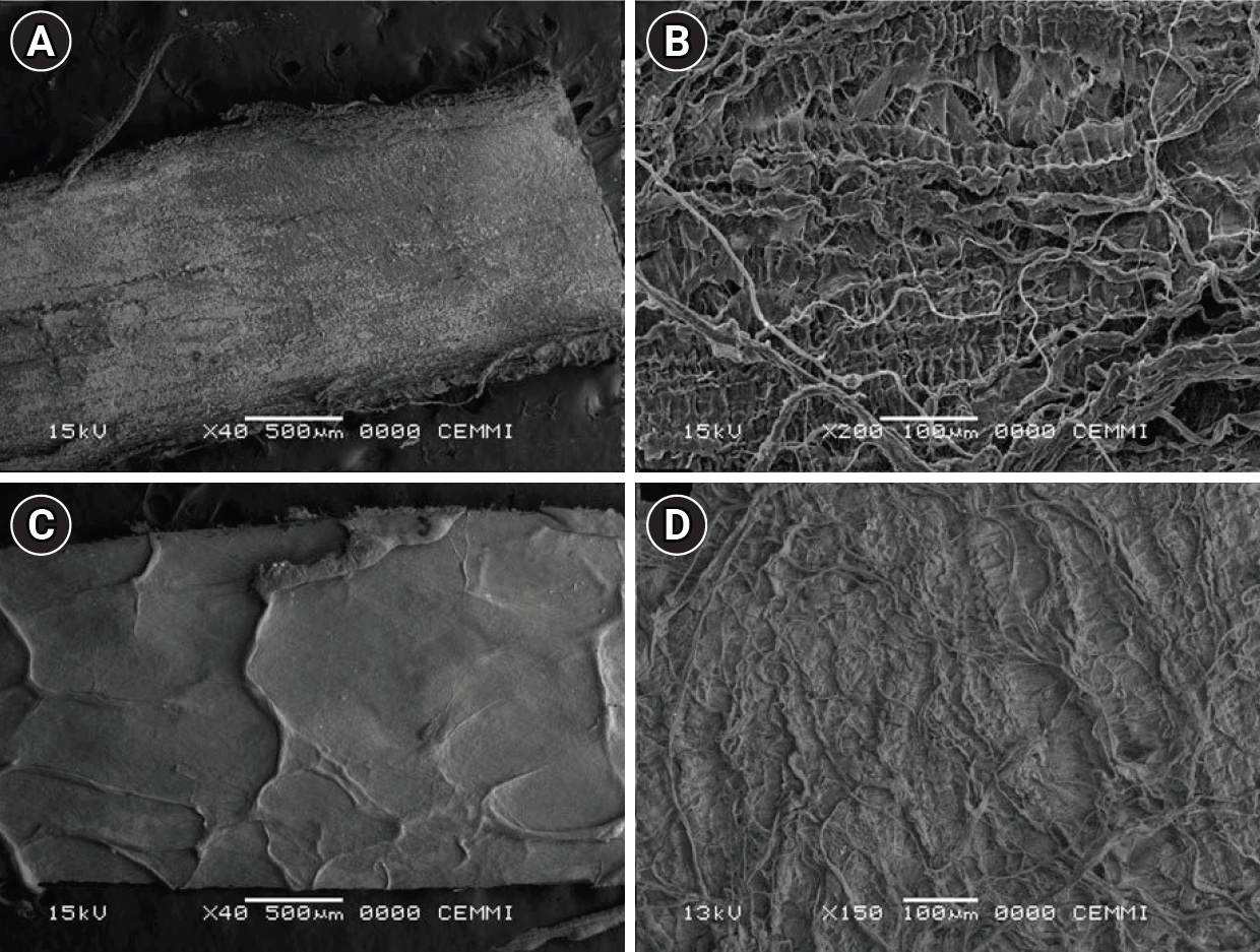
This study evaluated the biocompatibility of a new xenogeneic resorbable membrane derived from porcine esophagus membrane (Quirumatrix, Cells Tech Co.) and compared it with a porcine pericardium membrane (Straumann Jason, Straumann Holding AG.) traditionally used for guided tissue regeneration. The parameters investigated were the viability, migration, and adhesion of SaOS-2 osteoblast-like cells derived from osteosarcoma on both membranes.
Methods
The cells were cultured in 100 mm plates in RPMI 1640 medium (40 mL), supplemented. They were incubated at 37°C in a humidified atmosphere with 95% air and 5% to 10% CO2. Cell morphology and adhesion were evaluated using phase contrast optical microscopy and scanning electron microscope. Cell viability and proliferation were evaluated using a fluorometric resazurin reduction assay, with fluorescence intensity measured at 48, 72, and 96 hours. Cell migration was evaluated using staining with Alexa Fluor 555 Phalloidin (Cell Signaling Technology) and DAPI, with a reference line. Cell migration was analyzed by measuring displacement within the delineated area using an Axio Imager M2 fluorescence microscope (Carl Zeiss). Each membrane was photographed. The statistical analysis was performed using GraphPad Prism ver. 10.2.3 (GraphPad Software). A p-value <0.05 was considered significant between experimental groups.
Results
Both membranes were shown to be biocompatible. The porcine pericardium membrane showed greater cell adhesion and proliferation compared to the porcine esophagus membrane. Cell migration was significantly greater in the Jason membrane.
Conclusions
The results revealed that both evaluated membranes are biocompatible and non-cytotoxic; further research is needed to understand their long-term behavior, interactions with other types of cells, and performance in specific therapeutic situations. -
Citations
Citations to this article as recorded by- Fluoride-reinforced 50S8P sol–gel bioactive glass for potential enamel remineralisation
Qichen Sun, Siti Noor Fazliah Mohd Noor, Siti Fatimah Samsurrijal, Nawal Radhiah Abdul Rahman
Biomedical Physics & Engineering Express.2026; 12(5): 055014. CrossRef
- Fluoride-reinforced 50S8P sol–gel bioactive glass for potential enamel remineralisation
- 2,061 View
- 123 Download
- 1 Web of Science
- 1 Crossref
- Fracture resistance of regenerated immature teeth in different simulated stages of root development: an in vitro cyclic loading study
- Kyveli-Artemis Polydora, Konstantinos Kodonas, Anastasia Fardi, Christos Gogos
- Restor Dent Endod 2026;51(2):e21. Published online April 28, 2026
- DOI: https://doi.org/10.5395/rde.2026.51.e21
-
Abstract
PDF
 Supplementary Material
 - Objectives
This in vitro study aimed to assess the fracture resistance of simulated stages of root maturation following regenerative endodontic treatment using a cyclic loading method.
Methods
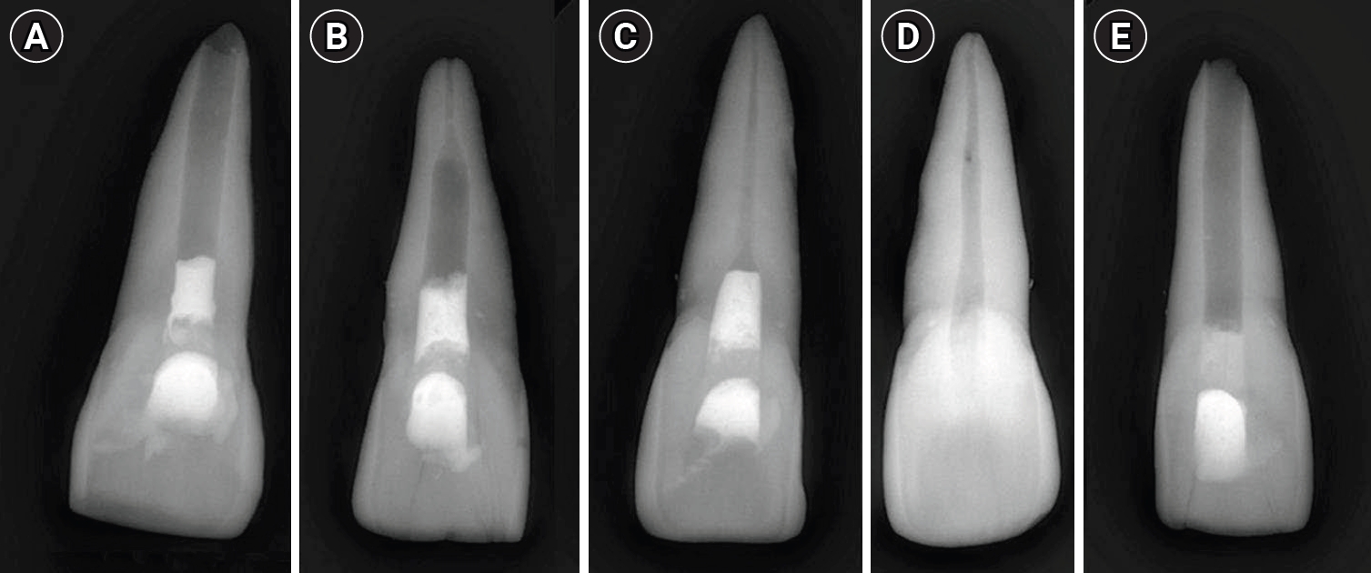
Ninety extracted maxillary central incisors were randomly allocated into three experimental groups representing different stages of root development, following revitalization: Group A for completely immature teeth immediately after treatment; Group B for teeth with apical closure, and Group C for teeth with apical closure and wall thickening. Two control groups were also included: Group D for intact teeth and Group E for simulated immature teeth without the bioceramic material. Following simulation of immature apices and treatment with a bioceramic material, all specimens were subjected to cyclic loading using a step-stress fatigue protocol until failure. The number of cycles to fracture and the peak load were recorded and statistically analyzed.
Results
Statistically significant differences in loading forces were observed between the negative control group (Group D) and Groups A, B, and E (p < 0.05). However, no statistically significant differences were detected among the experimental groups. These results indicate that apical closure and dentinal wall thickening alone did not substantially improve mechanical reinforcement under cyclic loading conditions.
Conclusions
Although intact teeth exhibited superior mechanical performance, apical closure and wall thickening alone were insufficient to enhance reinforcement under cyclic loading.
- 1,845 View
- 163 Download
- Interplay of hypoxia, angiogenesis, and macrophages in pulp and periapical lesions: an immunohistochemical cross-sectional study
- Puja Chatterjee, Mala Kamboj, Shweta Mittal, Anjali Narwal, Anju Devi
- Restor Dent Endod 2026;51(2):e22. Published online April 23, 2026
- DOI: https://doi.org/10.5395/rde.2026.51.e22
-
Abstract
PDF
- Objectives
This study evaluated and correlated the immune expression of hypoxia and angiogenesis with macrophages in periapical granuloma (PG), radicular cyst (RC), and healthy pulp (HP).
Methods
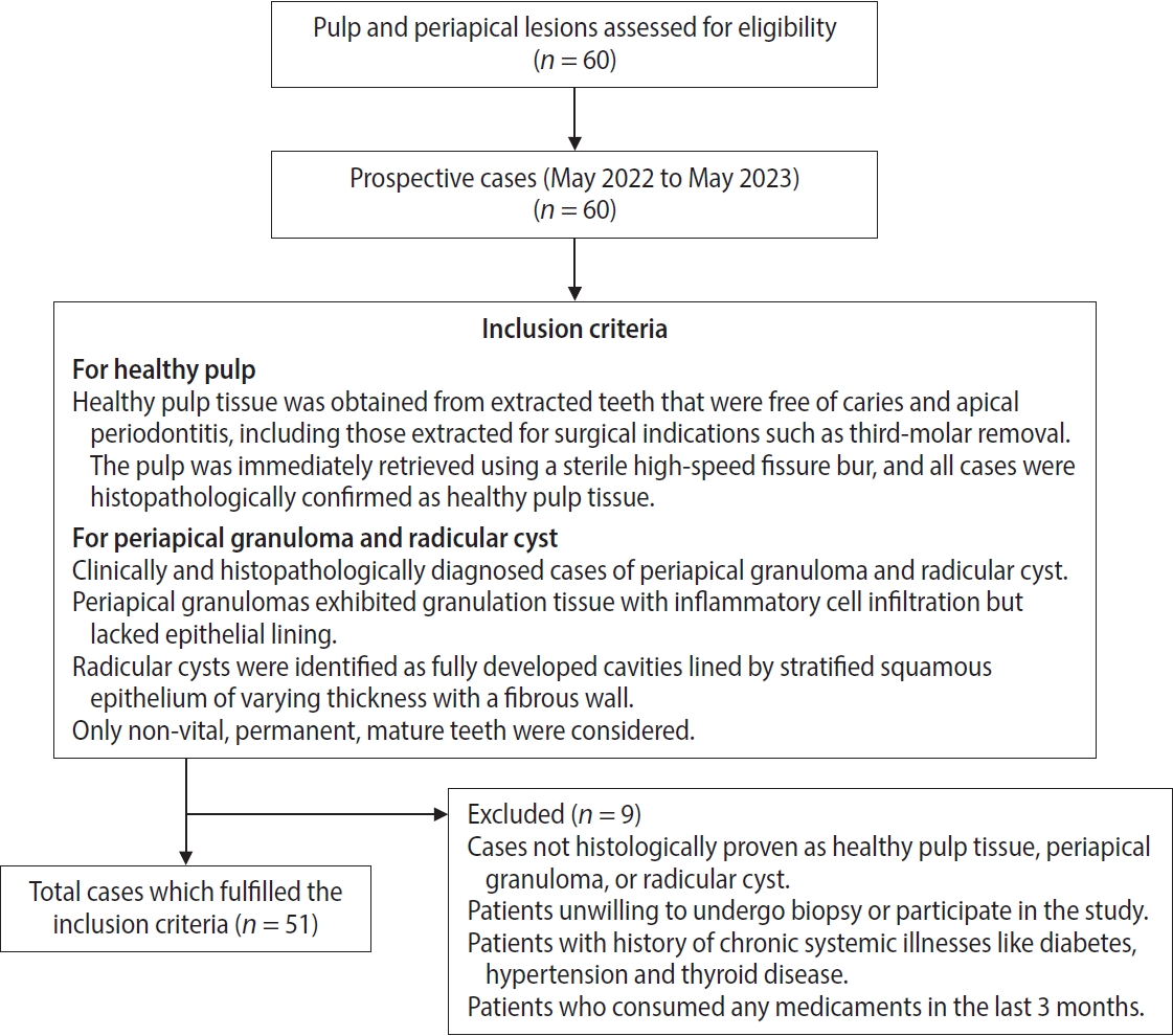
An observational study was performed on 51 tissue blocks equally divided among the groups, stained immunohistochemically for hypoxia-inducible factor (HIF)-1α, vascular endothelial growth factor (VEGF), and CD68, and the mean expression was calculated. Data were analyzed using Kruskal-Wallis, Mann-Whitney, Spearman correlation tests (p < 0.001), and multiple linear regression analysis (p ≤ 0.05).
Results
HIF-1α expression was highest in PG than RC and HP (p < 0.001). Significant differences were found between HP, PG, and RC (both p < 0.001). VEGF expression was highest in RC than in PG and HP (p < 0.001), with significant differences between HP and both PG and RC (p < 0.001); pairwise comparisons were significant between all groups (p < 0.001, p < 0.001, p = 0.018). Correlation analysis showed significant correlations between VEGF and CD68 in HP and PG (p = 0.007 and p = 0.028, respectively). Linear regression showed that study groups were significantly associated with mean scores of HIF-1α, VEGF, and CD68 (p = 0.002, p = 0.001, p < 0.001).
Conclusions
HIF-1α, VEGF, and CD68 showed increased expression in PGs and RCs, suggesting an association between hypoxic conditions, enhanced angiogenic activity, and macrophage presence within the periapical inflammatory microenvironment. Future studies exploring HIF-1α and VEGF inhibitors as potential treatment modalities for periapical lesions are warranted. -
Citations
Citations to this article as recorded by- Radiologic and Immunohistochemical Correlation of Programmed Death 1/Programmed Death Ligand 1 and CD4+ T Cells in Periapical Granuloma and Radicular Cyst: An Observational Study
Astha Upadhyay, Mala Kamboj, Shweta Mittal, Anjali Narwal, Anju Devi, Adarsh Kumar
Journal of Endodontics.2026;[Epub] CrossRef
- Radiologic and Immunohistochemical Correlation of Programmed Death 1/Programmed Death Ligand 1 and CD4+ T Cells in Periapical Granuloma and Radicular Cyst: An Observational Study
- 1,554 View
- 66 Download
- 1 Crossref
- Effect of high irradiance and short exposure curing time on the fracture toughness of bulk-fill resin-based composite: an in vitro study
- Beatriz Ometto Sahadi, Tainah Oliveira Rifane, Carolina Bosso André, Vitaliano Gomes Araújo-Neto, Richard Thomas Bengt Price, Marcelo Giannini
- Restor Dent Endod 2026;51(2):e23. Published online April 20, 2026
- DOI: https://doi.org/10.5395/rde.2026.51.e23
-
Abstract
PDF
- Objectives
This study aimed to determine the effect of high irradiance and short exposure time on the fracture toughness of bulk-fill resin-based composites (RBCs).
Methods
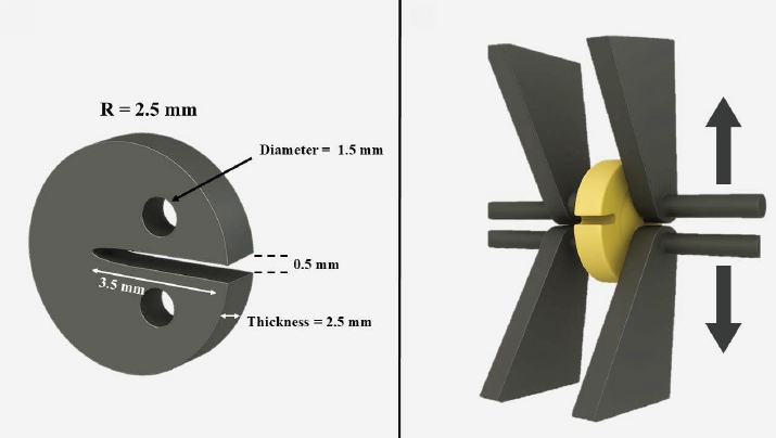
Three RBCs were tested: Tetric PowerFill (TPF; Ivoclar Vivadent), Opus Bulk Fill APS (OBF; FGM Dental Group), and Filtek One Bulk Fill (FOB; Solventum). Sixty single-edge-notched disc specimens were prepared using a fracture toughness mold. Each group consisted of 20 samples, divided into two subgroups (n = 10). The RBCs were lightcured either for 3 seconds in high-irradiance mode (‘3s cure’) or for the manufacturer-recommended times (TPF, 10 seconds; OBF, 30 seconds; FOB, 20 seconds) in ‘high power’ mode using the Bluephase PowerCure (Ivoclar Vivadent). The peak spectral wavelength was measured using a spectrophotometer. Specimens were tested on a universal testing machine, and data were analyzed by two-way analysis of variance and Bonferroni test (α = 0.05).
Results
Radiant exposure values (J/cm²) were 9.5 for the 3-second mode and 12.4, 24.8, and 37.1 for 10, 20, and 30 seconds (high power mode), respectively. FOB (4.22 and 3.79 MPa∙m0.5 for 20 and 3 seconds) had the highest mean fracture toughness, while OBF showed the lowest (2.01 and 2.10 MPa∙m0.5 for 30 and 3 seconds). TPF produced intermediate results (2.72 and 2.70 MPa∙m0.5 for 10 and 3 seconds). Exposure time did not affect TPF and OBF, while the 3-second exposure significantly reduced the fracture toughness for FOB.
Conclusions
The RBCs tested had different fracture toughness values regardless of exposure time. High irradiance and short exposure can reduce fracture toughness depending on the RBC tested.
- 1,460 View
- 105 Download
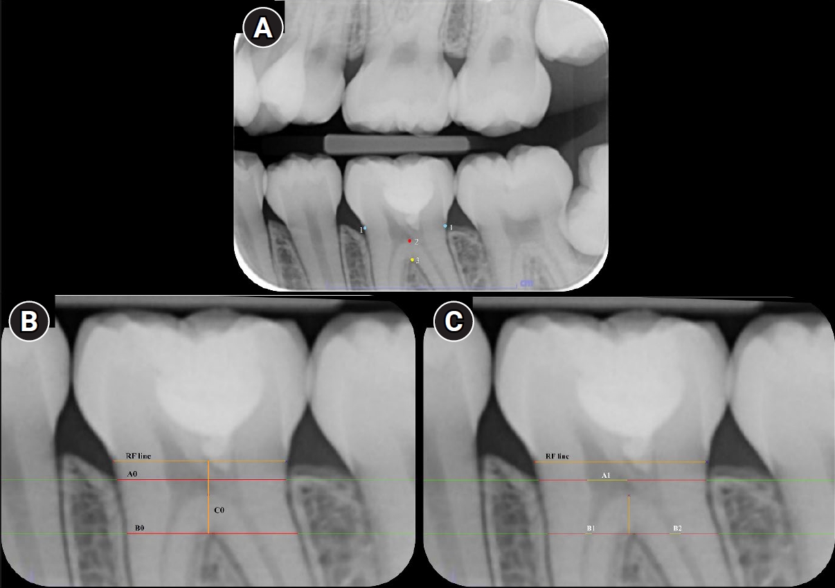
- Magnitude of pulp space narrowing over time and contributing factors in teeth with vital pulp therapy: a retrospective cohort study
- Akarapong Boontankun, Papimon Chompu‑inwai, Chanika Manmontri, Nattakan Chaipattanawan, Areerat Nirunsittirat, Phichayut Phinyo, Trasapong Thaiupathump
- Restor Dent Endod 2026;51(2):e24. Published online May 13, 2026
- DOI: https://doi.org/10.5395/rde.2026.51.e24
-
Abstract
PDFSupplementary Material
- Objectives
This study aimed to compare the magnitude of pulp space narrowing over time—measured as the change in pulp/tooth proportion from baseline—between mandibular molars treated with different types of vital pulp therapy (VPT) and their contralateral sound molars (controls). This study also investigated factors influencing the magnitude of pulp space narrowing in molars that have undergone VPT.
Methods
This retrospective cohort study involved the assessment of bitewing radiographs of VPT-treated molars and controls at baseline and follow-up. Using reference points and lines on the radiograph, pulp/tooth proportions were measured by examiners. The intraclass correlation coefficient (ICC) was used to report examiner reliability. The changes in pulp/tooth proportions from baselines were compared between subgroups using multilevel mixed effect linear regression and the Wald test.
Results
A total of 382 bitewing radiographs from 134 teeth were included. The follow-up period ranged from 6 to 84 months (mean, 27.12 ± 17.67 months). ICC values indicated good to excellent examiner reliability. Compared to the controls, changes in pulp/tooth proportion from baselines, indicating pulp space narrowing, were significantly greater in teeth with partial pulpotomy (at pulp chamber width) and coronal pulpotomy (at pulp canal width). Factors affecting the magnitude of pulp space narrowing included the more invasive type of VPT and the more severe preoperative diagnosis.
Conclusions
The magnitude of pulp space narrowing was greater in VPT-treated molars than in controls. The more invasive type of VPT and severe preoperative diagnosis were factors contributing to the magnitude of pulp space narrowing.
- 1,807 View
- 92 Download
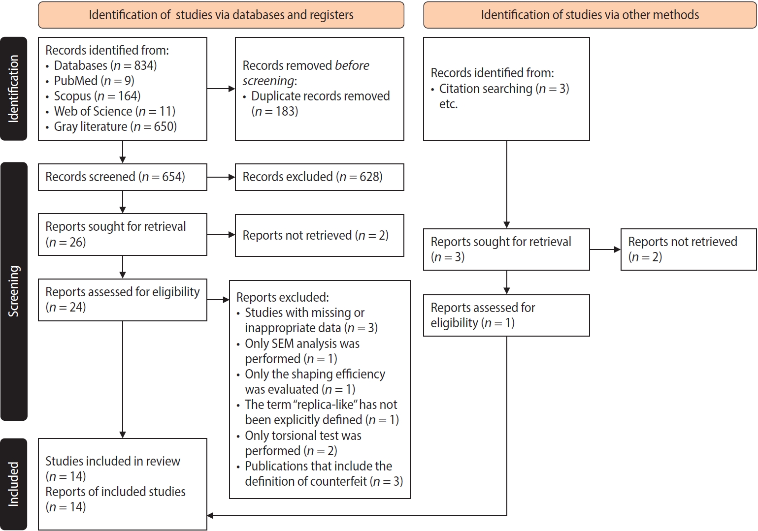
- Comparison of the cyclic fatigue resistance of original and replica-like files: a systematic review and meta-analysis
- Mert Unal, Fatih Cakici
- Restor Dent Endod 2026;51(2):e25. Published online May 19, 2026
- DOI: https://doi.org/10.5395/rde.2026.51.e25
-
Abstract
PDF
- Objectives
This systematic review and meta-analysis aimed to evaluate the existing literature and quantitatively analyze the cyclic fatigue resistance of original and replica-like nickel-titanium endodontic files.
Methods
A comprehensive search was conducted across four electronic databases up to July 6, 2025, following PRISMA guidelines (PROSPERO: CRD420251086699). The methodological quality of included studies was assessed using criteria adapted from previous in vitro systematic reviews. Publication bias was evaluated using Egger’s test and the trim-and-fill method. A random-effects meta-analysis was performed to compare the cyclic fatigue resistance of original and replica- like files using time to fracture (TTF) and number of cycles to fracture (NCF) as outcomes. These were expressed as standardized mean differences (SMDs) with 95% confidence intervals (CIs). Heterogeneity was analyzed using the I² statistic.
Results
A total of 14 studies involving 1,276 endodontic files (nine original and 31 replica-like types) were included. Based on TTF values, replica-like files showed significantly greater cyclic fatigue resistance than original files (SMD, –0.845; 95% CI, –1.268 to –0.423; p < 0.001). However, NCF-based analysis revealed no statistically significant difference (SMD, –1.532; 95% CI, –3.615 to 0.550; p = 0.149).
Conclusions
Replica-like files exhibited cyclic fatigue resistance comparable to original instruments and may be considered potential alternatives. However, due to high heterogeneity and methodological variability, these findings should be interpreted with caution.
- 1,185 View
- 58 Download
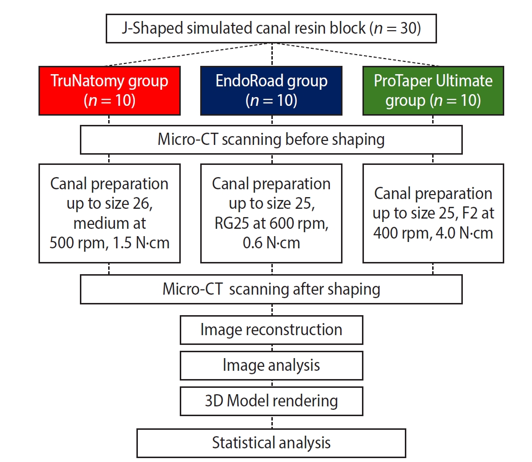
- In vitro assessment of geometric characteristics in canal preparation using nickel-titanium files used for minimal invasiveness: an experimental study
- EunJin Jang, Hyeon-Cheol Kim, WooCheol Lee
- Restor Dent Endod 2026;51(2):e26. Published online May 13, 2026
- DOI: https://doi.org/10.5395/rde.2026.51.e26
-
Abstract
PDF
- Objectives
This study aimed to assess geometric characteristics in canal preparation using nickel-titanium (NiTi) files used for minimal invasiveness.
Methods
Thirty J-shaped simulated canals in resin blocks were instrumented with either TruNatomy (TR; Dentsply Sirona), EndoRoad (ER; Maruchi), or ProTaper Ultimate (PTU; Dentsply Sirona). The simulated canal blocks were scanned using microcomputed tomography before and after instrumentation. The scanned images were reconstructed, and the canal surface area was measured from 0.5 to 6.5 mm from the apex. Three-dimensional representative models of each group were rendered. The data were statistically analyzed using one-way analysis of variance and Kruskal-Wallis test at 95% significance level.
Results
TR showed a superior ability to maintain the canal’s center. TR demonstrated comparable apical preparation to PTU. ER showed a smaller and limited apical preparation than other systems, with a tendency for canal preparation toward the inner side of the curvature. PTU featured the largest prepared apical size among the file groups and tended to straighten the curvature by preparing the canal more towards the outward side. The surface area instrumented using each NiTi file showed statistically significant differences among the three groups at all levels except 0.5, 2.0, and 3.5 mm from the apex (p < 0.05). There was no statistically significant difference between TR and PTU at a level of 0.5 mm from the apex (p > 0.05).
Conclusions
While PTU is suitable for general canal preparation to facilitate irrigation and intracanal medication, TR and ER excel in preserving canal centering with minimal concern for canal transportation by minimally invasive preparation.
- 1,207 View
- 49 Download
Case Report
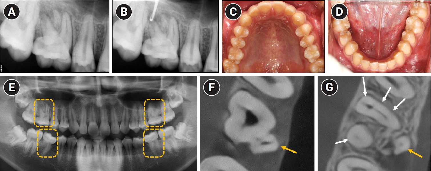
- Endodontic treatment of a molar-incisor malformation of the maxillary first molar: a case report
- Woo-Lim Kim, Se-Hee Park, Kyung-Mo Cho, Jin-Woo Kim
- Restor Dent Endod 2026;51(2):e27. Published online May 21, 2026
- DOI: https://doi.org/10.5395/rde.2026.51.e27
-
Abstract
PDF
- Molar-incisor malformation (MIM) is a developmental dental anomaly primarily affecting permanent first molars, often accompanied by structural irregularities such as cervical mineralized diaphragms (CMDs) and furcal channels. These anatomical complexities present significant challenges for endodontic treatment. This case report presents the endodontic management of a maxillary first molar diagnosed with MIM—a condition for which root canal treatment has rarely been reported. The affected tooth exhibited characteristic features of MIM, including underdeveloped roots, CMD, and an open furcal channel. Initial canal negotiation revealed four buccal canals, but the palatal canal could not be located via conventional access. A separate access approach enabled successful identification, disinfection, and obturation of the palatal canal. Follow-up imaging showed healing of the periapical lesion and favorable clinical outcomes. This case highlights the diagnostic and technical challenges in managing MIM-affected teeth and underscores the importance of advanced imaging, tailored access strategies, and careful material selection to achieve successful endodontic outcomes.
- 1,391 View
- 118 Download
 First
First Prev
Prev


