-
Influence of modeling agents on the surface properties of an esthetic nano-hybrid composite
-
Zeynep Bilge Kutuk, Ecem Erden, Damla Lara Aksahin, Zeynep Elif Durak, Alp Can Dulda
-
Restor Dent Endod 2020;45(2):e13. Published online January 29, 2020
-
DOI: https://doi.org/10.5395/rde.2020.45.e13
-
-
 Abstract Abstract
 PDF PDF PubReader PubReader ePub ePub
- Objective
The aim of this study was to evaluate the influence of different modeling agents on the surface microhardness (Vickers hardness number; VHN), roughness (Ra), and color change (ΔE) of a nano-hybrid composite with or without exposure to discoloration by coffee. Materials and MethodsSixty-four cylinder-shaped nano-hybrid composite specimens were prepared using a Teflon mold. The specimens' surfaces were prepared according to the following groups: group 1, no modeling agent; group 2, Modeling Liquid; group 3, a universal adhesive (G-Premio Bond); and group 4, the first step of a 2-step self-adhesive system (OptiBond XTR). Specimens were randomly allocated into 2 groups (n = 8) according to the storage medium (distilled water or coffee). VHN, Ra, and ΔE were measured at 24 hours, 1 week, and 6 weeks. The Kruskal-Wallis test followed by the Bonferroni correction for pairwise comparisons was used for statistical analysis (α = 0.05). ResultsStorage time did not influence the VHN of the nano-hybrid composite in any group (p > 0.05). OptiBond XTR Primer application affected the VHN negatively in all investigated storage medium and time conditions (p < 0.05). Modeling Liquid application yielded improved Ra values for the specimens stored in coffee at each time point (p < 0.05). Modeling Liquid application was associated with the lowest ΔE values in all investigated storage medium and time conditions (p < 0.05). ConclusionDifferent types of modeling agents could affect the surface properties and discoloration of nano-hybrid composites.
-
Citations
Citations to this article as recorded by  - The Impact of Modeling Liquids on Surface Roughness and Color Properties of Bulkfill Resin Composites After Simulated Tooth Brushing: An in Vitro Study. Part I
Camila Falconí‐Páez, Claudia González‐Vaca, Juliana Guarneri, Newton Fahl, Paulina Aliaga‐Sancho, Maria Lujan Mendez‐Bauer, Cesar Augusto Galvão Arrais, Andrés Dávila‐Sánchez
Journal of Esthetic and Restorative Dentistry.2025; 37(2): 514. CrossRef - Coating Agents for Resin Composites: Effect on Color Stability, Roughness, and Surface Micromorphology Subjected to Brushing Wear
FR Hojo, TC Martins, WF Vieira-Junior, FMG França, CP Turssi, RT Basting
Operative Dentistry.2025; 50(1): 101. CrossRef - Effect of modeling liquid application on color stability and surface roughness of single-shade composites
Melek Güven Bekdaş, Ihsan Hubbezoglu
BMC Oral Health.2025;[Epub] CrossRef - Influence of modeling liquids on the color adaptation and optical properties of single and simply shade resin composites
Bengü Doğu Kaya, Mehmet Buldur, Burcu Gözetici-Çil
Odontology.2025;[Epub] CrossRef - Effect of modeling liquids on Vickers microhardness, flexural strength and color stability of resin-based composites
Ahmed Alshawi, Benin Dikmen, Sevda Ozel Yildiz, Ugur Erdemir
Materials Research Express.2025; 12(11): 115402. CrossRef - Impact of combining dental composite brushes with modeling resins on the color stability and topographic features of composites
Abdulrahman A Balhaddad, Faisal Alharamlah, Alhanoof Aldossary, Wejdan Almutairi, Turki Alshehri, Mary Anne S Melo, Afnan O Al-Zain, Eman H Ismail
Journal of Applied Biomaterials & Functional Materials.2024;[Epub] CrossRef - Investigation of the Degree of Monomer Conversion in Dental Composites through Various Methods: An In Vitro Study
Musa Kazim Ucuncu, Ozge Celiksoz, Emine Sen, Yasemin Yucel Yucel, Bircan Dinc
Applied Sciences.2024; 14(11): 4406. CrossRef - EFEITO DOS LÍQUIDOS MODELADORES NA SUPERFÍCIE DA RESINA COMPOSTA – UMA REVISÃO DE LITERATURA
Samuel Silva Dias, Matheus Fernando Lopes, Jeffison Teles Dias, Caio Junji Tanaka, Jose Augusto Rodrigues
RECIMA21 - Revista Científica Multidisciplinar - ISSN 2675-6218.2024; 5(2): e524899. CrossRef - Effect of Instrument Lubricant on Mechanical Properties of Restorative Composite
G Pippin, D Tantbirojn, M Wolfgang, JS Nordin, A Versluis
Operative Dentistry.2024; 49(4): 475. CrossRef - Full analysis of the effects of modeler liquids on the properties of direct resin-based composites: a meta-analysis review of in vitro studies
Eduardo Trota Chaves, Lisia Lorea Valente, Eliseu Aldrighi Münchow
Clinical Oral Investigations.2023; 27(7): 3289. CrossRef - Influence of Modeling Liquids and Universal Adhesives Used as Lubricants on Color Stability and Translucency of Resin-Based Composites
Gaetano Paolone, Claudia Mazzitelli, Giacomo Zechini, Salvatore Scolavino, Cecilia Goracci, Nicola Scotti, Giuseppe Cantatore, Enrico Gherlone, Alessandro Vichi
Coatings.2023; 13(1): 143. CrossRef - Influence of Instrument Lubrication on Properties of Dental Composites
Juliusz Kosewski, Przemysław Kosewski, Agnieszka Mielczarek
European Journal of Dentistry.2022; 16(04): 719. CrossRef - Effect of Modelıng Liquid Use on Color and Whiteness Index Change of Composite Resins
Numan AYDIN, Serpil KARAOĞLANOĞLU, Bilge ERSÖZ
Cumhuriyet Dental Journal.2022; 25(Supplement): 119. CrossRef - Effects of Immediate Coating on Unset Composite with Different Bonding Agents to Surface Hardness
Nantawan Krajangta, Supissara Ninbanjong, Sunisa Khosook, Kanjana Chaitontuak, Awiruth Klaisiri
European Journal of Dentistry.2022; 16(04): 828. CrossRef - Modeling Liquids and Resin-Based Dental Composite Materials—A Scoping Review
Gaetano Paolone, Claudia Mazzitelli, Uros Josic, Nicola Scotti, Enrico Gherlone, Giuseppe Cantatore, Lorenzo Breschi
Materials.2022; 15(11): 3759. CrossRef - Shear Bond Strength of Composite Diluted with Composite-Handling Agents on Dentin and Enamel
Mijoo Kim, Deuk-Won Jo, Shahed Al Khalifah, Bo Yu, Marc Hayashi, Reuben H. Kim
Polymers.2022; 14(13): 2665. CrossRef - Effect of Modeling Resins on Microhardness of Resin Composites
Ezgi T. Bayraktar, Pinar Y. Atali, Bora Korkut, Ezgi G. Kesimli, Bilge Tarcin, Cafer Turkmen
European Journal of Dentistry.2021; 15(03): 481. CrossRef
-
1,803
View
-
29
Download
-
17
Crossref
-
Effect of various bleaching treatments on shear bond strength of different universal adhesives and application modes
-
Fatma Dilsad Oz, Zeynep Bilge Kutuk
-
Restor Dent Endod 2018;43(2):e20. Published online April 16, 2018
-
DOI: https://doi.org/10.5395/rde.2018.43.e20
-
-
 Abstract Abstract
 PDF PDF PubReader PubReader ePub ePub
- Objectives
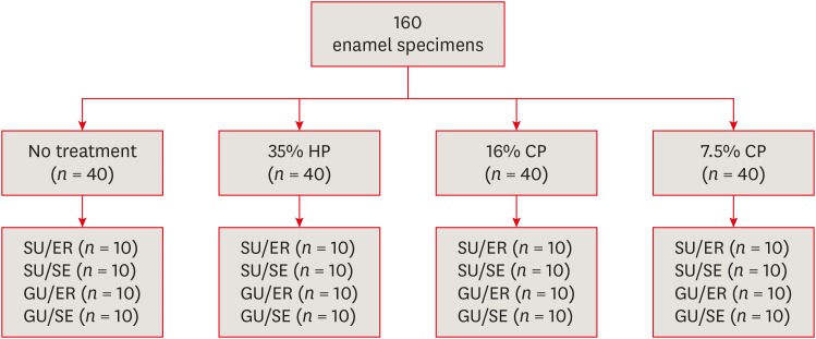
The aim of this in vitro study was to evaluate the bond strength of 2 universal adhesives used in different application modes to bleached enamel. Materials and MethodsExtracted 160 sound human incisors were used for the study. Teeth were divided into 4 treatment groups: No treatment, 35% hydrogen peroxide, 16% carbamid peroxide, 7.5% carbamid peroxide. After bleaching treatments, groups were divided into subgroups according to the adhesive systems used and application modes (n = 10): 1) Single Bond Universal, etch and rinse mode; 2) Single Bond Universal, self-etch mode; 3) Gluma Universal, etch and rinse mode; 4) Gluma Universal, self-etch mode. After adhesive procedures nanohybrid composite resin cylinders were bonded to the enamel surfaces. All specimens were subjected to shear bond strength (SBS) test after thermocycling. Data were analyzed using a 3-way analysis of variance (ANOVA) and Tukey post hoc test. ResultsNo significant difference were found among bleaching groups (35% hydrogen peroxide, 16% carbamid peroxide, 7.5% carbamid peroxide, and no treatment groups) in the mean SBS values. There was also no difference in SBS values between Single Bond Universal and Gluma Universal at same application modes, whereas self-etch mode showed significantly lower SBS values than etch and rinse mode (p < 0.05). ConclusionsThe bonding performance of the universal adhesives was enhanced with the etch and rinse mode application to bleached enamel and non-bleached enamel.
-
Citations
Citations to this article as recorded by  - Antioxidant effect on shear bond strength of resin composite to in-office versus home bleached enamel surface
Maha Mosaad Mohamed, Magda E. -A. Shalaby, Eman A. E. -G. Shebl
Tanta Dental Journal.2025; 22(3): 409. CrossRef - Effects of Time-Elapsed Bleaching on the Surface and Mechanical Properties of Dentin Substrate Using Hydrogen Peroxide-Free Nanohydroxyapatite Gel
Aftab Khan, Abdulaziz AlKhureif, Manal Almutairi, Abrar Nooh, Saeed Hassan, Yasser Alqahtani
International Journal of Nanomedicine.2024; Volume 19: 10307. CrossRef - Effect of sodium ascorbate on the shear bond strength of orthodontic brackets to bleached enamel using universal dental adhesive
Saeid Sadeghian, Kamyar Fathpour, Mahshid Biglari
Dental Research Journal.2023;[Epub] CrossRef - Quantitative Measurements of the Depth of Enamel Demineralization before and after Bleach: An In Vitro Study
Sara Naim, Gianrico Spagnuolo, Essam Osman, Syed Sarosh Mahdi, Gopi Battineni, Syed Saad B. Qasim, Mariangela Cernera, Hasna Rifai, Nada Jaafar, Elie Maalouf, Carina Mehanna Zogheib, Konstantinos Michalakis
BioMed Research International.2022;[Epub] CrossRef - DİŞ BEYAZLATMA İŞLEMİNİN LİTYUM DİSİLİKAT SERAMİĞİN BAĞLANMA DAYANIMINA ETKİSİ
Merve YILDIRAK, Rıfat GÖZNELİ
Atatürk Üniversitesi Diş Hekimliği Fakültesi Dergisi.2020; : 1. CrossRef - The Effect of Different Bleaching Protocols, Used with and without Sodium Ascorbate, on Bond Strength between Composite and Enamel
Maroun Ghaleb, Giovanna Orsini, Angelo Putignano, Sarah Dabbagh, Georges Haber, Louis Hardan
Materials.2020; 13(12): 2710. CrossRef - Influence of phototherapy on adhesive strength and microleakage of bleached enamel bonded to orthodontic brackets: An in-vitro study
Erum Khan, Ibrahim Alshahrani, Muhammad Abdullah Kamran, Abdulaziz Samran, Ali Alqerban, Saad Abdul Rehman
Photodiagnosis and Photodynamic Therapy.2019; 25: 344. CrossRef - Effect of Er: YAG Laser on Microtensile Bond Strength of Bleached Dentin to Composite
Mohsen Rezaei, Elham Aliasghar, Mohammad Bagher Rezvani, Nasim Chiniforush, Zohreh Moradi
Journal of Lasers in Medical Sciences.2019; 10(2): 117. CrossRef
-
1,883
View
-
18
Download
-
8
Crossref
-
Comparison of two different methods of detecting residual caries
-
Uzay Koç Vural, Zeynep Bilge Kütük, Esra Ergin, Filiz Yalçın Çakır, Sevil Gürgan
-
Restor Dent Endod 2017;42(1):48-53. Published online January 25, 2017
-
DOI: https://doi.org/10.5395/rde.2017.42.1.48
-
-
 Abstract Abstract
 PDF PDF PubReader PubReader ePub ePub
- Objectives
The aim of this study was to investigate the ability of the fluorescence-aided caries excavation (FACE) device to detect residual caries by comparing conventional methods in vivo. Materials and MethodsA total of 301 females and 202 males with carious teeth participated in this study. The cavity preparations were done by grade 4 (Group 1, 154 teeth), grade 5 (Group 2, 176 teeth), and postgraduate (Group 3, 173 teeth) students. After caries excavation using a handpiece and hand instruments, the presence of residual caries was evaluated by 2 investigators who were previously calibrated for visual-tactile assessment with and without magnifying glasses and trained in the use of a FACE device. The tooth number, cavity type, and presence or absence of residual caries were recorded. The data were analyzed using the Chi-square test, the Fisher's Exact test, or the McNemar test as appropriate. Kappa statistics was used for calibration. In all tests, the level of significance was set at p = 0.05. ResultsAlmost half of the cavities prepared were Class II (Class I, 20.9%; Class II, 48.9%; Class III, 20.1%; Class IV, 3.4%; Class V, 6.8%). Higher numbers of cavities left with caries were observed in Groups 1 and 2 than in Group 3 for all examination methods. Significant differences were found between visual inspection with or without magnifying glasses and inspection with a FACE device for all groups (p < 0.001). More residual caries were detected through inspection with a FACE device (46.5%) than through either visual inspection (31.8%) or inspection with a magnifying glass (37.6%). ConclusionsWithin the limitations of this study, the FACE device may be an effective method for the detection of residual caries.
-
Citations
Citations to this article as recorded by  - CariesXplainer: enhancing dental caries detection using Gradient-weighted Class Activation Mapping and transfer learning
Saira Asghar, Junaid Rashid, Anum Masood
Multimedia Tools and Applications.2025;[Epub] CrossRef - Comparative evaluation of caries detector dyes and laser fluorescence systems for intraoperative diagnosis during selective caries removal: a scoping review
Ana Iglesias-Poveda, Javier Flores-Fraile, Diego González-Gil, Joaquín López-Marcos
Frontiers in Dental Medicine.2025;[Epub] CrossRef - Fluorescence as a Quantitative Indicator of Cariogenic Bacteria During Chemo-Mechanical Caries Excavation with BRIX 3000 in Primary Teeth
Zornitsa Lazarova, Raina Gergova, Nadezhda Mitova
Journal of Functional Biomaterials.2025; 16(12): 453. CrossRef - Examining the Efficacy of a 405 nm Wavelength Diode Laser as a Diagnostic Tool in Routine Dental Practice
Marwan El Mobader, Samir Nammour
Cureus.2024;[Epub] CrossRef - Diş Çürüğünün Teşhisi ve Bu Amaçla Kullanılan Güncel Yöntemler
Oya BALA, Sümeyye KANLIDERE
Türk Diş Hekimliği Araştırma Dergisi.2023; 2(2): 219. CrossRef - Longitudinal study for dental caries calibration of dentists unexperienced in epidemiological surveys
Mariana NABARRETTE, Patrícia Rafaela dos SANTOS, Andréa Videira ASSAF, Glaucia Maria Bovi AMBROSANO, Marcelo de Castro MENEGHIM, Silvia Amélia Scudeler VEDOVELLO, Karine Laura CORTELLAZZI
Brazilian Oral Research.2023;[Epub] CrossRef - CURRENT CONCEPTS AND TECHNIQUES IN CARIES EXCAVATION: A REVIEW
Priyanka Aggarwal, Shivani Mathur, Tanya Batra
INTERNATIONAL JOURNAL OF SCIENTIFIC RESEARCH.2022; : 26. CrossRef - Current Strategies to Control Recurrent and Residual Caries with Resin Composite Restorations: Operator- and Material-Related Factors
Moataz Elgezawi, Rasha Haridy, Moamen A. Abdalla, Katrin Heck, Miriam Draenert, Dalia Kaisarly
Journal of Clinical Medicine.2022; 11(21): 6591. CrossRef - Rezidüel Çürük Tespitinde Kullanılan Geleneksel Yöntemin Farklı Yöntemlerle Klinik Olarak Doğrulanması
Fatma SAĞ GÜNGÖR
Selcuk Dental Journal.2021; 8(2): 402. CrossRef
-
1,556
View
-
10
Download
-
9
Crossref
|


