-
The application of “bone window technique” using piezoelectric saws and a CAD/CAM-guided surgical stent in endodontic microsurgery on a mandibular molar case
-
Ukseong Kim, Sunil Kim, Euiseong Kim
-
Restor Dent Endod 2020;45(3):e27. Published online May 21, 2020
-
DOI: https://doi.org/10.5395/rde.2020.45.e27
-
-
 Abstract Abstract
 PDF PDF PubReader PubReader ePub ePub
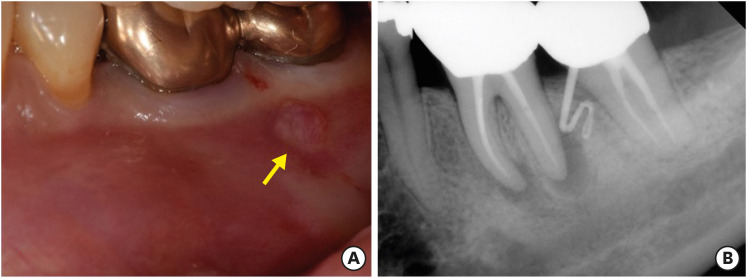
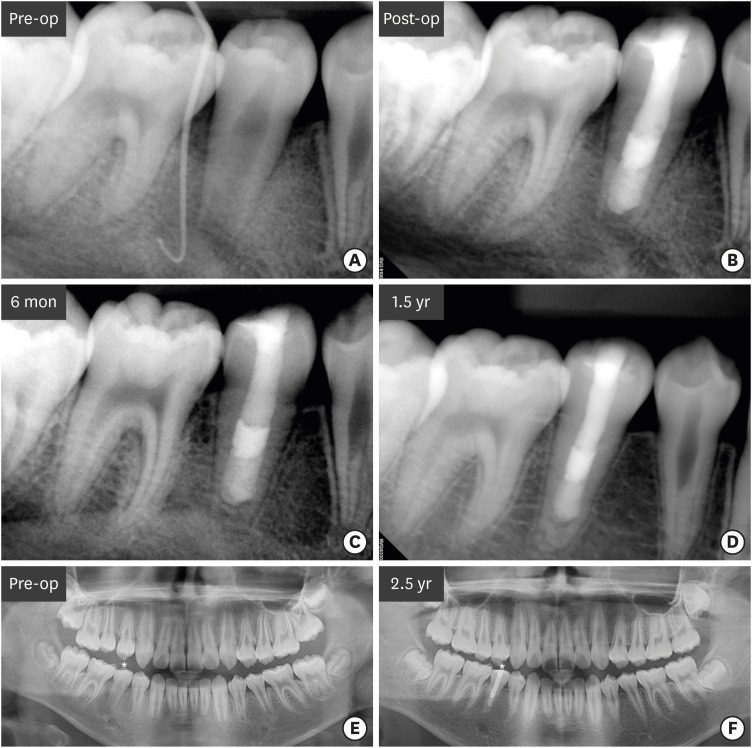
Apical surgery for a mandibular molar is still challenging for many reasons. This report describes the applications of computer-guided cortical ‘bone-window technique’ using piezoelectric saws that prevented any nerve damage in performing endodontic microsurgery of a mandibular molar. A 49-year-old woman presented with gumboil on tooth #36 (previously endodontically treated tooth) and was diagnosed with chronic apical abscess. Periapical lesions were confirmed using cone-beam computed tomography (CBCT). Endodontic microsurgery for the mesial and distal roots of tooth #36 was planned. Following the transfer of data of the CBCT images and the scanned cast to an implant surgical planning program, data from both devices were merged. A surgical stent was designed, on the superimposed three-dimensional model, to guide the preparation of a cortical window on the buccal side of tooth #36. Endodontic microsurgery was performed with a printed surgical template. Minimal osteotomy was required and preservation of the buccal cortical plate rendered this endodontic surgery less traumatic. No postoperative complications such as mental nerve damage were reported. Window technique guided by a computer-aided design/computer-aided manufacture based surgical template can be considerably useful in endodontic microsurgery in complicated cases. -
Citations
Citations to this article as recorded by  - Optimising Outcomes in Endodontic Microsurgery: Evidence, Uncertainties and Future Directions
Ukseong Kim, Euiseong Kim
International Endodontic Journal.2026; 59(5): 746. CrossRef - Accuracy of Guided Dual Technique in Esthetic Crown Lengthening: A Prospective Case‐Series Study
Meritxell Enfedaque‐Prat, Albert González‐Barnadas, Adrià Jorba‐García, Javi Vilarrasa, Jorge Toledano‐Serrabona, Rui Figueiredo, Eduard Valmaseda‐Castellón, Octavi Camps‐Font
Journal of Esthetic and Restorative Dentistry.2025; 37(6): 1284. CrossRef - Guided endodontics in the application of personalized mini-invasive treatment in clinical cases: a literature review
Shuangshuang Ren, Wanping Wang, Mingyue Cheng, Wenyue Tang, Yue Zhao, Leiying Miao
The Saudi Dental Journal.2025;[Epub] CrossRef - Accurately Defining the Location and Dimension of the Bony Lid Under the Guidance of Dynamic Navigation: Report on Three Cases
Kailiang Tang, Xiaole Zhang, Qibao Wang, Xinyu Zhao, Xijiao Yu, Yi Du
Australian Endodontic Journal.2025; 51(3): 785. CrossRef - Minimally Invasive Vertical Incision Subperiosteal Tunnelling Technique for Targeted Endodontic Surgery: Technical Overview and a Case Report
Francesc Abella Sans, Jaime Barragán Montes, Tomasz Zbozen, Nandini Suresh, Lalli Dharmarajan, Paul M. H. Dummer, Venkateshbabu Nagendrababu
International Endodontic Journal.2025; 58(11): 1799. CrossRef - Endodontic Microsurgery of Mandibular Molars with an Autonomous Robotic System
Haiying Zhang, Zi Yang, Mangnan Liu, Yaoxin Wang, Mei Fu, Benxiang Hou, Chen Zhang
Journal of Endodontics.2025; 51(12): 1830. CrossRef - Endodontic Microsurgery of a Mandibular Molar Using a Dynamic Navigation System (DNS) and Cortical Window Technique: A Case Report
Gustavo Castillo, Silvia Restrepo-Méndez, Oscar Zuluaga, Paola Escobar-Villegas
Journal of Endodontic Microsurgery.2024; 3: 1. CrossRef - The bone lid technique in endodontic microsurgery
Min Zhang, He Liu, Ya Shen
Asian Journal of Surgery.2024; 47(7): 3126. CrossRef - Guided Periradicular Surgery with Er,Cr:YSGG Laser Osteotomy: A Case Report
Julian Torres Celeita, Johanna Hernández la Rotta, Amdie Chirinos Salazar, Jorge Fandiño Rodríguez, Laura López Rincón, Mauren Orduz Solorzano, Diana Parra Galvis, Oscar Jiménez Peña
Journal of Endodontic Microsurgery.2024;[Epub] CrossRef - Piezoelectric Endodontic Microsurgery with Modified Cortical Window Technique: A Case Report
Rafael Fernández-Grisales, Wilder Rojas, Carolina Berruecos-Orozco
Journal of Endodontic Microsurgery.2023; 2: 34. CrossRef - The Impact of the Preferred Reporting Items for Case Reports in Endodontics (PRICE) 2020 Guidelines on the Reporting of Endodontic Case Reports
Sofian Youssef, Phillip Tomson, Amir Reza Akbari, Natalie Archer, Fayjel Shah, Jasmeet Heran, Sunmeet Kandhari, Sandeep Pai, Shivakar Mehrotra, Joanna M Batt
Cureus.2023;[Epub] CrossRef - Clinical and radiological outcomes of dynamic navigation in endodontic microsurgery: a prospective study
Chen Chen, Rui Zhang, Wei Zhang, Fangzhe Li, Zan Wang, Li Qin, Yun Chen, Zhuan Bian, Liuyan Meng
Clinical Oral Investigations.2023; 27(9): 5317. CrossRef - New-designed 3D printed surgical guide promotes the accuracy of endodontic microsurgery: a study of 14 upper anterior teeth
Dan Zhao, Weige Xie, Tianguo Li, Anqi Wang, Li Wu, Wen Kang, Lu Wang, Shiliang Guo, Xuna Tang, Sijing Xie
Scientific Reports.2023;[Epub] CrossRef - Failure case analysis during each stage of endodontic microsurgery: A retrospective study based on clinical databases
Changwoo Ryu, Sooil Shin, Yong-Bum Cho, Euiseong Kim, Minju Song
Saudi Endodontic Journal.2023; 13(2): 160. CrossRef - Piezoelectric Device and Dynamic Navigation System Integration for Bone Window-Guided Surgery
Frederico C. Martinho, Ina L. Griffin, Patricia A. Tordik
Journal of Endodontics.2023; 49(12): 1698. CrossRef - Bone Window Technique in Endodontic Microsurgery – Report of Two Cases
Spyros Floratos, Vasileios Molonis, Apostolos Tsolakis, Stylianos Kykalos, Konstantinos Kontzoglou
Journal of Endodontic Microsurgery.2022; 2: 24. CrossRef - An Update on Endodontic Microsurgery of Mandibular Molars: A Focused Review
Sun Mi Jang, Euiseong Kim, Kyung-San Min
Medicina.2021; 57(3): 270. CrossRef
-
3,267
View
-
61
Download
-
17
Crossref
-
Observation of an extracted premolar 2.5 years after mineral trioxide aggregate apexification using micro-computed tomography
-
Gayeon Lee, Chooryung Chung, Sunil Kim, Su-Jung Shin
-
Restor Dent Endod 2020;45(2):e4. Published online November 22, 2019
-
DOI: https://doi.org/10.5395/rde.2020.45.e4
-
-
Abstract
PDFPubReaderePub
Although numerous studies have been conducted on apexification using mineral trioxide aggregate (MTA), direct observation of extracted human teeth after the procedure has been rarely reported. This case report describes a mandibular premolar treated 2.5 years ago and extracted recently for orthodontic treatment. The tubercle of the right mandibular premolar of a 12-year-old boy with dens evaginatus was fractured and the pulp was exposed. The tooth was diagnosed with pulp necrosis and asymptomatic periapical abscess. During the first visit, copious irrigation was performed with 2.5% sodium hypochlorite. Calcium hydroxide paste was placed as an intracanal medicament. The sinus tract had disappeared at the second visit after 3 weeks. MTA was applied on to the bleeding point as a 4-mm-thick layer, followed by a 3-mm-thick gutta-percha filling and resin core build-up. After 2.5 years, the tooth and three other premolars were extracted for orthodontic treatment. The right and left mandibular premolars were scanned with micro-computed tomography to determine the root shape and canal anatomy. Irregular root growth was observed and the root outline of the right mandibular premolar differed from that of the contralateral tooth. Apexification with MTA leads to the formation of roots with irregular morphology, without any pulpal space. -
Citations
Citations to this article as recorded by - Incorporation of amoxicillin-loaded microspheres in mineral trioxide aggregate cement: an in vitro study
Fábio Rocha Bohns, Vicente Castelo Branco Leitune, Isadora Martini Garcia, Bruna Genari, Nélio Bairros Dornelles, Silvia Stanisçuaski Guterres, Fabrício Aulo Ogliari, Mary Anne Sampaio de Melo, Fabrício Mezzomo Collares
Restorative Dentistry & Endodontics.2020;[Epub] CrossRef
-
2,005
View
-
16
Download
-
1
Crossref
-
Development of a mouse model for pulp-dentin complex regeneration research: a preliminary study
-
Sunil Kim, Sukjoon Lee, Han-Sung Jung, Sun-Young Kim, Euiseong Kim
-
Restor Dent Endod 2019;44(2):e20. Published online May 7, 2019
-
DOI: https://doi.org/10.5395/rde.2019.44.e20
-
-
Abstract
PDFPubReaderePub
- Objectives
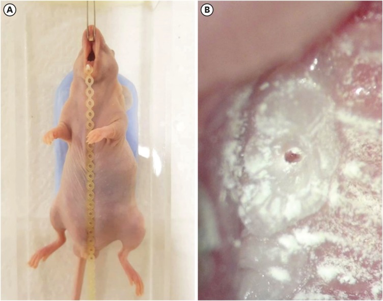
To achieve pulp-dentin complex regeneration with tissue engineering, treatment efficacies and safeties should be evaluated using in vivo orthotopic transplantation in a sufficient number of animals. Mice have been a species of choice in which to study stem cell biology in mammals. However, most pulp-dentin complex regeneration studies have used large animals because the mouse tooth is too small. The purpose of this study was to demonstrate the utility of the mouse tooth as a transplantation model for pulp-dentin complex regeneration research. Materials and MethodsExperiments were performed using 7-week-old male Institute of Cancer Research (ICR) mice; a total of 35 mice had their pulp exposed, and 5 mice each were sacrificed at 1, 2, 4, 7, 9, 12 and 14 days after pulp exposure. After decalcification in 5% ethylenediaminetetraacetic acid, the samples were embedded and cut with a microtome and then stained with hematoxylin and eosin. Slides were observed under a high-magnification light microscope. ResultsUntil 1 week postoperatively, the tissue below the pulp chamber orifice appeared normal. The remaining coronal portion of the pulp tissue was inflammatory and necrotic. After 1 week postoperatively, inflammation and necrosis were apparent in the root canals inferior to the orifices. The specimens obtained after experimental day 14 showed necrosis of all tissue in the root canals. ConclusionsThis study could provide opportunities for researchers performing in vivo orthotopic transplantation experiments with mice.
-
Citations
Citations to this article as recorded by - Is dental pulp inflammation capable of causing central inflammation, behavioral, and sensory alterations? A pre-clinical study
Iago Ramirez, Igor Bassi Ferreira Petean, Francisco Wanderley Garcia de Paula-Silva, Aline Aparecida Ferraresi Tiballi, Manoel Damião Sousa-Neto, Fabiane Carneiro Lopes-Olhê, Christie Ramos Andrade Leite-Panissi, Jardel Francisco Mazzi-Chaves
Archives of Oral Biology.2025; 177: 106320. CrossRef - PRIASE 2021 guidelines for reporting animal studies in Endodontology: explanation and elaboration
V. Nagendrababu, A. Kishen, P. E. Murray, M. H. Nekoofar, J. A. P. de Figueiredo, E. Priya, J. Jayaraman, S. J. Pulikkotil, A. Jakovljevic, P. M. H. Dummer
International Endodontic Journal.2021; 54(6): 858. CrossRef
-
2,621
View
-
13
Download
-
2
Crossref
|