-
Development of a mouse model for pulp-dentin complex regeneration research: a preliminary study
-
Sunil Kim, Sukjoon Lee, Han-Sung Jung, Sun-Young Kim, Euiseong Kim
-
Restor Dent Endod 2019;44(2):e20. Published online May 7, 2019
-
DOI: https://doi.org/10.5395/rde.2019.44.e20
-
-
 Abstract Abstract
 PDF PDF PubReader PubReader ePub ePub
- Objectives
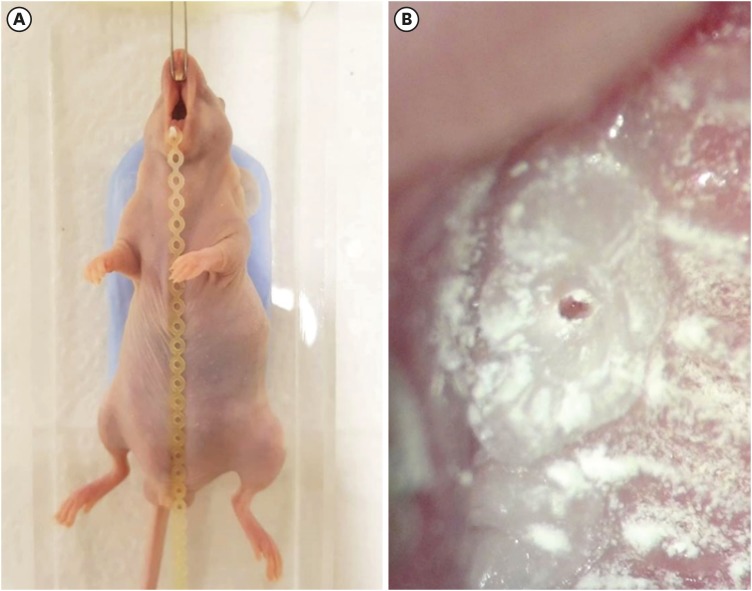
To achieve pulp-dentin complex regeneration with tissue engineering, treatment efficacies and safeties should be evaluated using in vivo orthotopic transplantation in a sufficient number of animals. Mice have been a species of choice in which to study stem cell biology in mammals. However, most pulp-dentin complex regeneration studies have used large animals because the mouse tooth is too small. The purpose of this study was to demonstrate the utility of the mouse tooth as a transplantation model for pulp-dentin complex regeneration research. Materials and MethodsExperiments were performed using 7-week-old male Institute of Cancer Research (ICR) mice; a total of 35 mice had their pulp exposed, and 5 mice each were sacrificed at 1, 2, 4, 7, 9, 12 and 14 days after pulp exposure. After decalcification in 5% ethylenediaminetetraacetic acid, the samples were embedded and cut with a microtome and then stained with hematoxylin and eosin. Slides were observed under a high-magnification light microscope. ResultsUntil 1 week postoperatively, the tissue below the pulp chamber orifice appeared normal. The remaining coronal portion of the pulp tissue was inflammatory and necrotic. After 1 week postoperatively, inflammation and necrosis were apparent in the root canals inferior to the orifices. The specimens obtained after experimental day 14 showed necrosis of all tissue in the root canals. ConclusionsThis study could provide opportunities for researchers performing in vivo orthotopic transplantation experiments with mice.
-
Citations
Citations to this article as recorded by  - PRIASE 2021 guidelines for reporting animal studies in Endodontology: explanation and elaboration
V. Nagendrababu, A. Kishen, P. E. Murray, M. H. Nekoofar, J. A. P. de Figueiredo, E. Priya, J. Jayaraman, S. J. Pulikkotil, A. Jakovljevic, P. M. H. Dummer
International Endodontic Journal.2021; 54(6): 858. CrossRef
-
218
View
-
3
Download
-
1
Crossref
-
Use of temporary filling material for index fabrication in Class IV resin composite restoration
-
Kun-Young Kim, Sun-Young Kim, Duck-Su Kim, Kyoung-Kyu Choi
-
Restor Dent Endod 2013;38(2):85-89. Published online May 28, 2013
-
DOI: https://doi.org/10.5395/rde.2013.38.2.85
-
-
 Abstract Abstract
 PDF PDF PubReader PubReader ePub ePub
When a patient with a fractured anterior tooth visits the clinic, clinician has to restore the tooth esthetically and quickly. For esthetic resin restoration, clinician can use 'Natural Layering technique' and an index for palatal wall may be needed. In this case report, we introduce pre-restoration index technique on a Class IV defect, in which a temporary filling material is used for easy restoration. Chair-side index fabrication for Class IV restoration is convenient and makes a single-visit treatment possible. -
Citations
Citations to this article as recorded by  - A digital workflow for layering composite resin restorations by using 3-dimensionally printed templates to replicate the contralateral tooth accurately and rapidly
Junjing Zhang, Lin Fan, Chenyang Xie, Junying Li, Yuqiang Zhang, Haiyang Yu
The Journal of Prosthetic Dentistry.2024; 131(5): 774. CrossRef - Combining a CAD-CAM composite resin palatal wall with a direct composite resin layering technique for the restoration of a large Class IV fracture: A clinical report
Jingjin Liu, Junling Zhang, Weicai Liu, Shanshan Liang
The Journal of Prosthetic Dentistry.2024;[Epub] CrossRef - Direct composite resin restoration of a class IV fracture by using 3D printing technology: A clinical report
Yi Gao, Jiyao Li, Bo Dong, Min Zhang
The Journal of Prosthetic Dentistry.2021; 125(4): 555. CrossRef - Esthetic rehabilitation of single anterior edentulous space using fiber-reinforced composite
Hyeon Kim, Min-Ju Song, Su-Jung Shin, Yoon Lee, Jeong-Won Park
Restorative Dentistry & Endodontics.2014; 39(3): 220. CrossRef
-
193
View
-
3
Download
-
4
Crossref
-
A maxillary canine with two separated root canals: a case report
-
Dong-Ryul Shin, Jin-Man Kim, Duck-Su Kim, Sun-Young Kim, Paul V Abbott, Sang-Hyuk Park
-
J Korean Acad Conserv Dent 2011;36(5):431-435. Published online September 30, 2011
-
DOI: https://doi.org/10.5395/JKACD.2011.36.5.431
-
-
 Abstract Abstract
 PDF PDF PubReader PubReader ePub ePub
Maxillary canines have less anatomical diversities than other teeth. They usually have a single root and root canal. This report describes an endodontic treatment of a maxillary canine with two separated root canals which have not been reported through the demonstration of radiography and computerized tomography (CT).
Even though appropriated endodontic treatment has been performed, the severe pain could happen due to lack of consideration of anatomical variations of the teeth. Therefore, the clinicians should be well aware of the possibility of anatomical variations in the root canal system during endodontic treatment even if the number of root canals is obvious such as in this case. -
Citations
Citations to this article as recorded by  - Management of an Unusual Maxillary Canine: A Rare Entity
Jaya Nagendra Krishna Muppalla, Krishnamurthy Kavuda, Rajani Punna, Amulya Vanapatla
Case Reports in Dentistry.2015; 2015: 1. CrossRef
-
244
View
-
2
Download
-
1
Crossref
-
The effect of tumor necrosis factor (TNF)-α to induce matrix metalloproteinase (MMPs) from the human dental pulp, gingival, and periodontal ligament cells
-
Eun-Mi Rhim, Sang-Hyuk Park, Duck-Su Kim, Sun-Young Kim, Kyoung-Kyu Choi, Gi-Woon Choi
-
J Korean Acad Conserv Dent 2011;36(1):26-36. Published online January 31, 2011
-
DOI: https://doi.org/10.5395/JKACD.2011.36.1.26
-
-
 Abstract Abstract
 PDF PDF PubReader PubReader ePub ePub
-
Objectives
In the present study, three kinds of tissues cells (pulp, gingiva, and periodontal ligament) were investigated if those cells express MMP and TIMP when they were stimulated with neuropeptides (substance P, CGRP) or proinflammatory cytokine, TNF-α.
Materials and Methods
The cells cultured from human dental pulp (PF), gingiva (GF) and periodontal ligament were (PDLF) stimulated with Mock, SP, TNF-α, and CGRP for 24 hrs and 48 hrs. for an RNase protection assay and Enzyme Linked Immunosorbent Assay.
Cells (PF, GF and PDLF) seeded in 100 mm culture dish were stimulated with SP (10-5, 10-8 M) or only with medium (Mock stimulation) for 4hrs and for 24 hrs for RNase Protection Assay, and they were stimulated with CGRP (10-5 M) and TNF-α (2 ng/mL) for 24 hrs and with various concentraion of TNF-α (2, 10, and 100 ng/mL) for Rnase Protection Assay with a human MMP-1 probe set including MMP 1, 2, 8, 7, 8, 9, 12, and TIMP 2, 3.
In addition, cells (PF, GF and PDLF) were stimulated with Mock and various concentraion of TNF-α (2, 10, and 100 ng/mL) for 24 hrs and with TNF-α (10 ng/mL) for 48 hrs, and the supernatents from the cells were collected for Enzyme Linked Immunosorbent Assay (ELISA) for MMP-1 and MMP-13.
Results
The expression of MMPs in PF, GF, PDLF after stimulation with SP and CGRP were not changed compared with Mock stimulation for 4 hrs and 24 hrs. The expression of MMP-1, -12, -13 24 hrs after stimulation with TNF-α were upregulated, however the expression of TIMP-3 in PF, GF, PDLF after stimulation with TNF-α were downregulated. TNF-α (2 ng/mL, 10 ng/mL, 100 ng/mL) increased MMP-1 and MMP-12 expression in PF dose dependently for 24 hrs.
Conclusions
TNF-α in the area of inflammation may play an important role in regulating the remodeling of dentin, cementum, and alveolar bone.
-
Citations
Citations to this article as recorded by  - Anti‐Inflammatory Effects of Melatonin and 5‐Methoxytryptophol on Lipopolysaccharide‐Induced Acute Pulpitis in Rats
Fatma Kermeoğlu, Umut Aksoy, Abdullah Sebai, Gökçe Savtekin, Hanife Özkayalar, Serkan Sayıner, Ahmet Özer Şehirli, Shuai CHEN
BioMed Research International.2021;[Epub] CrossRef - Cross-Talk between Ciliary Epithelium and Trabecular Meshwork Cells In-Vitro: A New Insight into Glaucoma
Natalie Lerner, Elie Beit-Yannai, Wayne Iwan Lee Davies
PLoS ONE.2014; 9(11): e112259. CrossRef
-
164
View
-
2
Download
-
2
Crossref
-
Real-time measurement of dentinal fluid flow during desensitizing agent application
-
Sun-Young Kim, Eun-Joo Kim, In-Bog Lee
-
J Korean Acad Conserv Dent 2010;35(5):313-320. Published online September 30, 2010
-
DOI: https://doi.org/10.5395/JKACD.2010.35.5.313
-
-
 Abstract Abstract
 PDF PDF PubReader PubReader ePub ePub
-
Objectives
The aim of this study was to examine changes in the dentinal fluid flow (DFF) during desensitizing agent application and to compare permeability after application among the agents.
Materials and Methods
A Class 5 cavity was prepared to exposure cervical dentin on an extracted human premolar which was connected to a sub-nanoliter fluid flow measuring device (NFMD) under 20 cm water pressure. DFF was measured from before application of desensitizing agent (Seal&Protect, SP; SuperSeal, SS; BisBlock, BB; Gluma desensitizer, GL; Bi-Fluoride 12, BF) through application procedure to 5 min after application.
Results
DFF rate after each desensitizing agent application was significantly reduced when compared to initial DFF rate before application (p < 0.05). SP showed a greater reduction in DFF rate than GL and BF did (p < 0.05). SS and BB showed a greater reduction in DFF rate than BF did (p < 0.05).
Conclusions
Characteristic DFF aspect of each desensitizing agent was shown in NFMD during the application procedure.
-
Citations
Citations to this article as recorded by  - CPNE7 Induces Biological Dentin Sealing in a Dentin Hypersensitivity Model
S.H. Park, Y.S. Lee, D.S. Lee, J.C. Park, R. Kim, W.J. Shon
Journal of Dental Research.2019; 98(11): 1239. CrossRef
-
127
View
-
1
Download
-
1
Crossref
-
A new method to measure the linear polymerization shrinkage of composites using a particle tracking method with computer vision
-
In-Bog Lee, Sun-Hong Min, Deog-Gyu Seo, Sun-Young Kim, Youngchul Kwon
-
J Korean Acad Conserv Dent 2010;35(3):180-187. Published online May 31, 2010
-
DOI: https://doi.org/10.5395/JKACD.2010.35.3.180
-
-
 Abstract Abstract
 PDF PDF PubReader PubReader ePub ePub
Since the introduction of restorative dental composites, their physical properties have been significantly improved. However, polymerization shrinkage is still a major drawback. Many efforts have been made to develop a low shrinking composite, and silorane-based composites have recently been introduced into the market. In addition, many different methods have been developed to measure the polymerization shrinkage.
In this study, we developed a new method to measure the linear polymerization shrinkage of composites without direct contact to a specimen using a particle tracking method with computer vision. The shrinkage kinetics of a commercial silorane-based composite (P90) and two conventional methacrylate-based composites (Z250 and Z350) were investigated and compared. The results were as follows:
The linear shrinkage of composites was 0.33-1.41%. Shrinkage was lowest for the silorane-based (P90) composite, and highest for the flowable Z350 composite.
The new instrument was able to measure the true linear shrinkage of composites in real time without sensitivity to the specimen preparation and geometry.
-
Citations
Citations to this article as recorded by  - Effect of layering methods, composite type, and flowable liner on the polymerization shrinkage stress of light cured composites
Youngchul Kwon, Jack Ferracane, In-Bog Lee
Dental Materials.2012; 28(7): 801. CrossRef
-
137
View
-
2
Download
-
1
Crossref
-
Real-time measurement of dentinal tubular fluid flow during and after amalgam and composite restorations
-
Sun-Young Kim, Byeong-Hoon Cho, Seung-Ho Baek, Bum-Sun Lim, In-Bog Lee
-
J Korean Acad Conserv Dent 2009;34(6):467-476. Published online November 30, 2009
-
DOI: https://doi.org/10.5395/JKACD.2009.34.6.467
-
-
 Abstract Abstract
 PDF PDF PubReader PubReader ePub ePub
The aim of this study was to measure the dentinal tubular fluid flow (DFF) during and after amalgam and composite restorations. A newly designed fluid flow measurement instrument was made. A third molar cut at 3 mm apical from the CEJ was connected to the flow measuring device under a hydrostatic pressure of 15 cmH2O. Class I cavity was prepared and restored with either amalgam (Copalite varnish and Bestaloy) or composite (Z-250 with ScotchBond MultiPurpose: MP, Single Bond 2: SB, Clearfil SE Bond: CE and Easy Bond: EB as bonding systems). The DFF was measured from the intact tooth state through restoration procedures to 30 minutes after restoration, and re-measured at 3 and 7days after restoration.
Inward fluid flow (IF) during cavity preparation was followed by outward flow (OF) after preparation. In amalgam restoration, the OF changed to IF during amalgam filling and slight OF followed after finishing.
In composite restoration, application CE and EB showed a continuous OF and air-dry increased rapidly the OF until light-curing, whereas in MP and SB, rinse and dry caused IF and OF, respectively. Application of hydrophobic bonding resin in MP and CE caused a decrease in flow rate or even slight IF. Light-curing of adhesive and composite showed an abrupt IF. There was no statistically significant difference in the reduction of DFF among the materials at 30 min, 3 and 7 days after restoration (P > 0.05). -
Citations
Citations to this article as recorded by  - Real-time measurement of dentinal fluid flow during desensitizing agent application
Sun-Young Kim, Eun-Joo Kim, In-Bog Lee
Journal of Korean Academy of Conservative Dentistry.2010; 35(5): 313. CrossRef
-
166
View
-
0
Download
-
1
Crossref
-
Slumping tendency and rheological property of flowable composites
-
In-Bog Lee, Sun-Hong Min, Sun-Young Kim, Byung-Hoon Cho, Seung-Ho Back
-
J Korean Acad Conserv Dent 2009;34(2):130-136. Published online March 31, 2009
-
DOI: https://doi.org/10.5395/JKACD.2009.34.2.130
-
-
 Abstract Abstract
 PDF PDF PubReader PubReader ePub ePub
The aim of this study was to develop a method for measuring the slumping resistance of flowable resin composites and to evaluate the efficacy using rheological methodology.
Five commercial flowable composites (Aelitefil flow:AF, Filtek flow:FF, DenFil flow:DF, Tetric flow:TF and Revolution:RV) were used. Same volume of composites in a syringe was extruded on a glass slide using a custom-made loading device. The resin composites were allowed to slump for 10 seconds at 25℃ and light cured. The aspect ratio (height/diameter) of cone or dome shaped specimen was measured for estimating the slumping tendency of composites. The complex viscosity of each composite was measured by a dynamic oscillatory shear test as a function of angular frequency using a rheometer. To compare the slumping tendency of composites, one way-ANOVA and Turkey's post hoc test was performed for the aspect ratio at 95% confidence level. Regression analysis was performed to investigate the relationship between the complex viscosity and the aspect ratio. The results were as follows.
1. Slumping tendency based on the aspect ratio varied among the five materials (AF < FF < DF < TF < RV).
2. Flowable composites exhibited pseudoplasticity in which the complex viscosity decreased with increasing frequency (shear rate). AF was the most significant, RV the least.
3. The slumping tendency was strongly related with the complex viscosity. Slumping resistance increased with increasing the complex viscosity.
The slumping tendency could be quantified by measuring the aspect ratio of slumped flowable composites. This method may be applicable to evaluate the clinical handling characteristics of flowable composites.
-
Is an oxygen inhibition layer essential for the interfacial bonding between resin composite layers?
-
Sun-Young Kim, Byeong-Hoon Cho, Seung-Ho Baek, In-Bog Lee
-
J Korean Acad Conserv Dent 2008;33(4):405-412. Published online July 31, 2008
-
DOI: https://doi.org/10.5395/JKACD.2008.33.4.405
-
-
 Abstract Abstract
 PDF PDF PubReader PubReader ePub ePub
This study was aimed to investigate whether an oxygen inhibition layer (OIL) is essential for the interfacial bonding between resin composite layers or not.
A composite (Z-250, 3M ESPE) was filled in two layers using two aluminum plate molds with a hole of 3.7 mm diameter. The surface of first layer of cured composite was prepared by one of five methods as followings, thereafter second layer of composite was filled and cured: Group 1 - OIL is allowed to remain on the surface of cured composite; Group 2 - OIL was removed by rubbing with acetone-soaked cotton; Group 3 - formation of the OIL was inhibited using a Mylar strip; Group 4 - OIL was covered with glycerin and light-cured; Group 5 (control) - composite was bulk-filled in a layer. The interfacial shear bond strength between two layers was tested and the fracture modes were observed. To investigate the propagation of polymerization reaction from active area having a photo-initiator to inactive area without the initiator, a flowable composite (Aelite Flow) or an adhesive resin (Adhesive of ScotchBond Multipurpose) was placed over an experimental composite (Exp_Com) which does not include a photoinitiator and light-cured. After sectioning the specimen, the cured thickness of the Exp_Com was measured.
The bond strength of group 2, 3 and 4 did not show statistically significant difference with group 1. Groups 3 and 4 were not statistically significant different with control group 5. The cured thicknesses of Exp_Com under the flowable resin and adhesive resin were 20.95 (0.90) um and 42.13 (2.09), respectively. -
Citations
Citations to this article as recorded by  - Finishing and Polishing of Composite Restoration: Assessment of Knowledge, Attitude and Practice Among Various Dental Professionals in India
Sankar Vishwanath, Sadasiva Kadandale, Senthil kumar Kumarappan, Anupama Ramachandran, Manu Unnikrishnan, Honap manjiri Nagesh
Cureus.2022;[Epub] CrossRef - Evaluation of Surface Roughness of Composite, Compomer and Carbomer After Curing Through Mylar Strip and Glycerin: A Comparative Study
Asli Topaloglu-Ak, Dilara Çayırgan, Melisa Uslu
Journal of Advanced Oral Research.2020; 11(1): 12. CrossRef - Effect of glycerin on the surface hardness of composites after curing
Hyun-Hee Park, In-Bog Lee
Journal of Korean Academy of Conservative Dentistry.2011; 36(6): 483. CrossRef
-
167
View
-
1
Download
-
3
Crossref
-
Shear bond strength of dentin bonding agents cured with a Plasma Arc curing light
-
Youngchul Kwon, Sun-Young Kim, Sae-Joon Chung, Young-Chul Han, In-Bog Lee, Ho-Hyun Son, Chung-Moon Um, Byeong-Hoon Cho
-
J Korean Acad Conserv Dent 2008;33(3):213-223. Published online May 31, 2008
-
DOI: https://doi.org/10.5395/JKACD.2008.33.3.213
-
-
 Abstract Abstract
 PDF PDF PubReader PubReader ePub ePub
The objective of this study was to compare dentin shear bond strength (DSBS) of dentin bonding agents (DBAs) cured with a plasma arc (PAC) light curing unit (LCU) and those cured with a light emitting diode (LED) LCU. Optical properties were also analyzed for Elipar freelight 2 (3M ESPE); LED LCU, Apollo 95E (DMT Systems); PAC LCU and VIP Junior (Bisco); Halogen LCU. The DBAs used for DSBS test were Scotchbond Multipurpose (3M ESPE), Singlebond 2 (3M ESPE) and Clearfil SE Bond (Kuraray). After DSBS testing, fractured specimens were analyzed for failure modes with SEM.
The total irradiance and irradiance between 450 nm and 490 nm of the LCUs were different. LED LCU showed narrow spectral distribution around its peak at 462 nm whereas PAC and Halogen LCU showed a broad spectrum. There were no significant differences in mean shear bond strength among different LCUs (P > 0.05) but were significant differences among different DBAs (P < 0.001) -
Citations
Citations to this article as recorded by  - Temperature changes under demineralized dentin during polymerization of three resin-based restorative materials using QTH and LED units
Sayed-Mostafa Mousavinasab, Maryam Khoroushi, Mohammadreza Moharreri, Mohammad Atai
Restorative Dentistry & Endodontics.2014; 39(3): 155. CrossRef
-
147
View
-
1
Download
-
1
Crossref
-
Development of nano-fluid movement measuring device and its application to hydrodynamic analysis of dentinal fluid
-
In-Bog Lee, Min-Ho Kim, Sun-Young Kim, Juhea Chang, Byung-Hoon Cho, Ho-Hyun Son, Seung-Ho Back
-
J Korean Acad Conserv Dent 2008;33(2):141-147. Published online March 31, 2008
-
DOI: https://doi.org/10.5395/JKACD.2008.33.2.141
-
-
 Abstract Abstract
 PDF PDF PubReader PubReader ePub ePub
This study was aimed to develop an instrument for real-time measurement of fluid conductance and to investigate the hydrodynamics of dentinal fluid. The instrument consisted of three parts; (1) a glass capillary and a photo sensor for detection of fluid movement, (2) a servo-motor, a lead screw and a ball nut for tracking of fluid movement, (3) a rotary encoder and software for data processing.
To observe the blocking effect of dentinal fluid movement, oxalate gel and self-etch adhesive agent were used. BisBlock (Bisco) and Clearfil SE Bond (Kuraray) were applied to the occlusal dentin surface of extracted human teeth. Using this new device, the fluid movement was measured and compared between before and after each agent was applied.
The instrument was able to measure dentinal fluid movement with a high resolution (0.196 nL) and the flow occurred with a rate of 0.84 to 15.2 nL/s before treatment. After BisBlock or Clearfil SE Bond was used, the fluid movement was decreased by 39.8 to 89.6%. -
Citations
Citations to this article as recorded by  - Nanoleakage of apical sealing using a calcium silicate-based sealer according to canal drying methods
Yoon-Joo Lee, Kyung-Mo Cho, Se-Hee Park, Yoon Lee, Jin-Woo Kim
Restorative Dentistry & Endodontics.2024;[Epub] CrossRef - CPNE7 Induces Biological Dentin Sealing in a Dentin Hypersensitivity Model
S.H. Park, Y.S. Lee, D.S. Lee, J.C. Park, R. Kim, W.J. Shon
Journal of Dental Research.2019; 98(11): 1239. CrossRef - Effect of oral health-related factors on oral health knowledge, attitude, and practice of college students
Su Bin Lee, Jeong Weon Yoon, Mi Gyung Seong, Min Kyung Lee, Ye Hwang Kim, Jung Hwa Lee
Journal of Korean Academy of Oral Health.2018; 42(4): 124. CrossRef - Real-time measurement of dentinal fluid flow during desensitizing agent application
Sun-Young Kim, Eun-Joo Kim, In-Bog Lee
Journal of Korean Academy of Conservative Dentistry.2010; 35(5): 313. CrossRef - Real-time measurement of dentinal tubular fluid flow during and after amalgam and composite restorations
Sun-Young Kim, Byeong-Hoon Cho, Seung-Ho Baek, Bum-Sun Lim, In-Bog Lee
Journal of Korean Academy of Conservative Dentistry.2009; 34(6): 467. CrossRef
-
167
View
-
1
Download
-
5
Crossref
-
Dentin bond strength of bonding agents cured with Light Emitting Diode
-
Sun-Young Kim, In-Bog Lee, Byeong-Hoon Cho, Ho-Hyun Son, Mi-Ja Kim, Chang-In Seok, Chung-Moon Um
-
J Korean Acad Conserv Dent 2004;29(6):504-514. Published online January 14, 2004
-
DOI: https://doi.org/10.5395/JKACD.2004.29.6.504
-
-
 Abstract Abstract
 PDF PDF PubReader PubReader ePub ePub
- ABSTRACT
This study compared the dentin shear bond strengths of currently used dentin bonding agents that were irradiated with an LED (Elipar FreeLight, 3M-ESPE) and a halogen light (VIP, BISCO). The optical characteristics of two light curing units were evaluated. Extracted human third molars were prepared to expose the occlusal dentin and the bonding procedures were performed under the irradiation with each light curing unit. The dentin bonding agents used in this study were Scotchbond Multipurpose (3M ESPE), Single Bond (3M ESPE), One-Step (Bisco), Clearfil SE bond (Kuraray), and Adper Prompt (3M ESPE). The shear test was performed by employing the design of a chisel-on-iris supported with a Teflon wall. The fractured dentin surface was observed with SEM to determine the failure mode.
The spectral appearance of the LED light curing unit was different from that of the halogen light curing unit in terms of maximum peak and distribution. The LED LCU (maximum peak in 465 ㎚) shows a narrower spectral distribution than the halogen LCU (maximum peak in 487 ㎚). With the exception of the Clearfil SE bond (P < 0.05), each 4 dentin bonding agents showed no significant difference between the halogen light-cured group and the LED light-cured group in the mean shear bond strength (P > 0.05).
The results can be explained by the strong correlation between the absorption spectrum of cam-phoroquinone and the narrow emission spectrum of LED.
|


