-
Prevalence of salivary microbial load and lactic acid presence in diabetic and non-diabetic individuals with different dental caries stages
-
Monika Mohanty, Shashirekha Govind, Shakti Rath
-
Restor Dent Endod 2024;49(1):e4. Published online January 12, 2024
-
DOI: https://doi.org/10.5395/rde.2024.49.e4
-
-
 Abstract Abstract
 PDF PDF PubReader PubReader ePub ePub
- Objectives
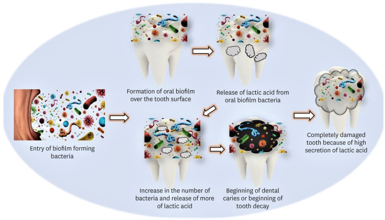
This study aims to correlate caries-causing microorganism load, lactic acid estimation, and blood groups to high caries risk in diabetic and non-diabetic individuals and low caries risk in healthy individuals. Materials and MethodsThis study includes 30 participants divided into 3 groups: Group A, High-risk caries diabetic individuals; Group B, High-risk caries non-diabetic individuals; and Group C, Low-risk caries individuals. The medical condition, oral hygiene, and caries risk assessment (American Dental Association classification and International Caries Detection and Assessment System scoring) were documented. Each individual’s 3 mL of saliva was analyzed for microbial load and lactic acid as follows: Part I: 2 mL for microbial quantity estimation using nutrient agar and blood agar medium, biochemical investigation, and carbohydrate fermentation tests; Part II: 0.5 mL for lactic acid estimation using spectrophotometric analysis. Among the selected individuals, blood group correlation was assessed. The χ2 test, Kruskal-Wallis test, and post hoc analysis were done using Dunn’s test (p < 0.05). ResultsGroup A had the highest microbial load and lactic acid concentration, followed by Groups B and C. The predominant bacteria were Lactobacilli (63.00 ± 15.49) and Streptococcus mutans (76.00 ± 13.90) in saliva. Blood Group B is prevalent in diabetic and non-diabetic high-risk caries patients but statistically insignificant. ConclusionsDiabetic individuals are more susceptible to dental caries due to high microbial loads and increased lactic acid production. These factors also lower the executing tendency of neutrophils, which accelerates microbial accumulation and increases the risk of caries in diabetic individuals.
-
Citations
Citations to this article as recorded by  - Oral Health Disparities in Type 2 Diabetes: Examining the Elevated Risk for Dental Caries—A Comparative Study
José Frias-Bulhosa, Maria Conceição Manso, Carla Lopes Mota, Paulo Melo
Dentistry Journal.2025; 13(6): 258. CrossRef - Exploring the photosensitizing potential of Nanoliposome Loaded Improved Toluidine Blue O (NLITBO) Against Streptococcus mutans: An in-vitro feasibility study
Swagatika Panda, Lipsa Rout, Neeta Mohanty, Anurag Satpathy, Bhabani Sankar Satapathy, Shakti Rath, Divya Gopinath, Geelsu Hwang
PLOS ONE.2024; 19(10): e0312521. CrossRef - Altered salivary microbiota associated with high-sugar beverage consumption
Xiaozhou Fan, Kelsey R. Monson, Brandilyn A. Peters, Jennifer M. Whittington, Caroline Y. Um, Paul E. Oberstein, Marjorie L. McCullough, Neal D. Freedman, Wen-Yi Huang, Jiyoung Ahn, Richard B. Hayes
Scientific Reports.2024;[Epub] CrossRef
-
3,990
View
-
97
Download
-
3
Web of Science
-
3
Crossref
-
Enhanced visualization of the root canal morphology using a chitosan-based endo-radiopaque solution
-
Shashirekha Govind, Amit Jena, Satabdi Pattanaik, Mahaprasad Anarasi, Satyajit Mohapatra, Vinay Shivagange
-
Restor Dent Endod 2021;46(3):e33. Published online June 4, 2021
-
DOI: https://doi.org/10.5395/rde.2021.46.e33
-
-
Abstract
PDFPubReaderePub
- Objectives
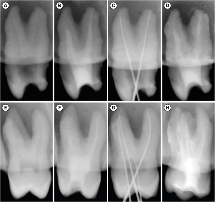
This study aimed to investigate the efficacy of ionic and non-ionic-based contrast media (in vitro study) and the combinatorial effect of chitosan-based endo-radiopaque solution (CERS) (in vivo study) for visualization of the root canal anatomy. Materials and MethodsIn vitro study (120 teeth): The root canal of maxillary premolars and molars (in vitro group 1 and 2 respectively, n = 60 each) were analyzed using 4 different contrast media (subgroups: Omnipaque 350, Iopamidol, Xenetix 350, and Urografin 76; n = 15 each) in combination with 5.25% sodium hypochlorite (NaOCl). Based on the results of the in vitro study, in vivo study (80 teeth) was done to compare Xenetix 350 + 5.25% NaOCl with CERS (in vivo group 1 and 2 respectively, n = 40 each) on maxillary and mandibular premolars and molars. Two endodontists used radiovisiography to assess the depth of ingress and identify the aberrant root anatomy after access cavity preparation, and after initial cleaning and shaping of canals. Kruskal-Wallis test was used for in vitro comparison (p < 0.05), and Wilcoxon signed-rank test and Mann-Whitney U test for in vivo analysis (p < 0.01). ResultsIn vitro study, Xenetix 350 + 5.25% NaOCl facilitated a significant higher visualization (p < 0.05). For in vivo study, CERS had a statistically significant depth of ingress (p < 0.01), and was efficient in identifying the aberrant root canal anatomy of premolars and molars. ConclusionsCERS facilitates better visualization of the root canal anatomy of human premolars and molars.
-
Citations
Citations to this article as recorded by - Influence of irrigating solutions on the hydration of calcium silicate-based dental biomaterials: An in vitro study
Pradeep M. Divya, Amit Jena, Saumyakanta Mohanty, Govind Shashirekha, Rashmi Rekha Mallick, Priyanka Sarangi
Journal of Conservative Dentistry and Endodontics.2025; 28(8): 758. CrossRef - Improving Endodontic Radiograph Interpretation with TV-CLAHE for Enhanced Root Canal Detection
Barbara Obuchowicz, Joanna Zarzecka, Michał Strzelecki, Marzena Jakubowska, Rafał Obuchowicz, Adam Piórkowski, Elżbieta Zarzecka-Francica, Julia Lasek
Journal of Clinical Medicine.2025; 14(15): 5554. CrossRef - Efficacy of sonic and ultrasonic activation on irrigant penetration in different tapered preparations: An in vitro study
M. Rama Sowmya, Kavalipurapu Venkata Teja, Pradeep Solete, Sahil Choudhari, S Delphine Priscilla Antony, Mohammed Mustafa
Endodontology.2024; 36(4): 370. CrossRef - Analysis of the value of visualized root canal technique in the clinical treatment of endodontics
Nana SUN, Nannan WANG, Xin QIAN
Panminerva Medica.2023;[Epub] CrossRef
-
2,342
View
-
23
Download
-
2
Web of Science
-
4
Crossref
|