-
Does minimally invasive canal preparation provide higher fracture resistance of endodontically treated teeth? A systematic review of in vitro studies
-
Sıla Nur Usta, Emmanuel João Nogueira Leal Silva, Seda Falakaloğlu, Mustafa Gündoğar
-
Restor Dent Endod 2023;48(4):e34. Published online October 17, 2023
-
DOI: https://doi.org/10.5395/rde.2023.48.e34
-
-
 Abstract Abstract
 PDF PDF PubReader PubReader ePub ePub
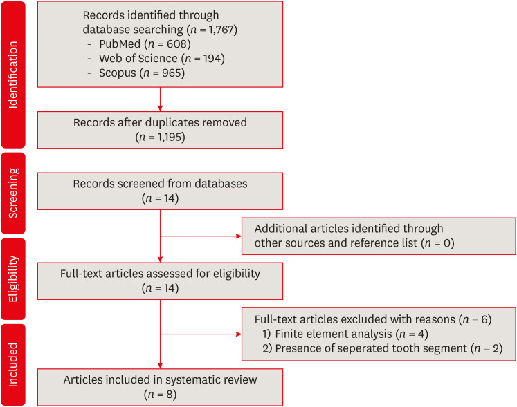
This systematic review aimed to investigate whether minimally invasive root canal preparation ensures higher fracture resistance compared to conventional root canal preparation in endodontically treated teeth (ETT). A comprehensive search strategy was conducted on the “PubMed, Web of Science, and Scopus” databases, alongside reference and hand searches, with language restrictions applied. Two independent reviews selected pertinent laboratory studies that explored the effect of minimally invasive root canal preparation on fracture resistance, in comparison to larger preparation counterparts. The quality of the studies was assessed, and the risk of bias was categorized as low, moderate, or high. The electronic search yielded a total of 1,767 articles. After applying eligibility criteria, 8 studies were included. Given the low methodological quality of these studies and the large variability of fracture resistance values, the impact of reduced apical size and/or taper on the fracture resistance of the ETT can be considered uncertain. This systematic review could not reveal sufficient evidence regarding the effect of minimally invasive preparation on increasing fracture resistance of ETT, primarily due to the inherent limitations of the studies and the moderate risk of bias. -
Citations
Citations to this article as recorded by  - Impact of conservative versus conventional instrumentation on the release of inflammatory mediators and post‐operative pain in mandibular molars with asymptomatic irreversible pulpitis: A randomized clinical trial
Sıla Nur Usta, Ana Arias, Emre Avcı, Emmanuel João Nogueira Leal Silva
International Endodontic Journal.2025;[Epub] CrossRef - Micro‐computed tomography evaluation of minimally invasive root canal preparation in 3D‐printed C‐shaped canal
Nutcha Supavititpattana, Siriwan Suebnukarn, Panupat Phumpatrakom, Kamon Budsaba
Australian Endodontic Journal.2024; 50(3): 621. CrossRef - Ex vivo investigation on the effect of minimally invasive endodontic treatment on vertical root fracture resistance and crack formation
Andreas Rathke, Henry Frehse, Maria Bechtold
Scientific Reports.2024;[Epub] CrossRef
-
341
View
-
31
Download
-
3
Web of Science
-
3
Crossref
-
Influence of access cavity design on calcium hydroxide removal using different cleaning protocols: a confocal laser scanning microscopy study
-
Seda Falakaloğlu, Merve Yeniçeri Özata, Betül Güneş, Emmanuel João Nogueira Leal Silva, Mustafa Gündoğar, Burcu Güçyetmez Topal
-
Restor Dent Endod 2023;48(3):e25. Published online July 24, 2023
-
DOI: https://doi.org/10.5395/rde.2023.48.e25
-
-
 Abstract Abstract
 PDF PDF PubReader PubReader ePub ePub
- Objectives
The purpose of this study was to evaluate the influence of endodontic access cavities design on the removal of calcium hydroxide medication of the apical third of mandibular incisor root canal walls and dentinal tubules with different cleaning protocols: EDDY sonic activation, Er,Cr:YSGG laser-activated irrigation, or conventional irrigation with IrriFlex. Materials and MethodsSeventy-eight extracted human mandibular incisors were assigned to 6 experimental groups (n = 13) according to the endodontic access cavity and cleaning protocol for calcium hydroxide removal: traditional access cavity (TradAC)/EDDY; ultraconservative access cavity performed in the incisal edge (UltraAC.Inc)/EDDY; TradAC/Er,Cr:YSGG; UltraAC.Inc/Er,Cr:YSGG; TradAC/IrriFlex; or UltraAC.Inc/IrriFlex. Confocal laser scanning microscopy images were used to measure the non-penetration percentage, maximum residual calcium hydroxide penetration depth, and penetration area at 2 and 4 mm from the apex. Data were statistically analyzed using Shapiro-Wilk and WRS2 package for 2-way comparison of non-normally distributed parameters (depth of penetration, area of penetration, and percentage of non-penetration) according to cavity and cleaning protocol with the significance level set at 5%. ResultsThe effect of cavity and cleaning protocol interactions on penetration depth, penetration area and non-penetration percentage was not found statistically significant at 2 and 4 mm levels (p > 0.05). ConclusionsThe present study demonstrated that TradAC or UltraAC.Inc preparations with different cleaning protocols in extracted mandibular incisors did not influence the remaining calcium hydroxide at 2 and 4 mm from the apex.
-
Citations
Citations to this article as recorded by  - Effect of Apical Preparation Size and Preparation Taper on Smear Layer Removal Using Two Different Irrigation Needles: A Scanning Electron Microscopy Study
Rania Lebbos, Naji Kharouf, Deepak Mehta, Jamal Jabr, Cynthia Kamel, Roula El Hachem, Youssef Haikel, Marc Krikor Kaloustian
European Journal of Dentistry.2024;[Epub] CrossRef
-
361
View
-
22
Download
-
1
Web of Science
-
1
Crossref
-
Comparison of the cyclic fatigue resistance of VDW.ROTATE, TruNatomy, 2Shape, and HyFlex CM nickel-titanium rotary files at body temperature
-
Mustafa Gündoğar, Gülşah Uslu, Taha Özyürek, Gianluca Plotino
-
Restor Dent Endod 2020;45(3):e37. Published online June 22, 2020
-
DOI: https://doi.org/10.5395/rde.2020.45.e37
-
-
 Abstract Abstract
 PDF PDF PubReader PubReader ePub ePub
- Objectives
This study aims to compare the cyclic fatigue resistance of VDW.ROTATE, TruNatomy, 2Shape, and HyFlex CM nickel-titanium (NiTi) rotary files at body temperature. Materials and MethodsIn total, 80 VDW.ROTATE (25/0.04), TruNatomy (26/0.04), 2Shape (25/0.04), and HyFlex CM (25/0.04) NiTi rotary files (n = 20 in each group) were subjected to static cyclic fatigue testing at body temperature (37°C) in stainless-steel artificial canals prepared according to the size and taper of the instruments until fracture occurred. The number of cycles to fracture (NCF) was calculated, and the lengths of the fractured fragments were measured. The data were statistically analyzed using a 1-way analysis of variance and post hoc Tamhane tests at the 5% significance level (p < 0.05). ResultsThere were significant differences in the cyclic fatigue resistance among the groups (p < 0.05), with the highest to lowest NCF values of the files as follows: VDW.ROTATE, HyFlex CM, 2Shape, and TruNatomy. There was no significant difference in the lengths of the fractured fragments among the groups. The scanning electron microscope images of the files revealed typical characteristics of fracture due to cyclic fatigue. ConclusionsThe VDW.ROTATE files had the highest cyclic fatigue resistance, and the TruNatomy and 2Shape files had the lowest cyclic fatigue resistance in artificial canals at body temperature.
-
Citations
Citations to this article as recorded by  - Comparative Evaluation of Efficiency of Different Endodontic File Systems; Protaper Universal, MTWO, Protaper Next, Trunatomy, I-Race in Terms of Remaining Dentin Thickness: An In vitro CBCT Analysis
Anju Retnakaran, Faisal M. A. Gaffoor, Rethi Gopakumar, C Sabari Girish, N. C Sajeena, N Gokul Krishna
Journal of Pharmacy and Bioallied Sciences.2024; 16(Suppl 2): S1409. CrossRef - Analysis of Cutting Capacity, Surface Finishing, and Mechanical Properties of NiTi Instruments 25/.04: ROTATE and LOGIC 2
Ridalton Carlos de Morais, Juliana Delatorre Bronzato, Adriana de-Jesus-Soares, Marcos Frozoni, Victor Talarico Leal Vieira
Journal of Endodontics.2024; 50(7): 982. CrossRef - Effect of Temperature on the Cyclic Fatigue Resistance and Phase Transformation Behavior of Three Different NiTi Endodontic Instruments
Esra İrem Yi̇ği̇t, İrem Çetinkaya
Cureus.2024;[Epub] CrossRef - In Vitro Evaluation of the Dynamic Cyclic Fatigue Resistance of a New TruNatomy Glider File after Different Cycles of Use
Lorena Ferreira Rego, Juliana Delatorre Bronzato, Alana Pinto Carôso Souza, Adriana de-Jesus-Soares, Marcos Frozoni
Journal of Endodontics.2024; 50(5): 619. CrossRef - Effect of Sodium Hypochlorite and Hypochlorous Acid Solutions on the Cyclic Fatigue Resistance of Waveonegold, K3XF and Hyflex-EDM: A Study of Metallurgical Properties
D. A. Bozkurt, M. Akman, H. B. Karadag, Z. Ovalioglu, Ö.Küçük Keleş
Strength of Materials.2023; 55(1): 191. CrossRef - Effectiveness of conservative instrumentation in root canal disinfection
Sıla Nur Usta, Carmen Solana, Matilde Ruiz-Linares, Pilar Baca, Carmen María Ferrer-Luque, Monica Cabeo, Maria Teresa Arias-Moliz
Clinical Oral Investigations.2023; 27(6): 3181. CrossRef - Cyclic fatigue resistance of different nickel‐titanium instruments in single and double curvature at room and body temperatures: A laboratory study
Giusy Rita Maria La Rosa, Maria Laura Leotta, Francesco Saverio Canova, Virginia Rosy Romeo, Gabriele Cervino, Luigi Generali, Eugenio Pedullà
Australian Endodontic Journal.2023; 49(3): 592. CrossRef - Influence of NiTi Wire Diameter on Cyclic and Torsional Fatigue Resistance of Different Heat-Treated Endodontic Instruments
Eugenio Pedullà, Francesco Saverio Canova, Giusy Rita Maria La Rosa, Alfred Naaman, Franck Diemer, Luigi Generali, Walid Nehme
Materials.2022; 15(19): 6568. CrossRef - Impact of Different Access Cavity Designs and Ni–Ti Files on the Elimination of Enterococcus faecalis from the Root Canal System: An In Vitro Study
Gizem Andac, Atakan Kalender, Buket Baddal, Fatma Basmaci
Applied Sciences.2022; 12(4): 2049. CrossRef - Comparison of canal transportations and centering ability of rotary instrument systems with different heat-treated NiTi alloys: An in vitro CBCT study
Mukadder İnci BAŞER KOLCU, Gülter Devrim KAKİ
Turkish Journal of Health Science and Life.2022; 5(2): 81. CrossRef - Comparative evaluation of cutting efficiency, cyclic fatigue, corrosion resistance, and autoclave cycle effects of three different file systems: An in-vitro micro-CT and metallurgy analysis
KondasV Venkatesh, EldhoJ Varghese
Journal of International Oral Health.2022; 14(6): 551. CrossRef - Influence of different heat treatments and temperatures on the cyclic fatigue resistance of endodontic instruments with the same design
Walid Nehme, Alfred Naaman, Franck Diemer, Maria Laura Leotta, Giusy Rita Maria La Rosa, Eugenio Pedullà
Clinical Oral Investigations.2022; 27(4): 1793. CrossRef - Analysis of cyclic fatigue resistance of ProTaper Universal and ProTaper Next rotary instruments
Nenad Stosic, Jelena Popovic, Marija Andjelkovic-Apostolovic, Aleksandar Mitic, Radomir Barac, Marija Nikolic, Marko Igic
Stomatoloski glasnik Srbije.2022; 69(3): 109. CrossRef - Influência do hipoclorito de sódio na resistência à fadiga cíclica em instrumentos rotatórios endodônticos de memória controlada de NiTi: uma avaliação experimental
Marcelo Leite MESQUITA, Carlos Eduardo da Silveira BUENO, Alexandre Sigrist DE MARTIN, Rina Andrea PELEGRINE, Carlos Eduardo FONTANA
Revista de Odontologia da UNESP.2022;[Epub] CrossRef - Comparison of Cyclic Fatigue Resistance of Novel TruNatomy Files with Conventional Endodontic Files: An In Vitro SEM Study
Sabari Murugesan, Vinoth Kumar, Bharath Naga Reddy, Syed Nahid Basheer, Rajeswary Kumar, Saravanan Selvaraj
The Journal of Contemporary Dental Practice.2022; 22(11): 1243. CrossRef - Cyclic Fatigue of TruNatomy Nickel-Titanium Rotary Instrument in Single and Double Curvature Canals: A Comparative Study
Sarah A Rashid, Hikmet A AI-Gharrawi
World Journal of Dentistry.2021; 12(1): 28. CrossRef
-
327
View
-
12
Download
-
16
Crossref
|


