-
In vitro experimental study comparing continuous and intermittent irrigation protocols: influence of sodium hypochlorite volume and contact time on tissue dissolution
-
Alfredo Iandolo, Dina Abdellatif, Davide Mancino, Gwenael Rolin, Camille Coussens, Aurelian Louvrier, Felipe G Belladonna, Edouard Euvrard, Emmanuel João Nogueira Leal da Silva
-
Restor Dent Endod 2025;50(4):e36. Published online October 15, 2025
-
DOI: https://doi.org/10.5395/rde.2025.50.e36
-
-
 Abstract Abstract
 PDF PDF PubReader PubReader ePub ePub
- Objectives
This study aimed to evaluate whether continuous irrigation with larger volumes or allowing sodium hypochlorite (NaOCl) resting time is more critical for pulp tissue dissolution using a controlled artificial root canal system.
Methods
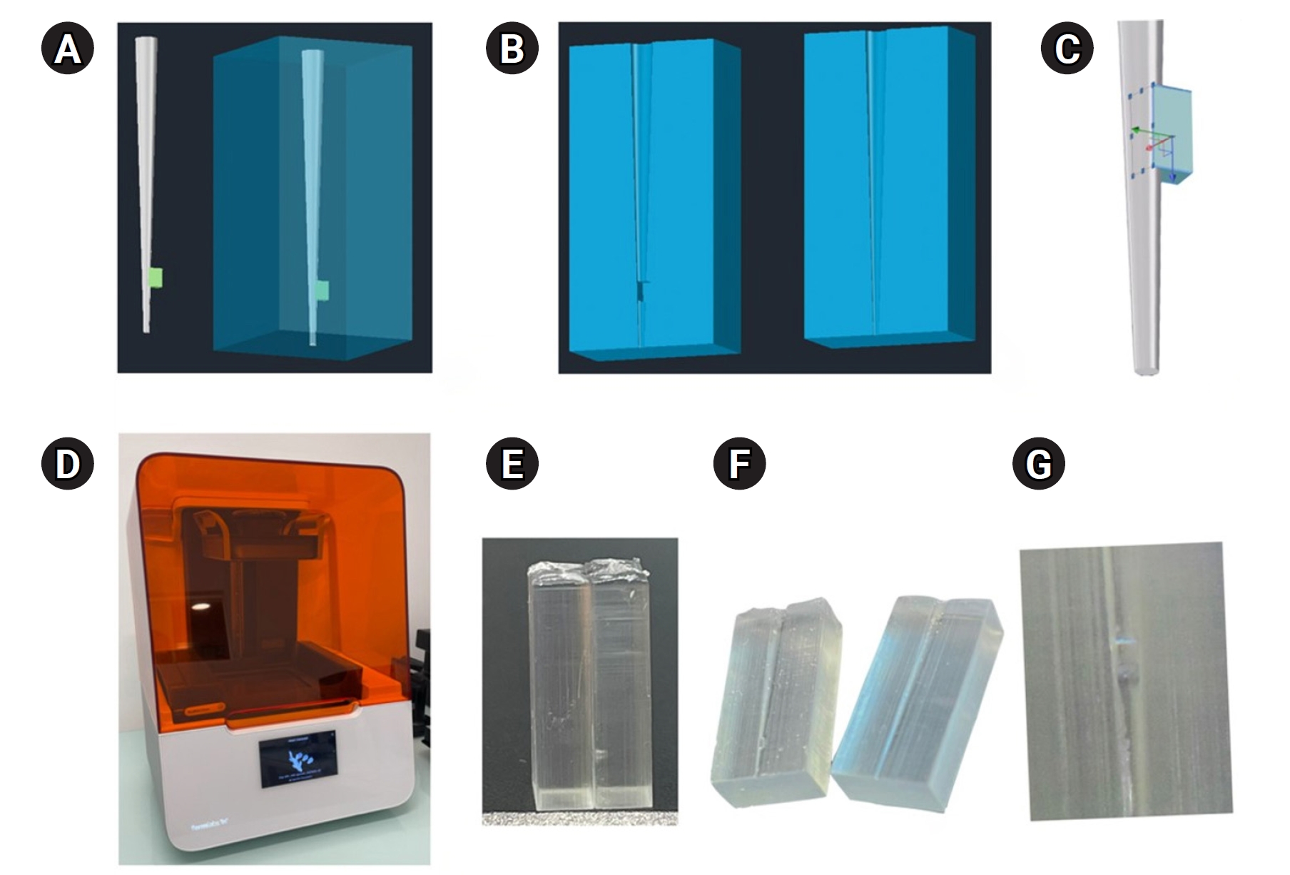
A three-dimensional printed artificial root canal with a lateral canal in the apical third was fabricated. Standardized bovine pulp tissue specimens were inserted, and three irrigation protocols were tested: group A (continuous NaOCl irrigation at 1 mL/min via syringe pump), group B (intermittent NaOCl irrigation with 0.1 mL and a 3-minute resting period), and group C (control, saline irrigation). The time for complete dissolution and the total NaOCl volume were recorded.
Results
Complete dissolution occurred in groups A and B, with significant differences in NaOCl volume and time (p < 0.05). In group A, complete dissolution was consistently observed after the 6th irrigation cycle, corresponding to a total NaOCl volume of 6.0 ± 0.66 mL per test. The average time required for complete dissolution in this group was 6 ± 0.66 minutes. In group B, complete dissolution occurred after the 4th cycle, with a total NaOCl volume of 0.4 ± 0.06 mL per test and a mean dissolution time of 12.6 ± 1.8 minutes.
Conclusions
NaOCl volume and exposure time significantly influence pulp tissue dissolution.
-
Analysis of thermal profiles on tooth structure and insert during one-piece or adapter-coupled ultrasonic insert use: an in vitro experimental study
-
Gabriela Loewen Brotto, Bruno Monguilhott Crozeta, Bruno Marques-da-Silva, Alysson Nunes Diógenes, Emmanuel João Nogueira Leal da Silva, Flávia Sens Fagundes Tomazinho
-
Restor Dent Endod 2025;50(3):e24. Published online July 11, 2025
-
DOI: https://doi.org/10.5395/rde.2025.50.e24
-
-
Abstract
PDFPubReaderePub
- Objectives
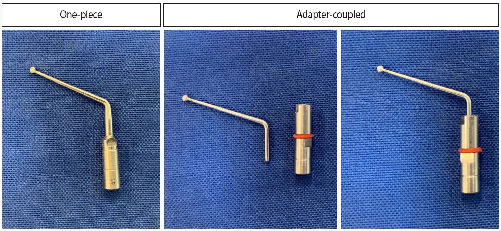
This in vitro study aimed to evaluate temperature variation on the external surface of mandibular molars and within ultrasonic inserts when using adapter-coupled versus one-piece inserts.
Methods
Twenty-four extracted human mandibular molars were divided into two groups based on the type of ultrasonic insert used: adapter-coupled and one-piece inserts. Temperature on the external surface of each tooth was measured with a thermocouple probe positioned in the furcation area, capturing data continuously. The temperature of the ultrasonic inserts was monitored in real-time using a thermal imaging camera. Measurements were taken in a controlled environment without cooling for over 120 seconds. Statistical analysis was conducted using analysis of variance (ANOVA) and two-way ANOVA with repeated measures to evaluate temperature variations between groups and over time, with significance set at 5%.
Results
In the external tooth surface temperature measurements, no significant differences were observed between the groups during the initial 15 seconds (p = 0.185) and 30 seconds (p = 0.067). However, significant differences emerged at 60 seconds (p = 0.025), 90 seconds (p = 0.024), and 120 seconds (p = 0.020), with the one-piece insert group demonstrating higher temperatures in the furcation region. Thermal imaging of the inserts revealed a significant difference at all time points (p < 0.001), with adapter-coupled inserts showing greater heating.
Conclusions
The use of ultrasonic inserts leads to a gradual rise in temperature on the external tooth surface. One-piece inserts generated higher temperatures on the tooth, while adapter-coupled inserts exhibited greater heating within the insert.
-
Does the use of different root canal sealers and adhesive resin cements impact the bond strength of glass fiber posts?
-
Ália Regina Neves de Paula Porto, Rudá França Moreira, Felipe Gonçalves Belladonna, Victor Talarico Leal Vieira, Emmanuel João Nogueira Leal da Silva
-
Restor Dent Endod 2025;50(3):e29. Published online August 29, 2025
-
DOI: https://doi.org/10.5395/rde.2025.50.e29
-
-
Abstract
PDFPubReaderePub
- Objectives
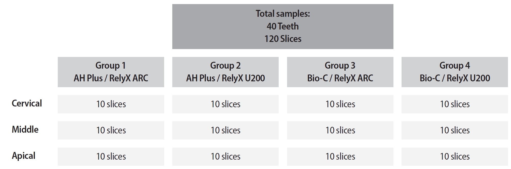
This study aimed to assess the influence of two endodontic sealers on the bond strength of glass fiber posts using conventional and self-adhesive resin cement through a push-out test. Methods: Forty central human incisors were randomly divided into four groups (n = 10) based on sealer (epoxy resin- based or calcium silicate-based) and cement (conventional and self-adhesive resin) types: AH Plus (Dentsply De- Trey)/RelyX ARC (3M ESPE), AH Plus/RelyX U200 (3M ESPE), Bio-C Sealer (Angelus)/RelyX ARC, and Bio-C Sealer/RelyX U200. After canal filling and post cementation, roots were sectioned to obtain one specimen per root third. A pushout test and failure pattern assessment were conducted, with bond strength analyzed using the one-way analysis of variance and Tukey test. Results: AH Plus/RelyX ARC showed the highest bond strength values, with a significant difference in the middle third. The most common failure was mixed (55%), while adhesive failures made up 45%, with 23.5% at the cement/post interface and 21.5% at the cement/dentin interface. Conclusions: AH Plus/RelyX ARC provided the highest bond strength values for glass fiber posts to dentin.
-
Citations
Citations to this article as recorded by  - Effect of Endodontic Sealers on the Bond Strength of Glass Fibre Posts: A Systematic Review
Thiago Bessa Marconato Antunes, Juliana D. Bronzato, Vanessa Gallego Arias Pecorari, Jennifer Santos Pereira, Talita Tartari, Adriana de Jesus Soares, Brenda P. F. A. Gomes, Marina Angélica Marciano
Australian Endodontic Journal.2026;[Epub] CrossRef
-
2,694
View
-
164
Download
-
1
Web of Science
-
1
Crossref
-
Success rate of direct pulp capping on permanent teeth using bioactive materials: a systematic review and meta-analysis of randomized clinical trials
-
Karem Paula Pinto, Gabriela Ribeiro da Silva, Cláudio Malizia Alves Ferreira, Luciana Moura Sassone, Emmanuel João Nogueira Leal da Silva
-
Restor Dent Endod 2024;49(4):e34. Published online September 6, 2024
-
DOI: https://doi.org/10.5395/rde.2024.49.e34
-
-
Abstract
PDF
 Supplementary MaterialPubReaderePub Supplementary MaterialPubReaderePub
This systematic review and meta-analysis aimed to evaluate the success rate of direct pulp capping (DPC) on permanent teeth, comparing the use of MTA with calcium hydroxide and calcium silicate-based cements. A systematic search was carried out in 4 databases until July 2023. The selection was based on PICOS criteria and only randomized clinical trials were included. The risk of bias was assessed using RoB-2 tool, and meta-analyses were performed using RevMan 5.3 software. The overall quality of evidence was determined using the GRADE tool. Thirteen studies were included. Meta-analyses indicated significantly higher success rate for DPC using MTA compared to calcium hydroxide, while no significant difference was observed between MTA and Biodentine, showing a success rate from 80% to 100% even after 3 years of follow-up. Five studies were classified as having high risk of bias and the GRADE assessment revealed low certainty of evidence. DPC is highly effective for permanent teeth when using MTA or Biodentine. There is a need for future well-designed randomized clinical trials to evaluate the efficacy of DPC using newer bioceramic materials. -
Citations
Citations to this article as recorded by - Physicochemical effects of nano type-B bone substitute on pulp protective cement formulations
Njwan Fadhel SHEHAB
Dental Materials Journal.2026; 45(1): 92. CrossRef - Photobiomodulation-assisted pulp capping using nano-hydroxyapatite and mineral trioxide aggregate: Report of two cases
Priya Pal, Rhythm Bains, Promila Verma, Vivek Kumar Bains
Journal of Healthcare Research and Education.2026; 2: 2. CrossRef - Histological Tissue Response to Calcium Silicate-Based Cements Assessed in Human Tooth Culture Models: A Systematic Review
Alberto Cabrera-Fernández, Hebertt Gonzaga dos Santos Chaves, Aránzazu Díaz-Cuenca, Juan J. Segura-Egea, Jenifer Martín-González, João Peça, Diana B. Sequeira, João Miguel Marques dos Santos
Journal of Functional Biomaterials.2026; 17(2): 78. CrossRef - Translational Pathways for Smart and Bioactive Dental Biomaterials: Biocompatibility Standards, Sterilisation, Sustainability and Regulation
Katarzyna Chojnacka, Marcin Mikulewicz
MedComm – Biomaterials and Applications.2026;[Epub] CrossRef - Decision-ready evidence for vital pulp therapy: a network meta-analysis of bioactive materials in mature permanent teeth
Firas Elmsmari, Reem B. Abdelsayed, Qamar Albasoumi, Tareq Aljafarawi, Swadheena Patro, Ajinkya M. Pawar
Frontiers in Dental Medicine.2026;[Epub] CrossRef - Comparative Evaluation of Platelet-Rich Fibrin and Mineral Trioxide Aggregate in the Direct Pulp Capping of Carious Exposures: A Randomized Clinical Trial
Geeta Asthana, Rajashree Tamuli, Sadhna Manglani, Saloni Mandhare, Ragini Kulkarni, Anooja Mathirat, Parneet Kaur, P Jyothirmayee, Surabhi Landge
Cureus.2026;[Epub] CrossRef - Indian Association of Conservative Dentistry and Endodontics consensus statement on deep caries management
Deepak Kumar Sharma, R. S. Mohan Kumar, Shishir Singh, Suparna Ganguly Saha, Meenal Nithin Gulve, Dipali Y. Shah, Sathish Abraham, Shruthi Nagaraja, Raksha Bhat
Journal of Conservative Dentistry and Endodontics.2025; 28(8): 714. CrossRef
-
22,411
View
-
691
Download
-
4
Web of Science
-
7
Crossref
-
Disinfectant effectiveness of chlorhexidine gel compared to sodium hypochlorite: a systematic review with meta-analysis
-
Theodoro Weissheimer, Karem Paula Pinto, Emmanuel João Nogueira Leal da Silva, Lina Naomi Hashizume, Ricardo Abreu da Rosa, Marcus Vinicius Reis Só
-
Restor Dent Endod 2023;48(4):e37. Published online October 26, 2023
-
DOI: https://doi.org/10.5395/rde.2023.48.e37
-
-
Abstract
PDFSupplementary MaterialPubReaderePub
This study aimed to compare the disinfectant ability of chlorhexidine (CHX) gel and sodium hypochlorite (NaOCl). Systematic searches were conducted from inception until December 8th, 2022 (MEDLINE/PubMed, Cochrane Library, Web of Science, Scopus, Embase, and Grey Literature databases). Only randomized clinical trials were included. The revised Cochrane risk of bias tools for randomized trials were used to assess the quality of studies. Meta-analyses were performed. The overall quality of evidence was assessed through the Grading of Recommendations Assessment, Development, and Evaluation tool. Six studies were included. Five had a low risk of bias and 1 had some concerns. Three studies assessed bacterial reduction. Two were included in the meta-analysis for bacterial reduction (mean difference, 75.03 [confidence interval, CI, −271.15, 421.22], p = 0.67; I2 = 74%); and 3 in the meta-analysis for cultivable bacteria after chemomechanical preparation (odds ratio, 1.03 [CI, 0.20, 5.31], P = 0.98; I2 = 49%). Five studies assessed endotoxin reduction. Three were included in a meta-analysis (mean difference, 20.59 [CI, −36.41, 77.59], p = 0.48; I2 = 74%). There seems to be no difference in the disinfectant ability of CHX gel and NaOCl, but further research is necessary. -
Citations
Citations to this article as recorded by - Regenerative Endodontic Procedures: Mapping and Critical Appraisal of Clinical Trial Evidence
Felipe Oliveira Nunes, Eduardo Borges Sollim, Carolynne Ferreira dos Santos, Maria Karolina Martins Ferreira, João Daniel Mendonça Moura, Juliana Melo Brandão, Manoel Damião Sousa-Neto, Paulo Jorge Palma, Rafael Rodrigues Lima
Journal of Endodontics.2026;[Epub] CrossRef - The conundrum of current endodontic disinfection strategies in microbial load reduction: a scoping review
Shymaa Shaaban, Abdelrahman Thabet, Semha Elsayed Elnaggar, Farah Tarek Barakat, Rana Hegazi, Hams Abdelrahman, Rania Elbackly, Hisham Elnawam
BMC Oral Health.2026;[Epub] CrossRef - Pre-Clinical Laboratory Evaluation of Chlorhexidine for Disinfection of Semi-Critical Respiratory Equipment in Nursing Practice
Rustiana Tasya Ariningpraja, Marina Ulfa, Lukky Jayadi, Andini Maslukha, Nuraeni Effendy
Journal of Applied Nursing and Health.2026; 8(1): 310. CrossRef - Bactericidal Effects of Ultraviolet-C Light-Emitting Diode Prototype Device Through Thin Optical Fiber
Mi-Jeong Jeon, Yu-Sung Choi, Deog-Gyu Seo
Applied Sciences.2025; 15(8): 4504. CrossRef - Effectiveness of Irrigation Protocols in Endodontic Therapy: An Umbrella Review
Manuel J. Orozco-Gallego, Eliana L. Pineda-Vélez, Wilder J. Rojas-Gutiérrez, Martha L. Rincón-Rodríguez, Andrés A. Agudelo-Suárez
Dentistry Journal.2025; 13(6): 273. CrossRef - In Vitro Evaluation of Disinfectants on Gutta-Percha Cones: Antimicrobial Efficacy Against Enterococcus faecalis and Candida albicans
Tringa Kelmendi, Donika Bajrami Shabani, Aida Meto, Hani Ounsi
Journal of Clinical Medicine.2025; 14(19): 6846. CrossRef - Preparing porcine lens to mimic human lens capsule
Yajing Pei, Shaofeng Han, Mingfeng Lu, Yang Yang, Ke Ma
Journal of Cataract & Refractive Surgery.2024; 50(9): 963. CrossRef - Comparative Evaluation of Disinfection Protocols for Dental Impressions in Prosthodontics
Subhash Sonkesriya, Ghanshyam Gaur, Akanksha Maheshwari, Arun Kumar Ashahiya, Simran Kaur Aulakh, Amit Kumar, Bhumika Kamal Badiyani
Cureus.2024;[Epub] CrossRef
-
7,480
View
-
152
Download
-
6
Web of Science
-
8
Crossref
-
Shaping ability and apical debris extrusion after root canal preparation with rotary or reciprocating instruments: a micro-CT study
-
Emmanuel João Nogueira Leal da Silva, Sara Gomes de Moura, Carolina Oliveira de Lima, Ana Flávia Almeida Barbosa, Waleska Florentino Misael, Mariane Floriano Lopes Santos Lacerda, Luciana Moura Sassone
-
Restor Dent Endod 2021;46(2):e16. Published online February 25, 2021
-
DOI: https://doi.org/10.5395/rde.2021.46.e16
-
-
Abstract
PDFPubReaderePub
- Objectives
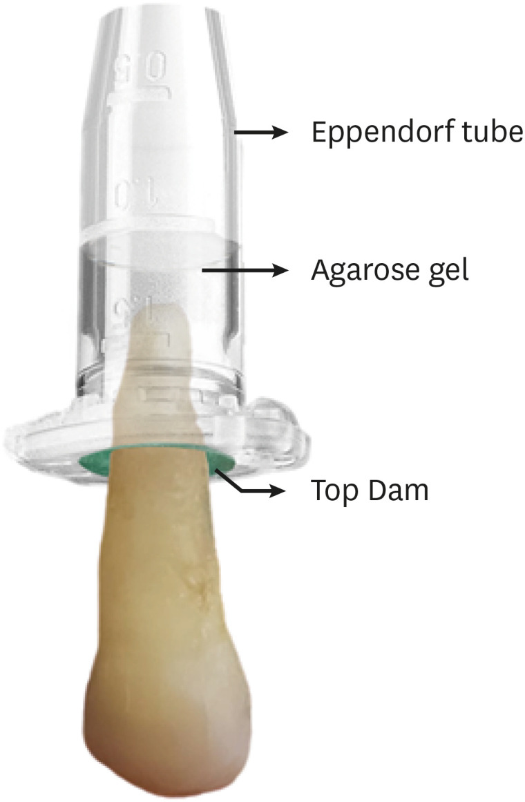
The aim of this study was to evaluate the shaping ability of the TruShape and Reciproc Blue systems and the apical extrusion of debris after root canal instrumentation. The ProTaper Universal system was used as a reference for comparison. Materials and MethodsThirty-three mandibular premolars with a single canal were scanned using micro-computed tomography and were matched into 3 groups (n = 11) according to the instrumentation system: TruShape, Reciproc Blue and ProTaper Universal. The teeth were accessed and mounted in an apparatus with agarose gel, which simulated apical resistance provided by the periapical tissue and enabled the collection of apically extruded debris. During root canal preparation, 2.5% sodium hypochlorite was used as an irrigant. The samples were scanned again after instrumentation. The percentage of unprepared area, removed dentin, and volume of apically extruded debris were analyzed. The data were analyzed using 1-way analysis of variance and the Tukey test for multiple comparisons at a 5% significance level. ResultsNo significant differences in the percentage of unprepared area were observed among the systems (p > 0.05). ProTaper Universal presented a higher percentage of dentin removal than the TruShape and Reciproc Blue systems (p < 0.05). The systems produced similar volumes of apically extruded debris (p > 0.05). ConclusionsAll systems caused apically extruded debris, without any significant differences among them. TruShape, Reciproc Blue, and ProTaper Universal presented similar percentages of unprepared area after root canal instrumentation; however, ProTaper Universal was associated with higher dentin removal than the other systems.
-
Citations
Citations to this article as recorded by - Endomotor integration, file kinematics impact on apical debris extrusion in severely curved canals
Anshika Saxena, Vineeta Nikhil
Endodontology.2026; 38(1): 87. CrossRef - Comparison of post-operative pain prevalence after single visit endodontic treatment with two NiTi rotary files - a randomized clinical trial
M. E. Khallaf, Yousra Aly, Amira Ibrahim Mohamed
Scientific Reports.2025;[Epub] CrossRef - Evaluation of Silver-Ion-Coated Rotary Nickel Titanium Files - An In Vitro Study
Jhanvi H. Sadaria, Kondas V. Venkatesh, Dhanasekaran Sihivahanan
Indian Journal of Dental Research.2025; 36(3): 344. CrossRef - A quantitative comparison of apically extruded debris during root canal preparation using NiTi full-sequence rotary and single-file rotary systems: An in vitro study
Pallavi Goel, R. Vikram, R. Anithakumari, M. S. Adarsha, M. E. Sudhanva
Endodontology.2024; 36(3): 235. CrossRef - Extrusion of Sodium Hypochlorite in Oval-Shaped Canals: A Comparative Study of the Potential of Four Final Agitation Approaches Employing Agarose-Embedded Mandibular First Premolars
Aalisha Parkar, Kulvinder Singh Banga, Ajinkya M. Pawar, Alexander Maniangat Luke
Journal of Clinical Medicine.2024; 13(10): 2748. CrossRef - Shaping Efficiency of Rotary and Reciprocating Kinematics of Engine-driven Nickel-Titanium Instruments in Moderate and Severely curved Root Canals Using Microcomputed Tomography: A Systematic Review of Ex Vivo Studies
Claudiu Călin, Ana-Maria Focșăneanu, Friedrich Paulsen, Andreea C. Didilescu, Tiberiu Niță
Journal of Endodontics.2024; 50(7): 907. CrossRef - Intracanal removal and apical extrusion of filling material after retreatment using rotary or reciprocating instruments: A new approach using human cadavers
Thamyres M. Monteiro, Victor O. Cortes‐Cid, Marilia F. V. Marceliano‐Alves, Andrea F. Campello, Luan F. Bastos, Ricardo T. Lopes, José F. Siqueira, Flávio R. F. Alves
International Endodontic Journal.2024; 57(1): 100. CrossRef - Assessment of debris extrusion on using automated irrigation device with conventional needle irrigation – An ex vivo study
Sahil Choudhari, Kavalipurapu Venkata Teja, Raja Kumar, Sindhu Ramesh
Saudi Endodontic Journal.2023; 13(3): 263. CrossRef - Postoperative pain perception and associated risk factors in children after continuous rotation versus reciprocating kinematics: A randomised prospective clinical trial
Ahmad Abdel Hamid Elheeny, Dania Ibrahem Sermani, Mahmoud Ahmed Abdelmotelb
Australian Endodontic Journal.2023; 49(S1): 345. CrossRef - A critical analysis of research methods and experimental models to study apical extrusion of debris and irrigants
Jale Tanalp
International Endodontic Journal.2022; 55(S1): 153. CrossRef - Quantitative evaluation of apically extruded debris using TRUShape, TruNatomy, and WaveOne Gold in curved canals
Nehal Nabil Roshdy, Reham Hassan
BDJ Open.2022;[Epub] CrossRef - Shaping ability of new reciprocating or rotary instruments with two cross‐sectional designs: An ex vivo study
Isabela G. Guedes, Renata C. V. Rodrigues, Marília F. Marceliano‐Alves, Flávio R. F. Alves, Isabela N. Rôças, José F. Siqueira
International Endodontic Journal.2022; 55(12): 1385. CrossRef
-
3,347
View
-
57
Download
-
8
Web of Science
-
12
Crossref
-
Maxillary first molar with 7 root canals diagnosed using cone-beam computed tomography
-
Evaldo Rodrigues, Antônio Henrique Braitt, Bruno Ferraz Galvão, Emmanuel João Nogueira Leal da Silva
-
Restor Dent Endod 2017;42(1):60-64. Published online August 29, 2016
-
DOI: https://doi.org/10.5395/rde.2017.42.1.60
-
-
Abstract
PDFPubReaderePub
Root canal anatomy is complex, and the recognition of anatomic variations could be a challenge for clinicians. This case report describes the importance of cone beam computed tomographyic (CBCT) imaging during endodontic treatment. A 23 year old woman was referred by her general dental practitioner with the chief complaint of spontaneous pain in her right posterior maxilla. From the clinical and radiographic findings, a diagnosis of symptomatic irreversible pulpitis was made and endodontic treatment was suggested to the patient. The patient underwent CBCT examination, and CBCT scan slices revealed seven canals: three mesiobuccal (MB1, MB2, and MB3), two distobuccal (DB1 and DB2), and two palatal (P1 and P2). Canals were successfully treated with reciprocating files and filled using single-cone filling technique. Precise knowledge of root canal morphology and its variation is important during root canal treatment. CBCT examination is an excellent tool for identifying and managing these complex root canal systems. -
Citations
Citations to this article as recorded by - KONİK IŞINLI BİLGİSAYARLI TOMOGRAFİ İLE DOĞRULANMIŞ OLAĞANDIŞI ÜST BİRİNCİ BÜYÜK AZI DİŞİN ENDODONTİK TEDAVİSİ
Didem Seda Gültekin, Funda Kont Çobankara
Journal of International Dental Sciences.2025; 11(1): 46. CrossRef - Clinical Significance of Mesiobuccal and Distobuccal Canal Variations in Maxillary Molars: A Case Series and a Mini Review
Mohsen Aminsobhani, Somayeh Majidi, Vlaho Brailo
Case Reports in Dentistry.2025;[Epub] CrossRef - An Unusual Case of Maxillary First Molar: A Case Report
Reetu Shrestha
International Journal of Innovative Science and Research Technology (IJISRT).2024; : 1330. CrossRef - Root canal therapy of maxillary first molar with seven canals diagnosed using cone beam computed tomography – a case report
Saini Rashmi, Saini V. Kumar
Tanta Dental Journal.2022; 19(3): 169. CrossRef - Four-Rooted Maxillary First Molars: A Systematic Review and Meta-Analysis
Gabriel Magnucki, Sven V. K. Mietling, Sreekanth Kumar Mallineni
International Journal of Dentistry.2021; 2021: 1. CrossRef - Endodontic treatment of various palatal roots in maxillary molars
Chengshi Wei, Keyi Li, Lili Shen, Guangliang Bai, Xiufen Tian
The Journal of the American Dental Association.2021; 152(12): 1044. CrossRef - Diversity of root canal morphology of maxillary first molars
Juhász Kincső-Réka, Kovács Mónika, Pop Mihai, Pop Silvia, Kerekes-Máthé Bernadette
Bulletin of Medical Sciences.2021; 94(1): 63. CrossRef - Endodontic Management of Maxillary First Molar with Seven Root Canals Diagnosed Using Cone-beam Computed Tomography: A Case Report
Ravindranath Megha, Venkatachalam Prakash
World Journal of Dentistry.2021; 12(1): 89. CrossRef - Endodontic management of the maxillary first molar with special root canals: A case report and review of the literature
Zhi-Hui Zhang, Hai-Lin Yao, Yan Zhang, Xiao Wang
World Journal of Clinical Cases.2020; 8(12): 2590. CrossRef - Management of a permanent maxillary first molar with unusual crown and root anatomy: a case report
Prateeksha Chowdhry, Pallavi Reddy, Mamta Kaushik
Restorative Dentistry & Endodontics.2018;[Epub] CrossRef - Usefulness of cone beam computed tomography in perplexing endodontic cases
Amandeep Kaur, Ajay Logani
Endodontology.2018; 30(2): 187. CrossRef - Endodontic management of a maxillary first molar with seven root canal systems evaluated using cone-beam computed tomography scanning
VijayReddy Venumuddala, Sridhar Moturi, SV Satish, BKalyan Chakravarthy, Sudhakar Malapati
Journal of International Society of Preventive and Community Dentistry.2017; 7(5): 297. CrossRef
-
3,126
View
-
16
Download
-
12
Crossref
-
Push-out bond strength of a self-adhesive resin cement used as endodontic sealer
-
Eduardo Diogo Gurgel-Filho, Felipe Coelho Lima, Vicente de Paula Aragão Saboia, Tauby de Souza Coutinho-Filho, Aline de Almeida Neves, Emmanuel João Nogueira Leal da Silva
-
Restor Dent Endod 2014;39(4):282-287. Published online August 20, 2014
-
DOI: https://doi.org/10.5395/rde.2014.39.4.282
-
-
Abstract
PDFPubReaderePub
- Objectives
The aim of the present study was to investigate the bond strength of RelyX Unicem (3M) to root canal dentin when used as an endodontic sealer. Materials and MethodsSamples of 24 single-rooted teeth were prepared with Gates Glidden drills and K3 files. After that, the roots were randomly assigned to three experimental groups (n = 8) according to the filling material, (1) AH Plus (Dentsply De Trey GmbH)/Gutta-Percha cone; (2) Epiphany SE (Pentron)/Resilon cone; (3) RelyX Unicem/Gutta-Percha cone. All roots were filled using a single cone technique associated to vertical condensation. After the filling procedures, each tooth was prepared for a push-out bond strenght test by cutting 1 mm-thick root slices. Loading was performed on a universal testing machine at a speed of 0.5 mm/min. One-way analysis of variance and Tukey test for multiple comparisons were used to compare the results among the experimental groups. ResultsEpiphany SE/Resilon showed significantly lower push-out bond strength than both AH Plus/Gutta-Percha and RelyX Unicem/Gutta-Percha (p < 0.05). There was no significant difference in bond strength between AH Plus/Gutta-Percha and RelyX Unicem/Gutta-Percha (p > 0.05). ConclusionsUnder the present in vitro conditions, bond strength to root dentin promoted by RelyX Unicem was similar to AH Plus. Epiphany SE/Resilon resulted in lower bond strength values when compared to both materials.
-
Citations
Citations to this article as recorded by - In vitro comparative evaluation of physicochemical and mechanical properties, cytocompatibility, and antimicrobial efficacy of various bioceramic root canal sealers
Fushi Wang, Jiaxing Li, Jingjing Wan, Siyuan Li, Shijia Tang, Li Wang, Liuyan Meng
Ceramics International.2026; 52(7): 9561. CrossRef - In-Vitro Comparative Adhesion Evaluation of Bioceramic and Dual-Cure Resin Endodontic Sealers Using SEM, AFM, Push-Out and FTIR
Radu Marcel Chisnoiu, Marioara Moldovan, Doina Prodan, Andrea Maria Chisnoiu, Dana Hrab, Ada Gabriela Delean, Alexandrina Muntean, Doina Iulia Rotaru, Ovidiu Pastrav, Mihaela Pastrav
Applied Sciences.2021; 11(10): 4454. CrossRef - Push-out Bond Strength of Fiber Posts Cemented Using New Universal Adhesives on Etched and Nonetched Intraradicular Dentin
Hani F Ounsi, Simone Grandini, Marco Ferrari, Valentina Spicciarelli, Giacomo Corsentino, Crystal Marruganti
The Journal of Contemporary Dental Practice.2020; 21(1): 91. CrossRef - Comparison of push-out bond strength of three different obturating systems to intraradicular dentin: An In vitro study
MohammedKhwaja Moinuddin, LKarthik Prasad, Nimeshika Ramachandruni, Shekar Kamishetty, RaviChandra Cherkupalli
Contemporary Clinical Dentistry.2019; 10(4): 631. CrossRef - The influence of methodological variables on the push‐out resistance to dislodgement of root filling materials: a meta‐regression analysis
F. M. Collares, F. F. Portella, S. B. Rodrigues, R. K. Celeste, V. C. B. Leitune, S. M. W. Samuel
International Endodontic Journal.2016; 49(9): 836. CrossRef - Effect of photon induced photoacoustic streaming (PIPS) on bond strength to dentine of two root canal filling materials
Ivana Miletić, Nicoletta Chieffi, Carlo Rengo, Marco Ferrari, Dan Nathanson, Anja Baraba
Lasers in Surgery and Medicine.2016; 48(10): 951. CrossRef
-
2,244
View
-
6
Download
-
6
Crossref
|