-
Comparative analysis of torsional and cyclic fatigue resistance of ProGlider, WaveOne Gold Glider, and TruNatomy Glider in simulated curved canal
-
Pedro de Souza Dias, Augusto Shoji Kato, Carlos Eduardo da Silveira Bueno, Rodrigo Ricci Vivan, Marco Antonio Hungaro Duarte, Pedro Henrique Souza Calefi, Rina Andréa Pelegrine
-
Restor Dent Endod 2023;48(1):e4. Published online December 8, 2022
-
DOI: https://doi.org/10.5395/rde.2023.48.e4
-
-
 Abstract Abstract
 PDF PDF PubReader PubReader ePub ePub
- Objectives
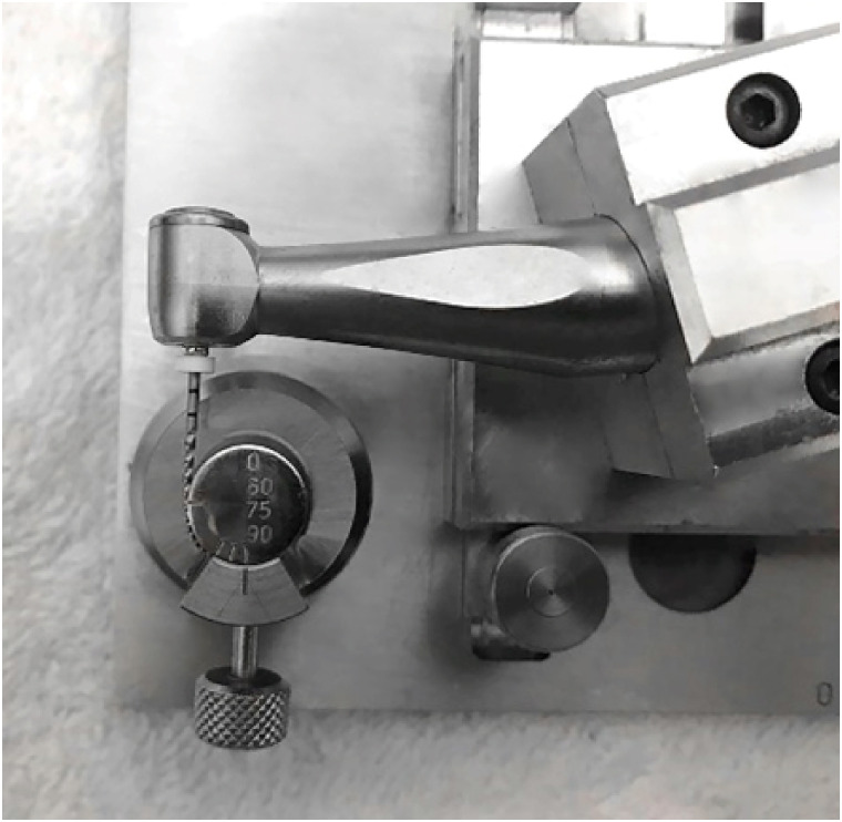
This study aimed to compare the torsional and cyclic fatigue resistance of ProGlider (PG), WaveOne Gold Glider (WGG), and TruNatomy Glider (TNG). Materials and MethodsA total of 15 instruments of each glide path system (n = 15) were used for each test. A custom-made device simulating an angle of 90° and a radius of 5 millimeters was used to assess cyclic fatigue resistance, with calculation of number of cycles to failure. Torsional fatigue resistance was assessed by maximum torque and angle of rotation. Fractured instruments were examined by scanning electron microscopy (SEM). Data were analyzed with Shapiro-Wilk and Kruskal-Wallis tests, and the significance level was set at 5%. ResultsThe WGG group showed greater cyclic fatigue resistance than the PG and TNG groups (p < 0.05). In the torsional fatigue test, the TNG group showed a higher angle of rotation, followed by the PG and WGG groups (p < 0.05). The TNG group was superior to the PG group in torsional resistance (p < 0.05). SEM analysis revealed ductile morphology, typical of the 2 fracture modes: cyclic fatigue and torsional fatigue. ConclusionsReciprocating WGG instruments showed greater cyclic fatigue resistance, while TNG instruments were better in torsional fatigue resistance. The significance of these findings lies in the identification of the instruments’ clinical applicability to guide the choice of the most appropriate instrument and enable the clinician to provide a more predictable glide path preparation.
-
Citations
Citations to this article as recorded by  - Comparative evaluation of the remaining dentin volume following instrumentation with rotary, reciprocating, and hand files during root canal treatment in primary molars: An ex vivo study
İrem Eren, Berkant Sezer
Journal of Dental Sciences.2024; 19(4): 2126. CrossRef - Screw-in force, torque generation, and performance of glide-path files with three rotation kinetics
Jee-Yeon Woo, Ji-Hyun Jang, Seok Woo Chang, Soram Oh
Odontology.2024; 112(3): 761. CrossRef - Evaluation of shaping ability of different glide path instruments: a micro-computed tomography study
Merve Yeniçeri Özata, Seda Falakaloğlu, Ali Keleş, Özkan Adıgüzel, Mustafa Gündoğar
BMC Oral Health.2023;[Epub] CrossRef
-
250
View
-
15
Download
-
3
Web of Science
-
3
Crossref
-
Fracture incidence of Reciproc instruments during root canal retreatment performed by postgraduate students: a cross-sectional retrospective clinical study
-
Liliana Machado Ruivo, Marcos de Azevedo Rios, Alexandre Mascarenhas Villela, Alexandre Sigrist de Martin, Augusto Shoji Kato, Rina Andrea Pelegrine, Ana Flávia Almeida Barbosa, Emmanuel João Nogueira Leal Silva, Carlos Eduardo da Silveira Bueno
-
Restor Dent Endod 2021;46(4):e49. Published online September 9, 2021
-
DOI: https://doi.org/10.5395/rde.2021.46.e49
-
-
 Abstract Abstract
 PDF PDF PubReader PubReader ePub ePub
- Objectives
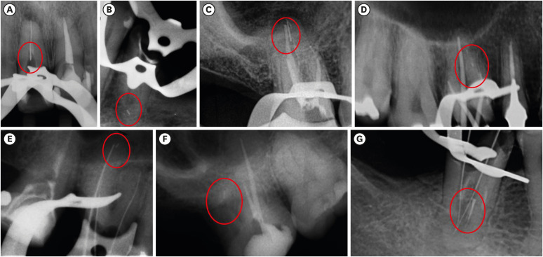
To evaluate the fracture incidence of Reciproc R25 instruments (VDW) used during non-surgical root canal retreatments performed by students in a postgraduate endodontic program. Materials and MethodsFrom the analysis of clinical record cards and periapical radiographs of root canal retreatments performed by postgraduate students using the Reciproc R25, a total of 1,016 teeth (2,544 root canals) were selected. The instruments were discarded after a single use. The general incidence of instrument fractures and its frequency was analyzed considering the group of teeth and the root thirds where the fractures occurred. Statistical analysis was performed using the χ2 test (p < 0.01). ResultsSeven instruments were separated during the procedures. The percentage of fracture in relation to the number of instrumented canals was 0.27% and 0.68% in relation to the number of instrumented teeth. Four fractures occurred in maxillary molars, 1 in a mandibular molar, 1 in a mandibular premolar and 1 in a maxillary incisor. A greater number of fractures was observed in molars when compared with the number of fractures observed in the other dental groups (p < 0.01). Considering all of the instrument fractures, 71.43% were located in the apical third and 28.57% in the middle third (p < 0.01). One instrument fragment was removed, one bypassed, while in 5 cases, the instrument fragment remained inside the root canal. ConclusionsThe use of Reciproc R25 instruments in root canal retreatments carried out by postgraduate students was associated with a low incidence of fractures.
-
Citations
Citations to this article as recorded by  - Reciprocating Torsional Fatigue and Mechanical Tests of Thermal-Treated Nickel Titanium Instruments
Victor Talarico Leal Vieira, Alejandro Jaime, Carlos Garcia Puente, Giuliana Soimu, Emmanuel João Nogueira Leal Silva, Carlos Nelson Elias, Gustavo de Deus
Journal of Endodontics.2025; 51(3): 359. CrossRef - Neodymium-Doped Yttrium Aluminum Perovskite (Nd:YAP) Laser in the Elimination of Endodontic Nickel-Titanium Files Fractured in Rooted Canals (Part 2: Teeth With Significant Root Curvature)
Amaury Namour, Marwan El Mobadder, Clément Cerfontaine, Patrick Matamba, Lucia Misoaga, Delphine Magnin , Praveen Arany, Samir Nammour
Cureus.2025;[Epub] CrossRef - Temperature-Dependent Effects on Cyclic Fatigue Resistance in Three Reciprocating Endodontic Systems: An In Vitro Study
Marcela Salamanca Ramos, José Aranguren, Giulia Malvicini, Cesar De Gregorio, Carmen Bonilla, Alejandro R. Perez
Materials.2025; 18(5): 952. CrossRef - Multimethod analysis of large‐ and low‐tapered single file reciprocating instruments: Design, metallurgy, mechanical performance, and irrigation flow
Emmanuel João Nogueira Leal Silva, Fernando Peña‐Bengoa, Natasha C. Ajuz, Victor T. L. Vieira, Jorge N. R. Martins, Duarte Marques, Ricardo Pinto, Mario Rito Pereira, Francisco Manuel Braz‐Fernandes, Marco A. Versiani
International Endodontic Journal.2024; 57(5): 601. CrossRef - Nd: YAP Laser in the Elimination of Endodontic Nickel-Titanium Files Fractured in Rooted Canals (Part 1: Teeth With Minimal Root Curvature)
Amaury Namour, Marwan El Mobadder, Patrick Matamba, Lucia Misoaga, Delphine Magnin , Praveen Arany, Samir Nammour
Cureus.2024;[Epub] CrossRef - Cyclic Fatigue of Different Reciprocating Endodontic Instruments Using Matching Artificial Root Canals at Body Temperature In Vitro
Sebastian Bürklein, Paul Maßmann, Edgar Schäfer, David Donnermeyer
Materials.2024; 17(4): 827. CrossRef - Endodontic Orthograde Retreatments: Challenges and Solutions
Alessio Zanza, Rodolfo Reda, Luca Testarelli
Clinical, Cosmetic and Investigational Dentistry.2023; Volume 15: 245. CrossRef - Design, metallurgy, mechanical properties, and shaping ability of 3 heat-treated reciprocating systems: a multimethod investigation
Emmanuel J. N. L. Silva, Jorge N. R. Martins, Natasha C. Ajuz, Henrique dos Santos Antunes, Victor Talarico Leal Vieira, Francisco Manuel Braz-Fernandes, Felipe Gonçalves Belladonna, Marco Aurélio Versiani
Clinical Oral Investigations.2023; 27(5): 2427. CrossRef - Noncontact 3D evaluation of surface topography of reciprocating instruments after retreatment procedures
Miriam Fatima Zaccaro-Scelza, Renato Lenoir Cardoso Henrique Martinez, Sandro Oliveira Tavares, Fabiano Palmeira Gonçalves, Marcelo Montagnana, Emmanuel João Nogueira Leal da Silva, Pantaleo Scelza
Brazilian Dental Journal.2022; 33(3): 38. CrossRef
-
293
View
-
6
Download
-
6
Web of Science
-
9
Crossref
-
Effect of ultrasonic cleaning on the bond strength of fiber posts in oval canals filled with a premixed bioceramic root canal sealer
-
Fernando Peña Bengoa, Maria Consuelo Magasich Arze, Cristobal Macchiavello Noguera, Luiz Felipe Nunes Moreira, Augusto Shoji Kato, Carlos Eduardo Da Silveira Bueno
-
Restor Dent Endod 2020;45(2):e19. Published online February 20, 2020
-
DOI: https://doi.org/10.5395/rde.2020.45.e19
-
-
 Abstract Abstract
 PDF PDF PubReader PubReader ePub ePub
- Objective
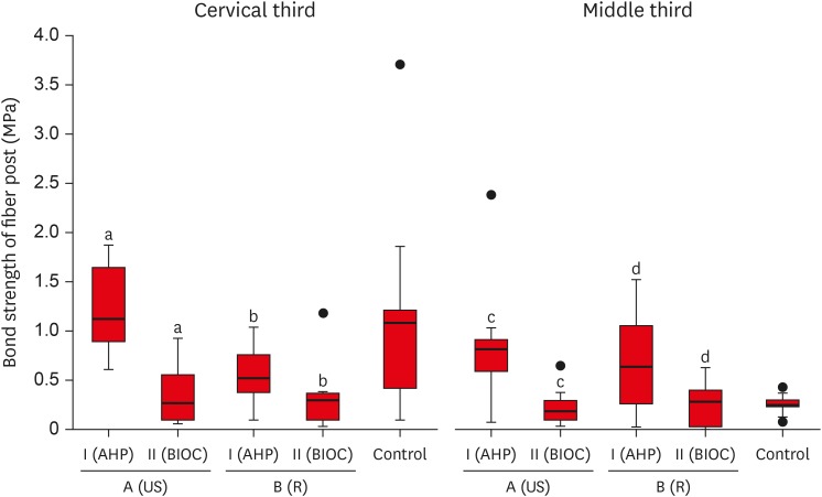
This study aimed to evaluate the effect of ultrasonic cleaning of the intracanal post space on the bond strength of fiber posts in oval canals filled with a premixed bioceramic (Bio-C Sealer [BIOC]) root canal sealer. Materials and MethodsFifty premolars were endodontically prepared and divided into 5 groups (n = 10), based on the type of root canal filling material used and the post space cleaning protocol. A1: gutta-percha + AH Plus (AHP) and post space preparation with ultrasonic cleaning, A2: gutta-percha + BIOC and post space preparation with ultrasonic cleaning, B1: gutta-percha + AHP and post space preparation, B2: gutta-percha + BIOC and post space preparation, C: control group. Fiber posts were cemented with a self-adhesive luting material, and 1 mm thick slices were sectioned from the middle and cervical third to evaluate the remaining filling material microscopically. The samples were subjected to a push-out test to analyze the bond strength of the fiber post, and the results were analyzed with the Shapiro-Wilk, Bonferroni, Kruskal-Wallis, and Mann-Whitney tests (p < 0.05). Failure modes were evaluated using optical microscopy. ResultsThe results showed that the fiber posts cemented in canals sealed with BIOC had lower bond strength than those sealed with AHP. The ultrasonic cleaning of the post space improved the bond strength of fiber posts in canals sealed with AHP, but not with BIOC. ConclusionsBIOC decreased the bond strength of fiber posts in oval canals, regardless of ultrasonic cleaning.
-
Citations
Citations to this article as recorded by  - Evaluation of different mechanical cleaning protocols associated with 2.5% sodium hypochlorite in the removal of residues from the post space
Matheus Sousa Vitória, Eran Nair Mesquita de Almeida, Antonia Patricia Oliveira Barros, Eliane Cristina Gulin de Oliveira, Joatan Lucas de Sousa Gomes Costa, Andrea Abi Rached Dantas, Milton Carlos Kuga
Journal of Conservative Dentistry and Endodontics.2024; 27(3): 274. CrossRef - Fiber post cemented using different adhesive strategies to root canal dentin obturated with calcium silicate-based sealer
Lalita Patthanawijit, Kallaya Yanpiset, Pipop Saikaew, Jeeraphat Jantarat
BMC Oral Health.2024;[Epub] CrossRef - Effect of endodontic sealers on push-out bond strength of CAD-CAM or prefabricated fiber glass posts
Andréa Pereira de Souza PINTO, Fabiana Mantovani Gomes FRANÇA, Roberta Tarkany BASTING, Cecilia Pedroso TURSSI, José Joatan RODRIGUES JÚNIOR, Flávia Lucisano Botelho AMARAL
Brazilian Oral Research.2023;[Epub] CrossRef - Effect of mechanical cleaning protocols in the fiber post space on the adhesive interface between universal adhesive and root dentin
Gabriela Mariana Castro‐Núnez, José Rodolfo Estruc Verbicário dos Santos, Joissi Ferrari Zaniboni, Wilfredo Gustavo Escalante‐Otárola, Thiago Soares Porto, Milton Carlos Kuga
Microscopy Research and Technique.2022; 85(6): 2131. CrossRef - Effect of bioceramic root canal sealers on the bond strength of fiber posts cemented with resin cements
Rafael Nesello, Isadora Ames Silva, Igor Abreu De Bem, Karolina Bischoff, Matheus Albino Souza, Marcus Vinícius Reis Só, Ricardo Abreu Da Rosa
Brazilian Dental Journal.2022; 33(2): 91. CrossRef - Effect of irrigation protocols on root canal wall after post preparation: a micro-CT and microhardness study
Camila Maria Peres de Rosatto, Danilo Cassiano Ferraz, Lilian Vieira Oliveira, Priscilla Barbosa Ferreira Soares, Carlos José Soares, Mario Tanomaru Filho, Camilla Christian Gomes Moura
Brazilian Oral Research.2021;[Epub] CrossRef
-
288
View
-
6
Download
-
6
Crossref
-
In vivo assessment of accuracy of Propex II, Root ZX II, and radiographic measurements for location of the major foramen
-
Fernanda Garcia Tampelini, Marcelo Santos Coelho, Marcos de Azevêdo Rios, Carlos Eduardo Fontana, Daniel Guimarães Pedro Rocha, Sergio Luiz Pinheiro, Carlos Eduardo da Silveira Bueno
-
Restor Dent Endod 2017;42(3):200-205. Published online May 16, 2017
-
DOI: https://doi.org/10.5395/rde.2017.42.3.200
-
-
 Abstract Abstract
 PDF PDF PubReader PubReader ePub ePub
- Objectives
The aim of this in vivo study was to assess the accuracy of 2 third-generation electronic apex locators (EALs), Propex II (Dentsply Maillefer) and Root ZX II (J. Morita), and radiographic technique for locating the major foramen (MF). Materials and MethodsThirty-two premolars with single canals that required extraction were included. Following anesthesia, access, and initial canal preparation with size 10 and 15 K-flex files and SX and S1 rotary ProTaper files, the canals were irrigated with 2.5% sodium hypochlorite. The length of the root canal was verified 3 times for each tooth using the 2 apex locators and once using the radiographic technique. Teeth were extracted and the actual WL was determined using size 15 K-files under a × 25 magnification. The Biostat 4.0 program (AnalystSoft Inc.) was used for comparing the direct measurements with those obtained using radiographic technique and the apex locators. Pearson's correlation analysis and analysis of variance (ANOVA) were used for statistical analyses. ResultsThe measurements obtained using the visual method exhibited the strongest correlation with Root ZX II (r = 0.94), followed by Propex II (r = 0.90) and Ingle's technique (r = 0.81; p < 0.001). Descriptive statistics using ANOVA (Tukey's post hoc test) revealed significant differences between the radiographic measurements and both EALs measurements (p < 0.05). ConclusionsBoth EALs presented similar accuracy that was higher than that of the radiographic measurements obtained with Ingle's technique. Our results suggest that the use of these EALs for MF location is more accurate than the use of radiographic measurements.
-
Citations
Citations to this article as recorded by  - How Do Different Image Modules Impact the Accuracy of Working Length Measurements in Digital Periapical Radiography? An In Vitro Study
Vahide Hazal Abat, Rabia Figen Kaptan
Diagnostics.2025; 15(3): 305. CrossRef - Influence of maintaining apical patency in post-endodontic pain
Snigdha Shubham, Manisha Nepal, Ravish Mishra, Kishor Dutta
BMC Oral Health.2021;[Epub] CrossRef
-
228
View
-
2
Download
-
2
Crossref
|


