-
Effect of cryotherapy duration on experimentally induced connective tissue inflammation in vivo
-
Jorge Vera, Mayra Alejandra Castro-Nuñez, María Fernanda Troncoso-Cibrian, Ana Gabriela Carrillo-Varguez, Edgar Ramiro Méndez Sánchez, Viviana Sarmiento, Lourdes Lanzagorta-Rebollo, Prasanna Neelakantan, Monica Romero, Ana Arias
-
Restor Dent Endod 2023;48(3):e29. Published online August 2, 2023
-
DOI: https://doi.org/10.5395/rde.2023.48.e29
-
-
 Abstract Abstract
 PDF PDF PubReader PubReader ePub ePub
- Objectives
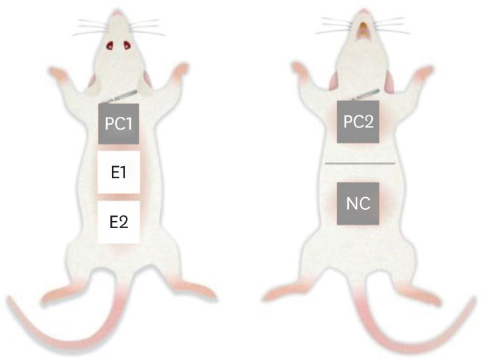
This study tested the hypothesis that cryotherapy duration influences lipopolysaccharide (LPS)-induced inflammation in a rat model. Materials and MethodsSix Wistar rats (Rattus norvegicus albinus) were used. Five sites were selected per animal and divided into 5 groups: a negative control group (NC), 2 positive control groups (PC1 and PC2), and 2 experimental groups (E1 and E2). Cryotherapy was applied for 1 minute (E1) or 5 minutes (E2). An acute inflammatory response was induced in the PC and E groups via subcutaneous administration of 0.5 mL/kg. In the PC2 group, a catheter was inserted without additional treatment. For the E1 and E2 groups, 2.5°C saline solution was administered through the implanted catheters for 1 and 5 minutes, respectively. The rats were sacrificed, and samples were obtained and processed for histological analysis, specifically examining the presence of polymorphonuclear neutrophils and hemorrhage. The χ2 test was used to compare the presence of acute inflammation across groups. Dependent variables were compared using the linear-by-linear association test. ResultsInflammation and hemorrhage varied significantly among the groups (p = 0.001). A significantly higher degree of acute inflammation was detected (p = 0.0002) in the PC and E1 samples than in the E2 group, in which cryotherapy was administered for 5 minutes. The PC and E1 groups also exhibited significantly greater numbers of neutrophils (p = 0.007), which were essentially absent in both the NC and E2 groups. ConclusionsCryotherapy administration for 5 minutes reduced the acute inflammation associated with LPS and catheter implantation.
-
Citations
Citations to this article as recorded by  - The impact of using cold irrigation on postoperative endodontic pain and substance P level: a randomized clinical trial
Reem Mohammed Amr Sharaf, Tariq Yehia Abdelrahman, Maram Farouk Obeid
Odontology.2025;[Epub] CrossRef - Cryotherapy as a supplementary aid to inferior alveolar nerve block in patients with symptomatic irreversible pulpitis: A randomized controlled trial
Setu Katyal, Poonam Bogra, Rajinder Bansal, Vishakha Grover, Saurabh Gupta, Saru Gupta
Medicine International.2025; 5(5): 1. CrossRef - Determining Efficacy of Intracanal Cryotherapy on Post Endodontic Pain in Irreversible Pulpitis
Anam Fayyaz Bashir, Ussamah Waheed Jatala, Moeen ud din Ahmad, Muhammad Talha Khan, Saima Razzaq Khan, Aisha Arshad Butt
Pakistan Journal of Health Sciences.2024; : 68. CrossRef
-
2,904
View
-
51
Download
-
1
Web of Science
-
3
Crossref
-
In vitro apical pressure created by 2 irrigation needles and a multisonic system in mandibular molars
-
Ronald Ordinola-Zapata, Joseph T. Crepps, Ana Arias, Fei Lin
-
Restor Dent Endod 2021;46(1):e14. Published online February 8, 2021
-
DOI: https://doi.org/10.5395/rde.2021.46.e14
-
-
 Abstract Abstract
 PDF PDF PubReader PubReader ePub ePub
- Objectives
The aim of this study was to evaluate the apical pressure generated by 2 endodontic irrigation needles and the GentleWave system in mandibular molars. Materials and MethodsThe mesial and distal root canals of 12 mandibular molars were irrigated with a 30-gauge close-end needle or with a 30-gauge open-end needle. Procedures were performed in the mesial and distal canals. The GentleWave procedure and irrigation at 1 mm from the apex in the distal roots using an open-end needle were used, respectively, as negative and positive controls. The apical pressure was measured using a data acquisition pressure setup. Apical pressure exerted by the different needles in the 2 different canal types was statistically compared using 2-way analysis of variance. ResultsSignificant differences were found in the apical pressure for both needles and the canal type. The lowest values were obtained with close-end needles and in mesial canals. Negative apical pressure values were obtained using GentleWave. ConclusionsThe needle and the canal type influenced the apical pressure. The GentleWave procedure produced negative apical pressure.
-
Citations
Citations to this article as recorded by  - Use of the gentlewave system in endodonticsUse of the gentlewave system in endodontics
Daiana Jacobi Lazzarotto, Mayara Colpo Prado, Lara Dotto, Rafael Sarkis-Onofre
Brazilian Journal of Oral Sciences.2025; 24: e254250. CrossRef - Bibliometric analysis of the GentleWave system: trends, collaborations, and research gaps
Raimundo Sales de Oliveira Neto, Thais de Moraes Souza, João Vitor Oliveira de Amorim, Thaine Oliveira Lima, Guilherme Ferreira da Silva, Rodrigo Ricci Vivan, Murilo Priori Alcalde, Marco Antonio Hungaro Duarte
Restorative Dentistry & Endodontics.2025; 50(2): e17. CrossRef - The effect of ultrasonic and multisonic irrigation on root canal microbial communities: An ex vivo study
Ki Hong Park, Ronald Ordinola‐Zapata, W. Craig Noblett, Bruno P. Lima, Christopher Staley
International Endodontic Journal.2024; 57(7): 895. CrossRef - Efficacy of the GentleWave System in the removal of biofilm from the mesial roots of mandibular molars before and after minimal instrumentation: An ex vivo study
Kwang Ho Kim, Céline Lévesque, Gevik Malkhassian, Bettina Basrani
International Endodontic Journal.2024; 57(7): 922. CrossRef - A critical analysis of research methods and experimental models to study irrigants and irrigation systems
Christos Boutsioukis, Maria Teresa Arias‐Moliz, Luis E. Chávez de Paz
International Endodontic Journal.2022; 55(S2): 295. CrossRef - Outcomes of the GentleWave system on root canal treatment: a narrative review
Hernán Coaguila-Llerena, Eduarda Gaeta, Gisele Faria
Restorative Dentistry & Endodontics.2022;[Epub] CrossRef
-
1,878
View
-
19
Download
-
6
Web of Science
-
6
Crossref
|


