-
Can different agents reduce the damage caused by bleaching gel to pulp tissue? A systematic review of basic research
-
Letícia Aparecida Silva Batista, Alexandre Henrique dos Reis-Prado, Hebertt Gonzaga dos Santos Chaves, Lara Cancella de Arantes, Luís Fernando Santos Alves Morgan, Carolina Bosso André, Thaís Yumi Suzuki, Francine Benetti
-
Restor Dent Endod 2023;48(4):e39. Published online November 6, 2023
-
DOI: https://doi.org/10.5395/rde.2023.48.e39
-
-
 Abstract Abstract
 PDF PDF Supplementary Material Supplementary Material PubReader PubReader ePub ePub
- Objectives
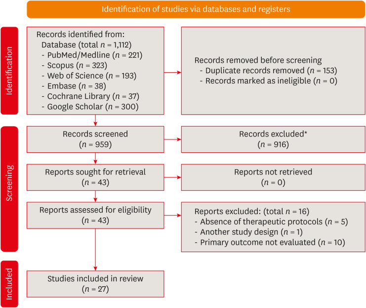
This study aimed to investigate the effectiveness of different topical/systemic agents in reducing the damage caused by bleaching gel to pulp tissue or cells. Materials and MethodsElectronic searches were performed in July 2023. In vivo and in vitro studies evaluating the effects of different topical or systemic agents on pulp inflammation or cytotoxicity after exposure to bleaching agents were included. The risk of bias was assessed. ResultsOut of 1,112 articles, 27 were included. Nine animal studies evaluated remineralizing/anti-inflammatories agents in rat molars subjected to bleaching with 35%–38% hydrogen peroxide (HP). Five of these studies demonstrated a significant reduction in inflammation caused by HP when combined with bioglass or MI Paste Plus (GC America), or following KF-desensitizing or Otosporin treatment (n = 3). However, orally administered drugs did not reduce pulp inflammation (n = 4). Cytotoxicity (n = 17) was primarily assessed using the 3-(4,5-dimethylthiazol-2-yl)-2,5-diphenyltetrazolium bromide assay on human dental pulp cells and mouse dental papilla Cell-23 cells. Certain substances, including sodium ascorbate, butein, manganese chloride, and peroxidase, were found to reduce cytotoxicity, particularly when applied prior to bleaching. The risk of bias was high in animal studies and low in laboratory studies. ConclusionsFew in vivo studies have evaluated agents to reduce the damage caused by bleaching gel to pulp tissue. Within the limitations of these studies, it was found that topical agents were effective in reducing pulp inflammation in animals and cytotoxicity. Further analyses with human pulp are required to substantiate these findings. Trial RegistrationPROSPERO Identifier: CRD42022337192
-
Citations
Citations to this article as recorded by  - 3D-Printed and Bioprinted Scaffolds in Regenerative Endodontics: A Systematic Review
Hebertt Gonzaga dos Santos Chaves, Diana B. Sequeira, Vilton Cardozo Moreira Dias, Alberto Cabrera-Fernández, João Peça, Francine Benetti, João Miguel Marques dos Santos
Applied Sciences.2026; 16(8): 3940. CrossRef - Clinical Study on the Efficacy of 35% Hydrogen Peroxide Gel According to Exposure Time (40 min vs. 20 min) by Spectrophotometry
Trinidad Rincón, Maria Portillo Muñoz, Maria Lobato, Ana María Martín Casado, Laryssa Mylenna Madruga Barbosa, Alessandro Loguercio, Cristina Gómez‐Polo
Journal of Esthetic and Restorative Dentistry.2026;[Epub] CrossRef - Clareamento dental e TikTok: avaliação da qualidade do conteúdo em mídia social
Rafaele T Costa, Thayna Silva do Carmo Tavares, André Walsh-Monteiro
Ciência ET Praxis.2025; 21(36): 111. CrossRef - Synthesis, characterization and evaluation of novel bleaching gels containing bioactive glass and nano-hydroxyapatite on hydrogen peroxide diffusion, bleaching efficacy and enamel protection
Adrieli Burey, Byron Carpio-Salvatierra, Michael Favoretto, María Luján Méndez Bauer, Viviane Hass, Alessandra Reis, Alessandro D. Loguercio, Paulo Vitor Farago
Clinical Oral Investigations.2025;[Epub] CrossRef - Cytotoxicity of Bleaching Products: A Systematic Review
Mireia Montaner, José Luis Sanz, Carmen Llena, María Melo, Clara Puig-Herreros, James Ghilotti
Applied Sciences.2024; 14(9): 3680. CrossRef
-
4,445
View
-
60
Download
-
4
Web of Science
-
5
Crossref
-
Effectiveness and safety of rotary and reciprocating kinematics for retreatment of curved root canals: a systematic review of in vitro studies
-
Lucas Pinho Simões, Alexandre Henrique dos Reis-Prado, Carlos Roberto Emerenciano Bueno, Ana Cecília Diniz Viana, Marco Antônio Húngaro Duarte, Luciano Tavares Angelo Cintra, Cleidiel Aparecido Araújo Lemos, Francine Benetti
-
Restor Dent Endod 2022;47(2):e22. Published online April 6, 2022
-
DOI: https://doi.org/10.5395/rde.2022.47.e22
-
-
Abstract
PDFPubReaderePub
- Objectives
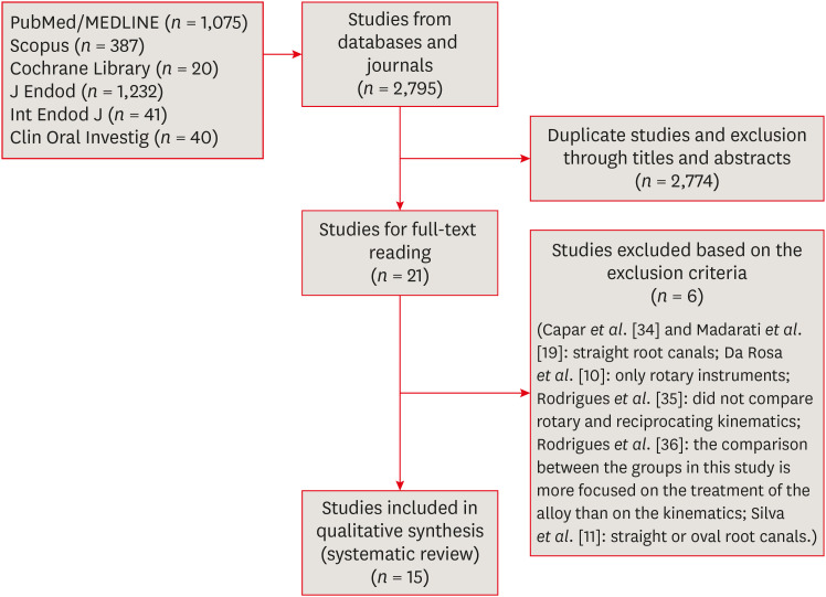
This systematic review (register-osf.io/wg7ba) compared the efficacy and safety of rotary and reciprocating kinematics in the removal of filling material from curved root canals. Materials and MethodsOnly in vitro studies evaluating both kinematics during retreatment were included. A systematic search (PubMed/MEDLINE, Scopus, and other databases, until January 2021), data extraction, and risk of bias analysis (Joanna Briggs Institute checklist) were performed. Efficacy in filling removal was the primary outcome. ResultsThe search resulted in 2,795 studies, of which 15 were included. Efficacy was measured in terms of the remaining filling material and the time required for this. Nine studies evaluated filling material removal, of which 7 found no significant differences between rotary and reciprocating kinematics. Regarding the time for filling removal, 5 studies showed no difference between both kinematics, 2 studies showed faster results with rotary systems, and other 2 showed the opposite. No significant differences were found in apical transportation, centering ability, instrument failure, dentin removed and extruded debris. A low risk of bias was observed. ConclusionsThis review suggests that the choice of rotary or reciprocating kinematics does not influence the efficacy of filling removal from curved root canals. Further studies are needed to compare the kinematics safety in curved root canals.
-
Citations
Citations to this article as recorded by - Apical Third Cleaning Efficiency of Hand, Rotary, and Reciprocating Root Canal Instrumentation: A Quantitative In Vitro Scanning Electron Microscopic Study
B. Sai Krishna, Sita Mahalakshmi Koppu, Dilip Katakam, Sahithi Nammaniwar, Ambika Belam, Akshita Balivada, Divakar K. P.
Cureus.2026;[Epub] CrossRef - Selective Nonsurgical Endodontic Retreatment: Relevant Aspects for Clinical Application—A Scoping Review
Tomás Manuel Braz Marinho, Luiz Renato Paranhos, Anne Caroline Brito Cabral dos Santos, Djessyca Miranda‐e‐Paulo, Rui Barbosa de Brito Júnior, João Marcos da Costa Ribeiro, Felipe de Souza Matos
Australian Endodontic Journal.2026;[Epub] CrossRef - EVALUATION OF MICROLEAKAGE AFTER ENDODONTIC FILLING IN TEETH WITH APICAL WIDENING: A SYSTEMATIC REVIEW
Isabella da Costa Ferreira, Gabriela da Costa Ferreira, Isabella Figueiredo de Assis Macedo, Gustavo Oliveira Campos, Isabella Faria da Cunha Peixoto, Ana Cecília Diniz Viana, Rodrigo Rodrigues Amaral, Warley Luciano Fonseca Tavares
ARACÊ .2025; 7(10): e8792. CrossRef - Fifteen years of engine‐driven nickel–titanium reciprocating instruments, what do we know so far? An umbrella review
Felipe Immich, Lucas Peixoto de Araújo, Rafaella Rodrigues da Gama, Wellington Luiz de Oliveira da Rosa, Evandro Piva, Giampiero Rossi‐Fedele
Australian Endodontic Journal.2024; 50(2): 409. CrossRef - Efficacy of Various Heat-treated Retreatment File Systems on the Apical Deformity and Canal Centering Ability in a Single-rooted Teeth using Nano CT
Swathi S, Pradeep Solete, Ganesh Jeevanandan, Delphine Priscilla Antony S, Kavalipurapu Venkata Teja, Dona Sanju
The Open Dentistry Journal.2024;[Epub] CrossRef - Micro-CT evaluation of the removal of root fillings using rotary and reciprocating systems supplemented by XP-Endo Finisher, the Self-Adjusting File, or Er,Cr:YSGG laser
Gülsen Kiraz, Bulem Üreyen Kaya, Mert Ocak, Muhammet Bora Uzuner, Hakan Hamdi Çelik
Restorative Dentistry & Endodontics.2023;[Epub] CrossRef - Influence of sodium hypochlorite on cyclic fatigue resistance of nickel–titanium instruments: A systematic review and meta-analysis of in vitro studies
Alexandre Henrique dos Reis-Prado, Lucas Guimarães Abreu, Lara Cancella de Arantes, Kiani dos Santos de Paula, Sabrina de Castro Oliveira, Juliana Goto, Ana Cecília Diniz Viana, Francine Benetti
Clinical Oral Investigations.2023; 27(11): 6291. CrossRef - Retreatment of XP-endo Shaper and R-Endo files in curved root canals
Hayam Y. Hassan, Fahd M. Hadhoud, Ayman Mandorah
BMC Oral Health.2023;[Epub] CrossRef - Advancing Endodontics through Kinematics
Shilpa Bhandi, Dario Di Nardo, Francesco Pagnoni, Rosemary Abbagnale
World Journal of Dentistry.2023; 14(6): 479. CrossRef
-
4,734
View
-
94
Download
-
7
Web of Science
-
9
Crossref
-
Biological assessment of a new ready-to-use hydraulic sealer
-
Francine Benetti, João Eduardo Gomes-Filho, India Olinta de Azevedo-Queiroz, Marina Carminatti, Letícia Citelli Conti, Alexandre Henrique dos Reis-Prado, Sandra Helena Penha de Oliveira, Edilson Ervolino, Elói Dezan-Júnior, Luciano Tavares Angelo Cintra
-
Restor Dent Endod 2021;46(2):e21. Published online March 24, 2021
-
DOI: https://doi.org/10.5395/rde.2021.46.e21
-
-
Abstract
PDFPubReaderePub
- Objectives
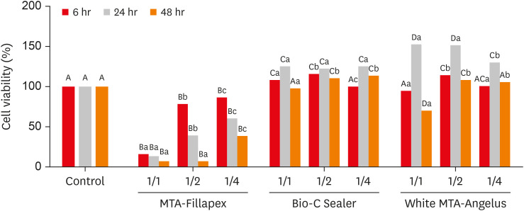
This study compared the cytotoxicity, biocompatibility, and tenascin immunolabeling of a new ready-to-use hydraulic sealer (Bio-C Sealer) with MTA-Fillapex and white MTA-Angelus. Materials and MethodsL929 fibroblasts were cultivated and exposed to undiluted and diluted material extracts. Polyethylene tubes with or without (the control) the materials were implanted into the dorsa of rats. At 7 days and 30 days, the rats were euthanized, and the specimens were prepared for analysis; inflammation and immunolabeling were measured, and statistical analysis was performed (p < 0.05). ResultsMTA-Fillapex exhibited greater cytotoxicity than the other materials at all time points (p < 0.05). The undiluted Bio-C Sealer exhibited greater cytocompatibility at 6 and 48 hours than white MTA-Angelus, with higher cell viability than in the control (p < 0.05). White MTA-Angelus displayed higher cell viability than the control at 24 hours, and the one-half dilution displayed similar results at both 6 and 48 hours (p < 0.05). At 7 days and 30 days, the groups exhibited moderate inflammation with thick fibrous capsules and mild inflammation with thin fibrous capsules, respectively (p > 0.05). At 7 days, moderate to strong immunolabeling was observed (p > 0.05). After 30 days, the control and MTA-Fillapex groups exhibited strong immunolabeling, the white MTA-Angelus group exhibited moderate immunolabeling (p > 0.05), and the Bio-C Sealer group exhibited low-to-moderate immunolabeling, differing significantly from the control (p < 0.05). ConclusionsBio-C Sealer and white MTA-Angelus exhibited greater cytocompatibility than MTA-Fillapex; all materials displayed adequate biocompatibility and induced tenascin immunolabeling.
-
Citations
Citations to this article as recorded by - Clinical and radiographic assessment of mineral trioxide aggregate with platelet rich fibrin as pulp capping biomaterials: a 12-month randomized trial
Rahma Ahmed Ibrahem Hafiz Abuhashema, Mona El Saied Essa, Shereen Hafez Ibrahim, Omaima Mohamed Safwat
Scientific Reports.2025;[Epub] CrossRef - Biocompatibility and bioactivity of bioceramic endodontic sealer: NeoSealer Flo
Evelin Carine Alves SILVA, Jéssica Arielli PRADELLI, Guilherme Ferreira da SILVA, Paulo Sérgio CERRI, Mario TANOMARU-FILHO, Juliane Maria GUERREIRO-TANOMARU
Journal of Applied Oral Science.2025;[Epub] CrossRef - Influence of photoactivation on tissue response to different dyes used in photodynamic therapy and laser ablation therapy
Luciano Tavares Angelo Cintra, Cristiane Cantiga-Silva, Henrique Augusto Banci, Flávio Duarte Faria, Nathália Evelyn da Silva Machado, Carolina de Barros Morais Cardoso, Pedro Henrique Chaves de Oliveira, Lucas Rodrigues de Araújo Estrela, Gustavo Sivieri
Journal of Photochemistry and Photobiology B: Biology.2024; 251: 112843. CrossRef - Bleaching effectiveness and cytotoxicity of new experimental formulation of niobium-based bleaching gel
Camila de Sousa Caneschi, Francine Benetti, Luiz Carlos Alves de Oliveira, Jadson Cláudio Belchior, Raquel Conceição Ferreira, Allyson Nogueira Moreira, Luís Fernando dos Santos Alves Morgan
Clinical Oral Investigations.2023; 27(4): 1613. CrossRef - Biological investigation of resinous endodontic sealers containing calcium hydroxide
Carlos Roberto Emerenciano Bueno, Francine Benetti, Marina Tolomei Sandoval Cury, Ana Maria Veiga Vasques, Leopoldo Cosme-Silva, Índia Olinta de Azevedo Queiroz, Ana Cláudia Rodrigues da Silva, Rogério de Castilho Jacinto, Luciano Tavares Angelo Cintra, E
PLOS ONE.2023; 18(7): e0287890. CrossRef - Tricalcium silicate cement sealers
Anita Aminoshariae, Carolyn Primus, James C. Kulild
The Journal of the American Dental Association.2022; 153(8): 750. CrossRef - Comparative evaluation of push-out bond strength of bioceramic and epoxy sealers after using various final irrigants: An in vitro study
Chandrasekhar Veeramachaneni, Swathi Aravelli, Sreeja Dundigalla
Journal of Conservative Dentistry.2022; 25(2): 145. CrossRef
-
3,177
View
-
28
Download
-
8
Web of Science
-
7
Crossref
|