Articles
- Page Path
- HOME > Restor Dent Endod > Volume 45(2); 2020 > Article
- Research Article Comparative evaluation of the effectiveness of ultrasonic tips versus the Terauchi file retrieval kit for the removal of separated endodontic instruments
-
Preeti Jain Pruthi
 , Ruchika Roongta Nawal
 , Sangeeta Talwar
 , Mahesh Verma
-
Restor Dent Endod 2020;45(2):e14.
DOI: https://doi.org/10.5395/rde.2020.45.e14
Published online: February 6, 2020
Department of Conservative Dentistry and Endodontics, Maulana Azad Institute of Dental Sciences, New Delhi, DL, India.
- Correspondence to Preeti Jain Pruthi, BDS, MDS. Senior Research Associate, Department of Conservative Dentistry and Endodontics, Maulana Azad Institute of Dental Sciences, MAMC complex, 2 Bahadur Shah Zafar Marg, New Delhi, DL 110002, India. drpreetijain82@gmail.com
Copyright © 2020. The Korean Academy of Conservative Dentistry
This is an Open Access article distributed under the terms of the Creative Commons Attribution Non-Commercial License (https://creativecommons.org/licenses/by-nc/4.0/) which permits unrestricted non-commercial use, distribution, and reproduction in any medium, provided the original work is properly cited.
- 3,601 Views
- 100 Download
- 23 Crossref
Abstract
-
Objective The aim of this study was to perform a comparative evaluation of the effectiveness of ultrasonic tips versus the Terauchi file retrieval kit (TFRK) for the removal of broken endodontic instruments.
-
Materials and Methods A total of 80 extracted human first mandibular molars with moderate root canal curvature were selected. Following access cavity preparation canal patency was established with a size 10/15 K-file in the mesiobuccal canals of all teeth. The teeth were divided into 2 groups of 40 teeth each: the P group (ProUltra tips) and the T group (TFRK). Each group was further subdivided into 2 smaller groups of 20 teeth each according to whether ProTaper F1 rotary instruments were fractured in either the coronal third (C constituting the PC and TC groups) or the middle third (M constituting the PM and TM groups). Instrument retrieval was performed using either ProUltra tips or the TFRK.
-
Results The overall success rate at removing the separated instrument was 90% in group P and 95% in group T (p > 0.05) The mean time for instrument removal was higher with the ultrasonic tips than with the TFRK (p > 0.05).
-
Conclusion Both systems are acceptable clinical tools for instrument retrieval but the loop device in the TFRK requires slightly more dexterity than is needed for the ProUltra tips.
INTRODUCTION
MATERIALS AND METHODS
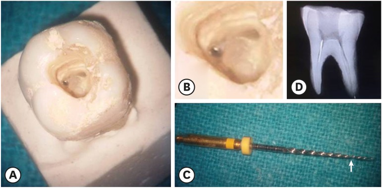
(A) Separated instrument in the coronal third of the mesiobuccal canal. (B) Magnified view of Figure 1A. (C) The arrow shows the separated portion of the ProTaper F1 rotary file. (D) Radiograph showing an instrument in the coronal third of the canal.

RESULTS
Intergroup comparison of the time (in minutes) taken for instrument retrieval (p > 0.05)
| Group | No. | Mean | Standard deviation | p-value | |
|---|---|---|---|---|---|
| PC vs. TC | 0.066 | ||||
| PC | 20 | 17.95 | 4.817 | ||
| TC | 20 | 15.35 | 3.801 | ||
| PM vs. TM | 0.310 | ||||
| PM | 20 | 46.38 | 7.482 | ||
| TM | 20 | 44.22 | 4.466 | ||
Intragroup comparison of the time (in minutes) taken for instrument retrieval (p < 0.05)
| Group | No. | Mean | Standard deviation | p-value | |
|---|---|---|---|---|---|
| PC vs. PM | 0.001* | ||||
| PC | 20 | 17.95 | 4.817 | ||
| PM | 20 | 46.38 | 7.482 | ||
| TC vs. TM | 0.001* | ||||
| TC | 20 | 15.35 | 3.801 | ||
| TM | 20 | 44.22 | 4.466 | ||
DISCUSSION
CONCLUSIONS
-
Conflict of Interest: No potential conflict of interest relevant to this article was reported.
-
Author Contributions:
Conceptualization: Pruthi PJ, Nawal RR, Talwar S, Verma M.
Investigation: Pruthi PJ, Nawal RR.
Methodology: Pruthi PJ, Nawal RR, Talwar S.
Validation: Pruthi PJ, Nawal RR, Talwar S, Verma M.
Visualization: Pruthi PJ, Nawal RR, Talwar S, Verma M.
Writing - original draft: Pruthi PJ.
Writing - review & editing: Nawal RR, Talwar S.
- 1. Thompson SA. An overview of nickel-titanium alloys used in dentistry. Int Endod J 2000;33:297-310.ArticlePubMed
- 2. Andreasen G, Wass K, Chan KC. A review of superelastic and thermodynamic nitinol wire. Quintessence Int 1985;16:623-626.PubMed
- 3. Wolcott S, Wolcott J, Ishley D, Kennedy W, Johnson S, Minnich S, Meyers J. Separation incidence of protaper rotary instruments: a large cohort clinical evaluation. J Endod 2006;32:1139-1141.ArticlePubMed
- 4. Iqbal MK, Kohli MR, Kim JS. A retrospective clinical study of incidence of root canal instrument separation in an endodontics graduate program: a PennEndo database study. J Endod 2006;32:1048-1052.ArticlePubMed
- 5. Di Fiore PM, Genov KA, Komaroff E, Li Y, Lin L. Nickel-titanium rotary instrument fracture: a clinical practice assessment. Int Endod J 2006;39:700-708.ArticlePubMed
- 6. Suter B, Lussi A, Sequeira P. Probability of removing fractured instruments from root canals. Int Endod J 2005;38:112-123.ArticlePubMed
- 7. Ward JR. The use of an ultrasonic technique to remove a fractured rotary nickel-titanium instrument from the apical third of a curved root canal. Aust Endod J 2003;29:25-30.ArticlePubMed
- 8. Murad M, Murray C. Impact of retained separated endodontic instruments during root canal treatment on clinical outcomes remains uncertain. J Evid Based Dent Pract 2011;11:87-88.ArticlePubMed
- 9. Terauchi Y, O'Leary L, Suda H. Removal of separated files from root canals with a new file-removal system: case reports. J Endod 2006;32:789-797.ArticlePubMed
- 10. Schneider SW. A comparison of canal preparations in straight and curved root canals. Oral Surg Oral Med Oral Pathol 1971;32:271-275.ArticlePubMed
- 11. Fu M, Zhang Z, Hou B. Removal of broken files from root canals by using ultrasonic techniques combined with dental microscope: a retrospective analysis of treatment outcome. J Endod 2011;37:619-622.ArticlePubMed
- 12. Hülsmann M. Methods for removing metal obstructions from the root canal. Endod Dent Traumatol 1993;9:223-237.ArticlePubMed
- 13. Cattoni M. Common failures in endodontics and their corrections. Dent Clin North Am 1963;7:383-399.
- 14. Feldman G, Solomon C, Notaro P, Moskowitz E. Retrieving broken endodontic instruments. J Am Dent Assoc 1974;88:588-591.ArticlePubMed
- 15. Roig-Greene JL. The retrieval of foreign objects from root canals: a simple aid. J Endod 1983;9:394-397.ArticlePubMed
- 16. Eleazer PD, O'Connor RP. Innovative uses for hypodermic needles in endodontics. J Endod 1999;25:190-191.ArticlePubMed
- 17. Johnson WB, Beatty RG. Clinical technique for the removal of root canal obstructions. J Am Dent Assoc 1988;117:473-476.ArticlePubMed
- 18. Friedman S, Stabholz A, Tamse A. Endodontic retreatment--case selection and technique. 3. Retreatment techniques. J Endod 1990;16:543-549.PubMed
- 19. Okiji T. Modified usage of the Masserann kit for removing intracanal broken instruments. J Endod 2003;29:466-467.ArticlePubMed
- 20. Ruddle CJ. Nonsurgical endodontic retreatment. J Calif Dent Assoc 2004;32:474-484.ArticlePubMed
- 21. Hülsmann M. Removal of fractured root canal instruments using the Canal Finder System. Dtsch Zahnarztl Z 1990;45:229-232.PubMed
- 22. Plotino G, Pameijer CH, Grande NM, Somma F. Ultrasonics in endodontics: a review of the literature. J Endod 2007;33:81-95.ArticlePubMed
- 23. Yu DG, Kimura Y, Tomita Y, Nakamura Y, Watanabe H, Matsumoto K. Study on removal effects of filling materials and broken files from root canals using pulsed Nd:YAG laser. J Clin Laser Med Surg 2000;18:23-28.ArticlePubMed
- 24. Ebihara A, Takashina M, Anjo T, Takeda A, Suda H. Removal of root canal obstructions using pulsed Nd:YAG laser. ICS Lasers in Dentistry 2003;1248:257-259.Article
- 25. Ormiga F, da Cunha Ponciano Gomes JA, de Araújo MC. Dissolution of nickel-titanium endodontic files via an electrochemical process: a new concept for future retrieval of fractured files in root canals. J Endod 2010;36:717-720.ArticlePubMed
- 26. Ruddle C. Microendodontics. Eliminating intracanal obstructions. Oral Health 1997;87:19-21.PubMed
- 27. Ward JR, Parashos P, Messer HH. Evaluation of an ultrasonic technique to remove fractured rotary nickel-titanium endodontic instruments from root canals: an experimental study. J Endod 2003;29:756-763.ArticlePubMed
- 28. Ward JR, Parashos P, Messer HH. Evaluation of an ultrasonic technique to remove fractured rotary nickel-titanium endodontic instruments from root canals: clinical cases. J Endod 2003;29:764-767.ArticlePubMed
- 29. Sornkul E, Stannard JG. Strength of roots before and after endodontic treatment and restoration. J Endod 1992;18:440-443.ArticlePubMed
- 30. Gerek M, Başer ED, Kayahan MB, Sunay H, Kaptan RF, Bayırlı G. Comparison of the force required to fracture roots vertically after ultrasonic and Masserann removal of broken instruments. Int Endod J 2012;45:429-434.ArticlePubMed
- 31. Terauchi Y, O'Leary L, Kikuchi I, Asanagi M, Yoshioka T, Kobayashi C, Suda H. Evaluation of the efficiency of a new file removal system in comparison with two conventional systems. J Endod 2007;33:585-588.ArticlePubMed
REFERENCES
Tables & Figures
REFERENCES
Citations

- Comparative evaluation of success rate and operator variability in loop.based versus ultrasonic retrieval of fractured endodontic instruments: An ex vivo study
Tanushree Saxena, Vivek Devidas Mahale, Manish Ranjan, Sanyuta Singh, E. Aparna Mohan, M. Hema
Saudi Endodontic Journal.2026; 16(1): 73. CrossRef - Comparative evaluation of time efficiency and dentin preservation in ultrasonic versus loop retrieval of separated endodontic files: An ex vivo study with pilot nano-computed tomography analysis
Tanushree Saxena, Vivek Devidas Mahale, Manish Ranjan, M. Hema, Sanyukta Singh, E. Aparna Mohan
Saudi Endodontic Journal.2026; 16(1): 90. CrossRef - Comparison of the pull-out force of different microtube-based methods in fractured endodontic instrument removal: An in-vitro study
Nasim Hashemi, Mohsen Aminsobhani, Mohammad Javad Kharazifard, Fatemeh Hamidzadeh, Pegah Sarraf
BMC Oral Health.2025;[Epub] CrossRef - Fracture resistance and volumetric dentin change after management of broken instrument using static navigation – An in vitro study
Shady Atef Adeeb Yassa, Mohamed Nabeel, Ahmed M. Ghobashy, Moataz B. Alkhawas
Journal of Conservative Dentistry and Endodontics.2025; 28(4): 319. CrossRef - Remoção de instrumento fraturado com a técnica do laço: relato de caso
Larissa Sousa Rangel, Ryhan Menezes Cardoso, Thayane Kelly Trajano da Silva, Robeci Alves Macêdo Filho, Andressa Cartaxo de Almeida, Mariana Camilly Tavares Ferreira, Thalles Gabriel Germano Lima, Diana Santana de Albuquerque
Caderno Pedagógico.2025; 22(7): e16332. CrossRef - Would It Necessarily Require Retrieving Endodontic Files on Every Instance? Implementing Separated Files with the Bypass Technique: Report of Three Cases
Mohit S. Zarekar, Apurva S. Satpute, Mohini S. Zarekar
Journal of Primary Care Dentistry and Oral Health.2025; 6(2): 118. CrossRef - Novel electromagnetic device to retrieve fractured stainless steel endodontic files: an in-vitro investigation
Ashraf Mohammed Alhumaidi, Mubashir Baig Mirza, Ahmed A. Alelyani, Raid A. Almnea, Amal S. Shaiban, Ahmed Altuwalah, Riyadh Alroomy, Ahmed Abdullah Al Malwi, Ahmad Jabali, Mohammed M. Al Moaleem
BMC Oral Health.2025;[Epub] CrossRef - Efficiency of Root Canal Treatment Using Loops While Endodontic Treatment: A Clinical Study
Chitharanjan Shetty, Kodithala Sravya, Abhilasha Bhawalkar, Alok Dubey, Tejaswi Kala, Prachi Sethy
Journal of Pharmacy and Bioallied Sciences.2025;[Epub] CrossRef - Efficiency of fractured file retrieval according to different nickel-titanium alloys and fragment lengths
Joon Hyuk Yoon, Yoshitsugu Terauchi, Jae-Hoon Kim, Sang Won Kwak, Hyeon-Cheol Kim
BMC Oral Health.2025;[Epub] CrossRef - Broken Instrument Removal Methods with a Minireview of the Literature
Mohsen Aminsobhani, Nasim Hashemi, Fatemeh Hamidzadeh, Pegah Sarraf, Giovanni Mergoni
Case Reports in Dentistry.2024;[Epub] CrossRef - Comprehensive Assessment of Cyclic Fatigue Strength in Five Multiple-File Nickel–Titanium Endodontic Systems
Jorge N. R. Martins, Emmanuel J. N. L. Silva, Duarte Marques, Francisco M. Braz Fernandes, Marco A. Versiani
Materials.2024; 17(10): 2345. CrossRef - Management of an Intracanal Separated Instrument in the Lower Right First Molar: A Case Report
Pratik Rathod, Aditya Patel, Anuja Ikhar, Manoj Chandak, Joyeeta Mahapatra, Tejas Suryawanshi, Jay Patil, Priti Mahale
Cureus.2024;[Epub] CrossRef - Predictive factors in the retrieval of endodontic instruments: the relationship between the fragment length and location
Ricardo Portigliatti, Eugenia Pilar Consoli Lizzi, Pablo Alejandro Rodríguez
Restorative Dentistry & Endodontics.2024;[Epub] CrossRef - Efficacy of two instrument retrieval techniques in removing separated rotary and reciprocating nickel-titanium files in mandibular molars – An in vitro study
S. Jitesh, Smita Surendran, Velmurugan Natanasabapathy
Journal of Conservative Dentistry and Endodontics.2024; 27(12): 1240. CrossRef - Effect of Heat Treatment on Mechanical Properties of Nickel-Titanium Instruments
Eunmi Kim, Jung-Hong Ha, Samuel O. Dorn, Ya Shen, Hyeon-Cheol Kim, Sang Won Kwak
Journal of Endodontics.2024; 50(2): 213. CrossRef - Efficacy of instrument removal techniques in root canal treatment: a literature review
Rómulo Guillermo López Torres, Jairo Romario Moreno Ochoa, Verónica Alejandra Salame Ortiz
Salud, Ciencia y Tecnología - Serie de Conferencias.2024;[Epub] CrossRef - Efficacy of the HBW Ultrasonic Ring for retrieval of fragmented manual or rotatory instruments
Jennifer Galván-Pacheco, Verónica Méndez-González, Ana González-Amaro, Heriberto Bujanda-Wong, Amaury Pozos-Guillén, Arturo Garrocho-Rangel
Journal of Oral Science.2023; 65(4): 278. CrossRef - Retrieving Fragments
Swayangprabha Sarangi, Manoj Ghanshyamdasji Chandak, Kajol Naresh Relan, Payal Sandeep Chaudhari, Pooja Chandak, Anuja Ikhar
Journal of Datta Meghe Institute of Medical Sciences University.2022; 17(2): 429. CrossRef - A novel approach for retrieval of separated endodontic instrument: Two case reports
Tanvi Kohli, Syed Shahid Hilal
IP Indian Journal of Conservative and Endodontics.2022; 7(3): 143. CrossRef - A novel endodontic extractor needle for separated instrument retrieval
Saaid Al Shehadat, Colin Alexander Murray, Sunaina Shetty Yadadi
Advances in Biomedical and Health Sciences.2022; 1(2): 116. CrossRef - Present status and future directions: Removal of fractured instruments
Yoshi Terauchi, Wagih Tarek Ali, Mohamed Mohsen Abielhassan
International Endodontic Journal.2022; 55(S3): 685. CrossRef - Ultrasonic Use in Endodontic Management Approach, Review Article
Bakheet Mohammed Al-Ghannam, Khalid Abdulmohsen Almuhrij, Rund Talal Basfar, Raghad Omar Alamoudi, Aseel Mohammed Alqahtani, Ahmed Atef Sait, Ahmed Loay Ghannam, Sultan Khalid Abdoun
World Journal of Environmental Biosciences.2021; 10(1): 61. CrossRef - The Time Taken for Retrieval of Separated Instrument and the Change in Root Canal Volume after Two Different Techniques Using Cbct
Balu Santhosh Kumar, Sridevi Krishnamoorthy, Sandhya Shanmugam, Angambakkam Rajasekharan PradeepKumar
Indian Journal of Dental Research.2021; 32(4): 489. CrossRef

Figure 1
Intergroup comparison of the time (in minutes) taken for instrument retrieval (p > 0.05)
| Group | No. | Mean | Standard deviation | p-value | |
|---|---|---|---|---|---|
| PC vs. TC | 0.066 | ||||
| PC | 20 | 17.95 | 4.817 | ||
| TC | 20 | 15.35 | 3.801 | ||
| PM vs. TM | 0.310 | ||||
| PM | 20 | 46.38 | 7.482 | ||
| TM | 20 | 44.22 | 4.466 | ||
PC, ProUltra tips and coronal third; TC, Terauchi file retrieval kit and coronal third; PM, ProUltra tips and middle third; TM, Terauchi file retrieval kit and middle third.
Intragroup comparison of the time (in minutes) taken for instrument retrieval (p < 0.05)
| Group | No. | Mean | Standard deviation | p-value | |
|---|---|---|---|---|---|
| PC vs. PM | 0.001* | ||||
| PC | 20 | 17.95 | 4.817 | ||
| PM | 20 | 46.38 | 7.482 | ||
| TC vs. TM | 0.001* | ||||
| TC | 20 | 15.35 | 3.801 | ||
| TM | 20 | 44.22 | 4.466 | ||
PC, ProUltra tips and coronal third; TC, Terauchi file retrieval kit and coronal third; PM, ProUltra tips and middle third; TM, Terauchi file retrieval kit and middle third.
*Statistical significance was determined at p < 0.05.
PC, ProUltra tips and coronal third; TC, Terauchi file retrieval kit and coronal third; PM, ProUltra tips and middle third; TM, Terauchi file retrieval kit and middle third.
PC, ProUltra tips and coronal third; TC, Terauchi file retrieval kit and coronal third; PM, ProUltra tips and middle third; TM, Terauchi file retrieval kit and middle third.
*Statistical significance was determined at

 KACD
KACD
 ePub Link
 Cite
Cite

