Articles
- Page Path
- HOME > Restor Dent Endod > Volume 28(4); 2003 > Article
- Original Article Distribution of oral pathogens in infections of endodontic origin
- Seung-Yoon Kim, Ho-Young Choi, Sang-Hyuk Park, Gi-Woon Choi
-
2003;28(4):-313.
DOI: https://doi.org/10.5395/JKACD.2003.28.4.303
Published online: July 31, 2003
Department of Conservative Dentistry, Division of Dentistry, Graduate School Kyung-Hee University, Korea.
- Corresponding author (gwchoi@khu.ac.kr)
Copyright © 2003 Korean Academy of Conservative Dentistry
- 1,162 Views
- 2 Download
- 3 Crossref
Abstract
-
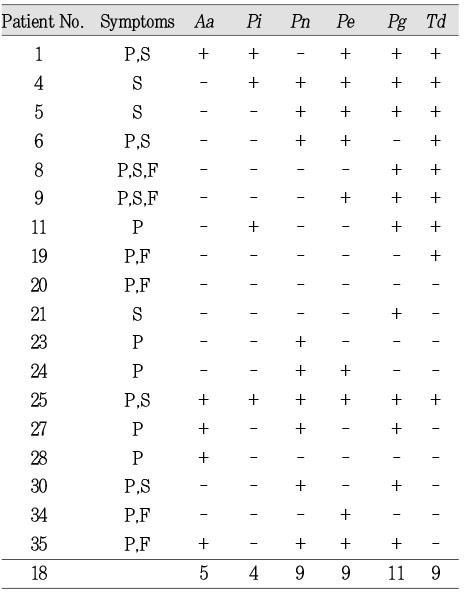
It has been documented that periodontopathic bacteria are also implicated in endodontic infections. 16S rDNA gene-directed PCR was to examine the prevalence of periodontopathic bacteria including Actinobacillus actinomycetemcomitans (Aa), Prevotella intermedia (Pi), Prevotella nigrescens (Pn), Porphyromonas gingivalis (Pg), Porphyromonas endodontalis (Pe), and Treponema denticola (Td) in the root canals of 36 endodontically infected teeth having apical lesions with or without clinical symptoms like pain, swelling, and fistula.
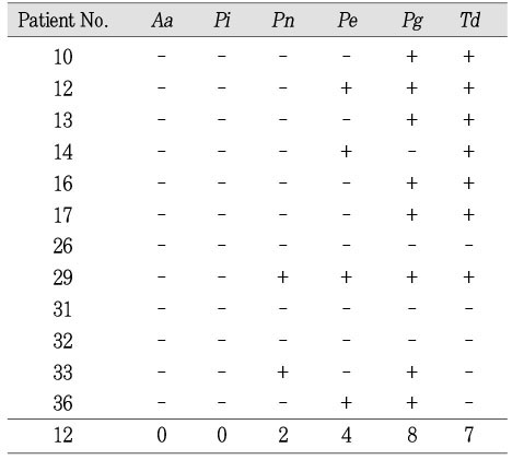
In 36 infected root canals, most frequently detected bacterial species was Pg (61.1%), followed by Td (52.8%) and Pe (38.9%).
Of 36 infected root canals, Aa was detected in 6 canals (16.7%) of the teeth, all of which showed clinical symptoms.
Of 36 infected root canals, Pi and Pn were found in 4 (13.9%) and 5 (33.3%), respectively. Notably, prevalence of Pn in the symptomatic teeth was 50.0%.
One of black-pigmented anaerobic bacteria (BPB) including Pi, Pn, Pe, and Pg was detected in all of the teeth that showed pain or especially swelling but not fistula. It was, however, found that prevalence of BPB in the asymptomatic teeth or the teeth with fistula was only 40%.
Pe and Pg were detected in the teeth regardless of the presence or absence of symptoms.
Td was detected in the teeth regardless of the presence or absence of symptoms.
High prevalence of BPB in the symptomatic teeth but low in the asymptomatic teeth suggests that BPB may play an important role in the pathogenesis of periapical lesions.
- 1. Miller WD. Microorganisms of the human mouth. 1890;Philadelphia: S.S. White Dental Co..
- 2. Dahlen G, Moller AJR. In: Slots J, Taubman MA, editors. Microbiology of endodontic infections. Contemporary oral microbiology and immunology. 1992;St. Louis: Mosby-Year Book, Inc.; 444-475.
- 3. Kettering JD, Torabinejad M. In: Cohen S, Burns RC, editors. Microbiology and immunology. Pathways of the pulp. 1994;St. Louis: Mosby-Year Book, Inc.; 363-376.
- 4. Sjogren U, et al. Influence of infection at the time of root filling on the outcome of endodontic treatment of teeth with apical periodontitis. Int Endod J. 1997;30: 297-306.PubMed
- 5. Haapasalo M. Black-pigmented Gram-negative anaerobes in endodontic infections. FEMS Immunol Med Microbiol. 1993;6: 213-218.ArticlePubMed
- 6. Sundqvist G. Bacteriological studies of necrotic dental pulps. Thesis. 1976;Umea University; Odontology, Dissertation No. 7.
- 7. Kobayashi T, Hayash A, Yoshikawa R, Okuda K, Hara K. The microbial flora from root canals and periodontal pockets of non-vital teeth associated with advanced periodontitis. Int Endod J. 1990;23: 100-106.ArticlePubMed
- 8. Van Winkelhoff AJ, Carlee AW, de Graaff J. Bacteroides endodontalis and other black-pigmented Bacteroides species in odontogenic abscesses. Infect Immun. 1985;49: 494-497.ArticlePubMedPMCPDF
- 9. Haapasalo M. Bacteroides spp. in dental root canal infections. Endod Dent Traumatol. 1989;5: 1-10.ArticlePubMed
- 10. Sundqvist G, Johansson E, Sjogren U. Prevalence of black-pigmented Bacteroides species in root canal infections. J Endod. 1989;15: 13-19.ArticlePubMed
- 11. Tanner A, Stillman N. Oral and dental infections with anaerobic bacteria: clinical features, predominant pathogens, and treatment. Clin Infect Dis. 1993;16: Suppl 4. S304-S309.ArticlePubMed
- 12. Baumgartner JC, Falkler WA Jr. Reactivity of IgG from explant cultures of periapical lesions with implicated microorganisms. J Endod. 1991;17: 207-212.ArticlePubMed
- 13. Tanner A, Stillman N. Oral and dental infections with anaerobic bacteria: clinical features, predominant pathogens and treatment. Clin Infect Dis. 1993;16: Suppl 4. S304-S309.ArticlePubMed
- 14. Haapasalo M, Ranta H, Ranta K, Shah H. Black-pigmented Bacteroides spp. in human apical periodontitis. Infect Immun. 1986;53: 149-153.ArticlePubMedPMCPDF
- 15. Chen H. The correlation of black-pigmented Bacteroides spp. to symptoms associated with apical periodontitis. Zhonghua Kou Qiang Yi Xue Za Zhi. 1991;26: 70-72.PubMed
- 16. Griffee MB, Patterson SS, Miller CH, Kafrawy AH, Newton CW. The relationship of Bacteroides melaninogenicus to symptoms associated with pulpal necrosis. Oral Surg Oral Med Oral Pathol. 1980;50: 457-461.ArticlePubMed
- 17. Yoshida M, Fukushima H, Yamamoto K, Ogawa K, Toda T, Sagawa H. Correlation between clinical symptoms and microorganisms isolated from root canals with periapical lesions. J Endod. 1987;13: 24-28.PubMed
- 18. Hahn CL, Falkler WA Jr, Minah GE. Correlation between thermal sensitivity and microorganisms isolated from deep carious dentin. J Endod. 1993;19: 26-30.ArticlePubMed
- 19. Hashioka K, Suzuki K, Yoshida T, Nakane A, Horiba N, Nakamura H. Relationship between clinical symptoms and enzyme-producing bacteria isolated from infected root canals. J Endod. 1994;20: 75-77.ArticlePubMed
- 20. Gomes BP, Drucker DB, Lilley JD. Associations of specific bacteria with some endodontic signs and symptoms. Int Endod J. 1994;27: 291-298.PubMed
- 21. Gomes BP, Lilley JD, Drucker DB. Clinical significance of dental root canal microflora. J Dent. 1996;24: 47-55.ArticlePubMed
- 22. Olsen GJ. Microbial ecology. Variation among the masses. Nature. 1990;345: 20.ArticlePubMedPDF
- 23. Ward DM, Weller R, Bateson MM. 16S rRNA sequences reveal numerous uncultured microorganisms in a natural community. Nature. 1990;345: 63-65.ArticlePubMedPDF
- 24. Amann RI, Ludwig W, Schleife KH. Phylogenetic identification and in situ detection of individual microbial cells without cultivation. Microbiol Rev. 1995;59: 143-169.ArticlePubMedPMCPDF
- 25. Siqueira JF Jr, Rocas IN, Souto R, de Uzeda M, Colombo AP. Checkerboard DNA-DNA hybridization analysis of endodontic infections. Oral Surg Oral Med Oral Pathol Oral Radiol Endod. 2000;89: 744-748.ArticlePubMed
- 26. Martin FE, Nadkarni MA, Jacques NA, Hunter N. Quantitative microbiological study of human carious dentine by culture and real-time PCR: association of anaerobes with histopathological changes in chronic pulpitis. J Clin Microbiol. 2002;40: 1698-1704.ArticlePubMedPMCPDF
- 27. Jung IY, Choi B, Kum KY, Yoo YJ, Yoon TC, Lee SJ, Lee CY. Identification of oral spirochetes at the species level and their association with other bacteria in endodontic infections. Oral Surg Oral Med Oral Pathol Oral Radiol Endod. 2001;92: 329-334.ArticlePubMed
- 28. Baumgartner JC, Watts CM, Xia T. Occurrence of Candida albicans in infections of endodontic origin. J Endod. 2000;26: 695-698.ArticlePubMed
- 29. Ashimoto A, Chen C, Bakker I, Slots J. Polymerase chain reaction detection of 8 putative periodontal pathogens in subgingival plaque of gingivitis and advanced periodontitis lesions. Oral Microbiol Immunol. 1996;11: 266-273.PubMed
- 30. Bogen G, Slots J. Black-pigmented anaerobic rods in closed periapical lesions. Int Endod J. 1999;32: 204-210.ArticlePubMedPDF
- 31. Conrads G, Gharbia SE, Gulabivala K, Lampert F, Shah HN. The use of a 16S rDNA directed PCR for the detection of endodontopathogenic bacteria. J Endod. 1997;23: 433-438.ArticlePubMed
- 32. Goncalves RB, Mouton C. Molecular detection of Bacteroides forsythus in infected root canals. J Endod. 1999;25: 336-340.ArticlePubMed
- 33. Siqueira JF Jr, Rocas IN, Favieri A, Santos KR. Detection of Treponema denticola in endodontic infections by 16S rRNA gene-directed polymerase chain reaction. Oral Microbiol Immunol. 2000;15: 335-337.ArticlePubMedPDF
- 34. Siqueira JF Jr, Rjcas IN, Oliveira JC, Santos KR. Detection of putative oral pathogens in acute periradicular abscesses by 16S rDNA-directed polymerase chain reaction. J Endod. 2001;27: 164-167.ArticlePubMed
- 35. Siqueira JF Jr, Rocas IN, Favieri A, Oliveira JC, Santos KR. Polymerase chain reaction detection of Treponema denticola in endodontic infections within root canals. Int Endod J. 2001;34: 280-284.ArticlePubMedPDF
- 36. Rupf S, Kannengiesser S, Merte K, Pfister W, Sigusch B, Eschrich K. Comparison of profiles of key periodontal pathogens in periodontium and endodontium. Endod Dent Traumatol. 2000;16: 269-275.ArticlePubMed
- 37. Socransky SS, Jaffajee AD, Cugini MA, Smith C, Kent RL Jr. Microbial complexes in subgingival plaque. J Clin Periodontol. 1998;25: 134-144.ArticlePubMed
- 38. Rocas IN, Siqueira JF Jr, Santos KR, Coelho AM. "Red complex" (Bacteroides forsythus, Porphyromonas gingivalis, and Treponema denticola) in endodontic infections: a molecular approach. Oral Surg Oral Med Oral Pathol Oral Radiol Endod. 2001;91: 468-471.ArticlePubMed
- 39. Sunde PT, Tronstad L, Eribe ER, Lind PO, Olsen I. Assessment of periradicular microbiota by DNA-DNA hybridization. Endod Dent Traumatol. 2000;16: 191-196.ArticlePubMed
- 40. Jansson L, Ehnevid H, Blomlof L, Weintraub A, Lindskog S. Endodontic pathogens in periodontal disease augmentation. J Clin Periodontol. 1995;22: 598-602.ArticlePubMed
- 41. Jansson L, Ehnevid H, Lindskog S, Blomlof L. The influence of endodontic infection on progression of marginal bone loss in periodontitis. J Clin Periodontol. 1995;22: 729-734.ArticlePubMed
- 42. Baumgartner JC, Watkins BJ, Bae KS, Xia T. Association of black-pigmented bacteria with endodontic infections. J Endod. 1999;25: 413-415.ArticlePubMed
- 43. Fouad AF, Barry J, Caimano M, Clawson M, Zhu Q, Carver R, Hazlett K, Radolf JD. PCR-based identification of bacteria associated with endodontic infections. J Clin Microbiol. 2002;40: 3223-3231.ArticlePubMedPMCPDF
- 44. Siqueira JF Jr, Rjcas IN, Oliveira JC, Santos KR. Molecular detection of black-pigmented bacteria in infections of endodontic origin. J Endod. 2001;27: 563-566.ArticlePubMed
- 45. Machado de Oliveira JC, Siqueira JF Jr, Alves GB, Hirata R Jr, Andrade AF. Detection of Porphyromonas endodontalis in infected root canals by 16S rRNA gene-directed polymerase chain reaction. J Endod. 2000;26: 729-732.ArticlePubMed
- 46. Slots J. Selective medium for isolation of Actinobacillus actinomycetemcomitans. J Clin Microbiol. 1982;15: 606-609.ArticlePubMedPMCPDF
- 47. Grenier D. Demonstration of a bimodal coaggregation reaction between Porphyromonas gingivalis and Treponema denticola. Oral Microbiol Immunol. 1992;7: 280-284.ArticlePubMed
- 48. Grenier D. Nutritional interactions between two suspected periodontopathogens, Treponema denticola and Porphyromonas gingivalis. Infect Immun. 1992;60: 5298-5301.ArticlePubMedPMCPDF
- 49. Simonson LG, McMahon KT, Childers DW, Morton HE. Bacterial synergy of Treponema denticola and Porphyromonas gingivalis in a multinational population. Oral Microbiol Immunol. 1992;7: 111-112.ArticlePubMed
- 50. Loesche WJ, Bretz WA, Kerschensteiner D, Stoll J, Socransky SS, Hujoel P, Lopatin DE. Development of a diagnostic test for anaerobic periodontal infections based on plaque hydrolysis of benzoyl-DL-arginine-naphthylamide. J Clin Microbiol. 1990;28: 1551-1559.ArticlePubMedPMCPDF
- 51. Kesavalu L, Holt SC, Ebersole JL. Virulence of a polymicrobic complex, Treponema denticola and Porphyromonas gingivalis, in a murine model. Oral Microbiol Immunol. 1998;13: 373-373.ArticlePubMed
- 52. Loesche WJ, Grossman NS. Periodontal disease as a specific, albeit chronic, infection: diagnosis and treatment. Clin Microbiol Rev. 2001;14: 727-752.ArticlePubMedPMCPDF
REFERENCES
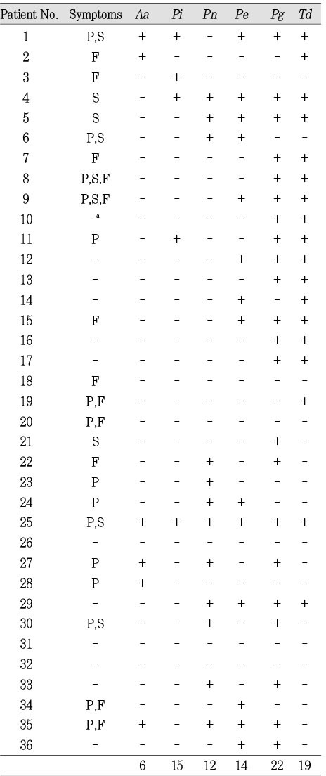
Abbreviations: P, Pain; S, Swelling; AA, alveolar abscess; F, fistula; Aa, A. actinomycetemcomitans; Pi, Pr. intermedia; Pn, Pr. nigrescens; Pe, P. endodontalis ; Pg, P. gingivalis ; Td, T. denticola ; a:-denotes no symptom
Tables & Figures
REFERENCES
Citations

- Isolation of Propionibacterium acnes among the microbiota of primary endodontic infections with and without intraoral communication
Sadia Ambreen Niazi, Hana Suleiman Al Kharusi, Shanon Patel, Kenneth Bruce, David Beighton, Federico Foschi, Francesco Mannocci
Clinical Oral Investigations.2016; 20(8): 2149. CrossRef - Antimicrobial Activity of Isothiocyanates (ITCs) Extracted from Horseradish (Armoracia rusticana) Root against Oral Microorganisms
HO-WON PARK, KYU-DUCK CHOI, IL-SHIK SHIN
Biocontrol Science.2013; 18(3): 163. CrossRef - Microbial profile of asymptomatic and symptomatic teeth with primary endodontic infections by pyrosequencing
Sang-Min Lim, Tae-Kwon Lee, Eun-Jeong Kim, Jun-Hong Park, Yoon Lee, Kwang-Shik Bae, Kee-Yeon Kum
Journal of Korean Academy of Conservative Dentistry.2011; 36(6): 498. CrossRef

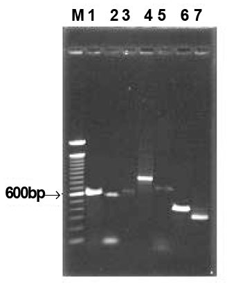
Fig. 1
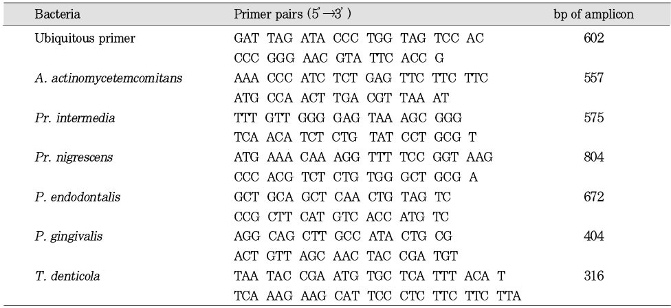
PCR primer pairs used in this study for detection of putative endodontopathogens
Distribution of bacteria in infection with apical lesions.
Abbreviations: P, Pain; S, Swelling; AA, alveolar abscess; F, fistula; Aa, A. actinomycetemcomitans; Pi, Pr. intermedia; Pn, Pr. nigrescens; Pe, P. endodontalis ; Pg, P. gingivalis ; Td, T. denticola ; a:-denotes no symptom
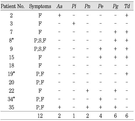
Distribution of bacteria in fistula*; Samples were obtained through the opening of the fistula, whereas the others from the infected the root canals.
Distribution of bacteria in necrotic root canal sample of apical lesions, having symptoms of pain and/or swelling
Distribution of bacteria in necrotic root canal samples of apical lesions without clinical symptoms
Abbreviations: P, Pain; S, Swelling; AA, alveolar abscess; F, fistula; Aa, A. actinomycetemcomitans; Pi, Pr. intermedia; Pn, Pr. nigrescens; Pe, P. endodontalis ; Pg, P. gingivalis ; Td, T. denticola ; a:-denotes no symptom

 KACD
KACD
 ePub Link
 Cite
Cite

