
Articles
- Page Path
- HOME > Restor Dent Endod > Volume 38(1); 2013 > Article
- Research Article Comparison of the centering ability of Wave·One and Reciproc nickel-titanium instruments in simulated curved canals
- Young-Jun Lim1, Su-Jung Park1, Hyeon-Cheol Kim2, Kyung-San Min3
-
2013;38(1):-25.
DOI: https://doi.org/10.5395/rde.2013.38.1.21
Published online: February 26, 2013
1Department of Conservative Dentistry, Wonkwang University School of Dentistry, Iksan, Korea.
2Department of Conservative Dentistry, Pusan National University School of Dentistry, Yangsan, Korea.
3Department of Conservative Dentistry, Chonbuk National University School of Dentistry, Jeonju, Korea.
- Correspondence to Kyung-San Min, DDS, PhD. Associate Professor, Department of Conservative Dentistry, Chonbuk National University School of Dentistry, 567 Baekjedae-ro, Deokjin-gu, Jeonju, Korea 561-756. TEL, +82-63-250-2219; FAX, +82-63-270-4004; endomin@gmail.com
• Received: October 6, 2012 • Revised: October 22, 2012 • Accepted: November 4, 2012
©Copyights 2013. The Korean Academy of Conservative Dentistry.
This is an Open Access article distributed under the terms of the Creative Commons Attribution Non-Commercial License (http://creativecommons.org/licenses/by-nc/3.0/) which permits unrestricted non-commercial use, distribution, and reproduction in any medium, provided the original work is properly cited.
- 2,359 Views
- 9 Download
- 29 Crossref
Abstract
-
Objectives The aim of this study was to evaluate the shaping ability of newly marketed single-file instruments, Wave·One (Dentsply-Maillefer) and Reciproc (VDW GmbH), in terms of maintaining the original root canal configuration and curvature, with or without a glide-path.
-
Materials and Methods According to the instruments used, the blocks were divided into 4 groups (n = 10): Group 1, no glide-path / Wave·One; Group 2, no glide-path / Reciproc; Group 3, #15 K-file / Wave·One; Group 4, #15 K-file / Reciproc. Pre- and post-instrumented images were scanned and the canal deviation was assessed. The cyclic fatigue stress was loaded to examine the cross-sectional shape of the fractured surface. The broken fragments were evaluated under the scanning electron microscope (SEM) for topographic features of the cross-section. Statistically analysis of the data was performed using one-way analysis of variance followed by Tukey's test (α = 0.05).
-
Results The ability of instruments to remain centered in prepared canals at 1 and 2 mm levels was significantly lower in Group 1 (p < 0.05). The centering ratio at 3, 5, and 7 mm level were not significantly different.
-
Conclusions The Wave·One file should be used following establishment of a glide-path larger than #15.
Introduction
The ultimate goal of root canal preparation is to clean and shape the root canal system while maintaining the original configuration. Over the years, many nickel-titanium (Ni-Ti) instruments have been developed to improve root canal preparation. They are available in various designs that differ in tip and taper design, rake angles, helical angles, pitch, and presence of radial lands.
Two brands of Ni-Ti instruments adopting the single-file system were recently introduced to the market and advocated the reciprocation concept: Wave·One (Dentsply-Maillefer, Ballaigues, Switzerland) and Reciproc (VDW GmbH, Munich, Germany). These files are made of a special Ni-Ti alloy called M-wire that is created by an innovative thermal-treatment process.1 This procedure has been developed using superelastic Ni-Ti wire blanks that contain substantial stable martensite under clinical conditions. The benefits of M-wire are increased flexibility of the instruments and resistance to cyclic fatigue.2
These files have a different mechanism of instrumentation compared to other previously developed files. The system is designed to be used with a dedicated reciprocating motion. The values of clockwise and counterclockwise rotations are different. A large rotating angle in the cutting direction (counter-clockwise) determines that the instrument advances in the canal and engages dentin to cut it, whereas a smaller angle in the opposite direction (clockwise) allows the file to be immediately disengaged and safely progress along the canal path, while reducing the screwing effect and file separation.3
In clinical practice, these Ni-Ti instruments carry a risk of fracture mainly because of flexural and torsional stresses.2,4,5 This risk may be reduced by performing coronal enlargement and preflaring manually to create a glide-path before using Ni-Ti instruments.3,6-8 However, the manufacturer of Reciproc instruments does not strictly recommend creating a glide-path when using the reciprocating instrumentation. In contrast, a glide-path of at least size 10 is recommended in the manufacturer's instructions for the use of Wave·One instruments. The glide-path is a smooth radicular tunnel from the canal orifice to the physiologic terminus.9 Blum and colleagues suggested creating a glide-path using small flexible stainless steel hand files to create or verify that within any portion of a root canal there is sufficient space for rotary instruments to follow.10 In general, a glide-path was prepared using #15 K-file to ensure sufficient space for the file to work and to avoid the risk of locking.
There are only limited studies available concerning the centering ability and preparation time of these recently introduced instruments by using reciprocating motion. Therefore, a comparison of these single-file systems with or without glide-path is necessary to assess the properties of these new files. The aim of this study was to evaluate the shaping ability of the newly marketed single-file instruments in terms of maintaining the original root canal configuration and curvature, with or without a glide-path.
Materials and Methods
Forty simulated curved root canals in clear resin blocks (Dentsply-Maillefer) were used for this study. An apical foramen size of 0.1 mm was confirmed, and each canal had a mean canal length of 17 mm. Each simulated canal was colored with red ink injected using a syringe. The blocks were divided into 4 groups according to the instruments used: Group 1, no glide-path / Wave·One (NW); Group 2, no glide-path / Reciproc (NR); Group 3, #15 K-file / Wave·One (KW); Group 4, #15 K-file / Reciproc (KR). For Groups 1 and 2, a glide-path was not established. For Groups 3 and 4, a #15 hand K-file was used after a #10 hand K-file had been used.
The Reciproc R25 instrument and Wave·One Primary file, both of which had a tip size of 0.25 mm and a 08 taper in the apical 3 mm, were selected. The files were operated with the Silver Reciproc motor (VDW GmbH) with their respective recommended settings: Reciproc with the "RECIPROC ALL" mode and Wave·One with the "WAVEONE ALL" mode. Reciproc and Wave·One were used in a reciprocating, slow in-and-out pecking motions. The flutes of the instruments were cleaned using gauze soaked with 70% ethyl alcohol after 3 in-and-out movements. Each instrument was discarded after use in 2 canals.
Pre- and post-instrumentation images were scanned and recorded. The images were superimposed using a computer software program (Photoshop 7.0, Adobe, San Jose, CA, USA). The ability of the instruments to remain centered in the canal was determined by calculating a centering ratio using perpendicular lines made by the canal axes at 1, 2, 3, 5, and 7 mm (Figure 1a). The centering ratio was calculated using the formula (X1-X2)/Y, where X1 represents the maximum extent of canal movement in one direction, X2 is the movement in the opposite direction, and Y is the diameter of the final canal preparation (Figure 1b). The data were analyzed using the SPSS program (version 10.0, SPSS GmbH, Munich, Germany). Changes in canal curvature and centering ratios at the 5 measuring points were statistically analyzed using one-way analysis of variance (ANOVA; α = 0.05) followed by Tukey's test.
The cyclic fatigue stress was loaded to examine the cross-sectional shape of fractured surface. In brief, an artificial canal block made of tempered steel with 0.6 mm apical diameter, 6.06 mm radius, and 45° angle of curvature, measured according to the method of Schneider, was incorporated into the blocks.11 A continuous up-and-down (4 mm in each direction at 0.5 second) pecking movement was incorporated to simulate the pecking motion in a real clinical situation. The files were operated in the VDW.SILVER motor (VDW) with each recommended setting: Reciproc files with the "RECIPROC ALL" mode and WaveOne with the "WAVEONE ALL" mode. Then, the broken fragments were evaluated under the SEM (S-4800 II; Hitachi High Technologies, Pleasanton, CA, USA) for topographic features of the fracture surfaces at various magnifications (180 - 200 times).
Results
As shown in Figure 2, at the 1 and 2 mm levels, the mean centering ratio was statistically significantly higher in Group 1 (p < 0.05). The centering ratio at the 3, 5, and 7 mm levels showed no statistically significant difference (p > 0.05).
The two reciprocating file systems used in this study have different cross sections, S-shaped and concave triangular shape for Reciproc R25, Wave·One, respectively (Figure 3).
Discussion
Recently, new systems that use reciprocating motion were introduced to the market, claiming to be able to shape root canals using a single file. These file systems make canal shaping simpler and faster. Reciprocation motion was proposed to increase the canal centering ability as well as to reduce the risk of root canal deformity.12-14 However, there have been little information about the centering ability of these files systems in curved root canals.
To assess the instrumentation of curved canals, clear resin blocks were used in this study. These were chosen because the shape, size, taper, and curvature of the experimental canals were standardized. The credibility of resin blocks as an ideal experimental model for the analysis of endodontic preparation and preparation techniques has been validated by Weine et al. and Dummer et al.15,16 However, there are limitations with the model, such as the different hardness between resin and dentin, and care should be exercised in the extrapolation of the present results to the use of these instruments in the clinical setting. A major drawback of using rotary instruments in resin blocks is the heat generated, which might soften the resin material and lead to binding of the cutting blade and separation of the instrument.17-19 Nevertheless, the use of simulated canals in resin blocks allows for the standardization of the research method and to exclude parameters that could influence the preparation outcome.
Proper shaping of the canal to create a continuously tapered funnel form is one of the most important objectives for root canal preparation. It facilitates irrigation and obturation of root canals.20 However, during preparation, some root canal aberrations are created, such as transportation, elbow, and apical zip. It has been shown that root canal instrumentation leads to changes in the working length by straightening of the curved canal during the course of the treatment.21 These aberrant results of root canal shaping make it difficult for clinicians to remove the infected tissue and properly obturate the root canal.22
The centering ratio can define the ability of instruments to remain centered in shaped canals. According to the formula, the centering ratio approaches zero as X1 and X2 become closer to the center. The lower the scores, the better are the instruments centered in the canal. In this study, the results of the assessment of the centering ratio in the 4 groups at 1 and 2 mm levels indicated that the ability of the instruments to remain centered in prepared canals was significantly lower in the no glide-path / Wave·One group. The tip size (diameter at D0) of Reciproc R25 and Wave·One primary were the same with each other. The two reciprocating file systems are made of the same alloy (M-wire) but have different cross sections, S-shaped and concave triangular shape for Reciproc R25 and Wave·One, respectively (Figure 3). The larger canal abberation achieved for the no glide-path/Wave·One group at 1 and 2 mm level might be due to the larger core diameter and greater number of spiraling flutes of Wave·One. The larger core diameter and greater number of spiraling flutes of the Wave·One instrument increases the stiffness of the tip, which results in more canal abberation.
Conclusions
Conclusively, the ability of the instruments to remain centered in the prepared canals at the 1 and 2 mm levels was significantly lower in Group 1 (no glide-path / Wave·One). However, the centering ratio at the 5 and 7 mm levels were not significantly different. The current study shows that both of the instrumentation systems possess an adequate centering ability. However, Wave·One should be used following establishment of a glide-path larger than #15.
- 1. Johnson E, Lloyd A, Kuttler S, Namerow K. Comparison between a novel nickel-titanium alloy and 508 nitinol on the cyclic fatigue life of ProFile 25/.04 rotary instruments. J Endod 2008;34:1406-1409.ArticlePubMed
- 2. Shen Y, Cheung GS, Bian Z, Peng B. Comparison of defects in ProFile and ProTaper systems after clinical use. J Endod 2006;32:61-65.ArticlePubMed
- 3. Plotino G, Grande NM, Testarelli L, Gambarini G. Cyclic fatigue of Reciproc and WaveOne reciprocating instruments. Int Endod J 2012;45:614-618.ArticlePubMed
- 4. Sattapan B, Nervo GJ, Palamara JE, Messer HH. Defects in rotary nickel-titanium files after clinical use. J Endod 2000;26:161-165.ArticlePubMed
- 5. Cheung GS, Peng B, Bian Z, Shen Y, Darvell BW. Defects in ProTaper S1 instruments after clinical use: fractographic examination. Int Endod J 2005;38:802-809.ArticlePubMed
- 6. Roland DD, Andelin WE, Browning DF, Hsu GH, Torabinejad M. The effect of preflaring on the rates of separation for 0.04 taper nickel titanium rotary instruments. J Endod 2002;28:543-545.ArticlePubMed
- 7. Peters OA, Peters CI, Schönenberger K, Barbakow F. ProTaper rotary root canal preparation: effects of canal anatomy on final shape analysed by micro CT. Int Endod J 2003;36:86-92.ArticlePubMedPDF
- 8. Berutti E, Negro AR, Lendini M, Pasqualini D. Influence of manual preflaring and torque on the failure rate of ProTaper rotary instruments. J Endod 2004;30:228-230.ArticlePubMed
- 9. West JD. The endodontic Glidepath: "Secret to rotary safety". Dent Today 2010;29:86-93.
- 10. Blum JY, Machtou P, Ruddle C, Micallef JP. Analysis of mechanical preparations in extracted teeth using ProTaper rotary instruments: value of the safety quotient. J Endod 2003;29:567-575.ArticlePubMed
- 11. Schneider SW. A comparison of canal preparations in straight and curved root canals. Oral Surg Oral Med Oral Pathol 1971;32:271-275.ArticlePubMed
- 12. Roane JB, Sabala CL, Duncanson MG Jr. The 'balanced force' concept for instrumentation of curved canals. J Endod 1985;11:203-211.ArticlePubMed
- 13. Roane JB, Sabala C. Clockwise or counterclockwise. J Endod 1984;10:349-353.ArticlePubMed
- 14. Southard DW, Oswald RJ, Natkin E. Instrumentation of curved molar root canals with the Roane technique. J Endod 1987;13:479-489.ArticlePubMed
- 15. Weine FS, Kelly RF, Lio PJ. The effect of preparation procedures on original canal shape and on apical foramen shape. J Endod 1975;1:255-262.ArticlePubMed
- 16. Dummer PM, Alodeh MH, al-Omari MA. A method for the construction of simulated root canals in clear resin blocks. Int Endod J 1991;24:63-66.ArticlePubMed
- 17. Kum KY, Spängberg L, Cha BY, Jung IY, Lee SJ, Lee CY. Shaping ability of three ProFile rotary instrumentation techniques in simulated resin root canals. J Endod 2000;26:719-723.ArticlePubMed
- 18. Thomson SA, Dummer PM. Shaping ability of ProFile.04 Taper Series 29 rotary nickel-titanium instruments in simulated root canals. Part 1. Int Endod J 1997;30:1-7.ArticlePubMed
- 19. Baumann MA, Roth A. Effect of experience on quality of canal preparation with rotary nickel-titanium files. Oral Surg Oral Med Oral Pathol Oral Radiol Endod 1999;88:714-718.ArticlePubMed
- 20. Schilder H. Cleaning and shaping the root canal. Dent Clin North Am 1974;18:269-296.ArticlePubMed
- 21. Davis RD, Marshall JG, Baumgartner JC. Effect of early coronal flaring on working length change in curved canals using rotary nickel-titanium versus stainless steel instruments. J Endod 2002;28:438-442.ArticlePubMed
- 22. Wu MK, Fan B, Wesselink PR. Leakage along apical root fillings in curved root canals. Part I: effects of apical transportation on seal of root fillings. J Endod 2000;26:210-216.ArticlePubMed
REFERENCES
Figure 1

(a) The picture indicates the points at which the canal width was measured after superimposition of pre- and post-operative images; (b) X1 represents the maximum extent of canal movement in one direction and X2 is the movement in the opposite direction. Y is the diameter of the final canal preparation.


Tables & Figures
REFERENCES
Citations
Citations to this article as recorded by 
- Centering Ability and Canal Transportation of Nickel-Titanium (NiTi) Single-File Systems With and Without Glide Path in Extracted Natural Teeth: A Systematic Review and Meta-Analysis
Indumathi Manoharan, Deblina Basu, Mathan Rajan
Cureus.2026;[Epub] CrossRef - Evaluation of the Centering Ability and Canal Transportation of Rotary File Systems in Different Kinematics Using CBCT
Nupur R Vasava, Shreya H Modi, Chintan Joshi, Mona C Somani, Sweety J Thumar, Aashray A Patel, Anisha D Parmar, Kruti M Jadawala
World Journal of Dentistry.2024; 14(11): 983. CrossRef - Comparative Evaluation of Root Canal Centering Ability of ProTaper, Mtwo, WaveOne, and Reciproc Using Cone-beam Computed Tomography: In Vitro Study
M Remya, Asha Joseph, Prabath Singh, Anju Varughese, Pallavi Chandran, Deepthy Subramanian, S Vijay Kumar
The Journal of Contemporary Dental Practice.2022; 23(6): 589. CrossRef - Shaping ability of ProTaper Gold and WaveOne Gold nickel-titanium rotary instruments in simulated S-shaped root canals
Lu Shi, Junling Zhou, Jie Wan, Yunfei Yang
Journal of Dental Sciences.2022; 17(1): 430. CrossRef - Comparison of canal transportation and centering ability of manual K-files and reciprocating files in glide path preparation: a micro-computed tomography study of constricted canals
Jing-Yi Liu, Zhi-Xiong Zhou, Wei-Ju Tseng, Bekir Karabucak
BMC Oral Health.2021;[Epub] CrossRef - Canal transportation and centering ability of root canals prepared using rotary and reciprocating systems with and without PathFiles in cone-beam computed tomography-based three-dimensional molar prototypes
MSruthi Sunildath, Josey Mathew, Liza George, RV Vineet, Priya Thomas, Dhanya John
Journal of Conservative Dentistry.2021; 24(3): 246. CrossRef - Shaping Ability of Reciproc R25 File and Mtwo System Used in Continuous and Reciprocating Motion
Vincenzo Campanella, Leonardo Gianni, Antonio Libonati, Gianni Gallusi
The Journal of Contemporary Dental Practice.2020; 21(2): 171. CrossRef - Canal shaping with a reciprocating system is easy to learn
E. Muñoz, L. Forner, S. Garcet, F. J. Rodríguez‐Lozano, C. Llena
International Endodontic Journal.2019; 52(8): 1244. CrossRef - Shaping Ability of HyFlex EDM and ProTaper Next Rotary Instruments in Curved Root Canals: A Micro-CT Study
Ahmed K Turkistani, Madiha M Gomaa, Lubna A Shafei, Loai Alsofi, Abdul Majeed, Emad AlShwaimi
The Journal of Contemporary Dental Practice.2019; 20(6): 680. CrossRef - Root canal volume change and transportation by Vortex Blue, ProTaper Next, and ProTaper Universal in curved root canals
Hyun-Jin Park, Min-Seock Seo, Young-Mi Moon
Restorative Dentistry & Endodontics.2018;[Epub] CrossRef - Comparison of shaping ability of ProTaper Next and 2Shape nickel–titanium files in simulated severe curved canals
Simone Staffoli, Taha Özyürek, Avi Hadad, Alex Lvovsky, Michael Solomonov, Hadas Azizi, Joe Ben Itzhak, Maurizo Bossù, Nicola M. Grande, Gianluca Plotino, Antonella Polimeni
Giornale Italiano di Endodonzia.2018; 32(2): 52. CrossRef - Comparing the Centering Ability of Different Pathfinding Systems and Their Effect on Final Instrumentation by Hyflex CM
Lu Shi, Shova Wagle
Journal of Endodontics.2017; 43(11): 1868. CrossRef - Rotary endodontics in primary teeth – A review
Sageena George, S. Anandaraj, Jyoti S. Issac, Sheen A. John, Anoop Harris
The Saudi Dental Journal.2016; 28(1): 12. CrossRef - Performance of Three Single Instrument Systems in the Preparation of Long Oval Canals
Beatriz Serrato Coelho, Rodrigo Otavio Jatahy Ferreira do Amaral, Denise Piotto Leonardi, Bruno Marques-da-Silva, Yara Teresinha Corrêa Silva-Sousa, Fredson Marcio Acris de Carvalho, Flares Baratto-Filho
Brazilian Dental Journal.2016; 27(2): 217. CrossRef - Quantitative transportation assessment in curved canals prepared with an off-centered rectangular design system
Emmanuel João Nogueira Leal SILVA, Vania Cristina Gomes VIEIRA, Michele Dias Nunes TAMEIRÃO, Felipe Gonçalves BELLADONNA, Aline de Almeida NEVES, Erick Miranda SOUZA, Gustavo DE-DEUS
Brazilian Oral Research.2016;[Epub] CrossRef - Influence of Cervical and Apical Enlargement Associated with the WaveOne System on the Transportation and Centralization of Endodontic Preparations
Rodrigo Otavio Jatahy Ferreira do Amaral, Denise Piotto Leonardi, Marilisa Carneiro Leão Gabardo, Beatriz Serrato Coelho, Kauhanna Vianna de Oliveira, Flares Baratto Filho
Journal of Endodontics.2016; 42(4): 626. CrossRef - Comparison of canal transportation in simulated curved canals prepared with ProTaper Universal and ProTaper Gold systems
Emmanuel João Nogueira Leal Silva, Brenda Leite Muniz, Frederico Pires, Felipe Gonçalves Belladonna, Aline Almeida Neves, Erick Miranda Souza, Gustavo De-Deus
Restorative Dentistry & Endodontics.2016; 41(1): 1. CrossRef - The Influence of Brushing Motion on the Cutting Behavior of 3 Reciprocating Files in Oval-shaped Canals
Shereen Alattar, Walid Nehme, Franck Diemer, Alfred Naaman
Journal of Endodontics.2015; 41(5): 703. CrossRef - Cyclic fatigue of instruments for endodontic glide path
Gianluca Gambarini, Gianluca Plotino, GianPaolo Sannino, Nicola Maria Grande, Alessio Giansiracusa, Lucila Piasecki, Ulisses Xavier da Silva Neto, Dina Al-Sudani, Luca Testarelli
Odontology.2015; 103(1): 56. CrossRef - Influence of the glide path on various parameters of root canal prepared with WaveOne reciprocating file using cone beam computed tomography
Anil Dhingra, Nidhi Nagar, Vipul Sapra
Dental Research Journal.2015; 12(6): 534. CrossRef - Apical Transportation, Centering Ability, and Cleaning Effectiveness of Reciprocating Single-file System Associated with Different Glide Path Techniques
Guilherme Moreira de Carvalho, Emílio Carlos Sponchiado Junior, Angela Delfina Bittencourt Garrido, Raphael Carlos Comelli Lia, Lucas da Fonseca Roberti Garcia, André Augusto Franco Marques
Journal of Endodontics.2015; 41(12): 2045. CrossRef - Influence of cervical preflaring on apical transportation in curved root canals instrumented by reciprocating file systems
Neisiana Barbieri, Denise Piotto Leonardi, Marina Samara Baechtold, Gisele Maria Correr, Marilisa Carneiro Leão Gabardo, João César Zielak, Flares Baratto-Filho
BMC Oral Health.2015;[Epub] CrossRef - Current Assessment of Reciprocation in Endodontic Preparation: A Comprehensive Review—Part II: Properties and Effectiveness
Gianluca Plotino, Hany Mohamed Aly Ahmed, Nicola Maria Grande, Stephen Cohen, Frédéric Bukiet
Journal of Endodontics.2015; 41(12): 1939. CrossRef - Influence of flexion angle of files on the decentralization of oval canals during instrumentation
Maria Antonieta Veloso Carvalho de OLIVEIRA, Letícia Duarte ALVES, Analice Giovani PEREIRA, Luís Henrique Araújo RAPOSO, João Carlos Gabrielli BIFFI
Brazilian Oral Research.2015; 29(1): 1. CrossRef - Quantitative Transportation Assessment in Simulated Curved Canals Prepared with an Adaptive Movement System
Emmanuel João Nogueira Leal Silva, Michele Dias Nunes Tameirão, Felipe Gonçalves Belladonna, Aline Almeida Neves, Erick Miranda Souza, Gustavo De-Deus
Journal of Endodontics.2015; 41(7): 1125. CrossRef - Efficacy of reciprocating and rotary systems for removing root filling material: A micro-computed tomography study
D. Helvacioglu-Yigit, A. Yilmaz, G. Kiziltas-Sendur, O. S. Aslan, P. V. Abbott
Scanning.2014; 36(6): 576. CrossRef - Effect of passive ultrasonic agitation during final irrigation on cleaning capacity of hybrid instrumentation
Marcilene Coelho Vinhorte, Eduardo Hideki Suzuki, Maíra Sousa de Carvalho, André Augusto Franco Marques, Emílio Carlos Sponchiado Júnior, Lucas da Fonseca Roberti Garcia
Restorative Dentistry & Endodontics.2014; 39(2): 104. CrossRef - Performance of RaCe Instrumentation System in Curved Root Canals: A Comprehensive Analysis by Three Study Methods
Denise Piotto Leonardi, Gilson Blitzkow Sydney, Mario Tanomaru Filho, Flares Baratto-Filho, Samantha Schaffer Pugsley Baratto, Paulo Sergio Cerri
Brazilian Dental Journal.2013; 24(3): 230. CrossRef - Endodontic treatment of mandibular molar with root dilaceration using Reciproc single-file system
Daniely Amorin Meireles, Mariana Mena Barreto Bastos, André Augusto Franco Marques, Lucas da Fonseca Roberti Garcia, Emílio Carlos Sponchiado
Restorative Dentistry & Endodontics.2013; 38(3): 167. CrossRef
 ePub Link
 Cite
CiteComparison of the centering ability of Wave·One and Reciproc nickel-titanium instruments in simulated curved canals
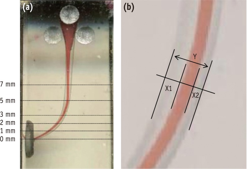
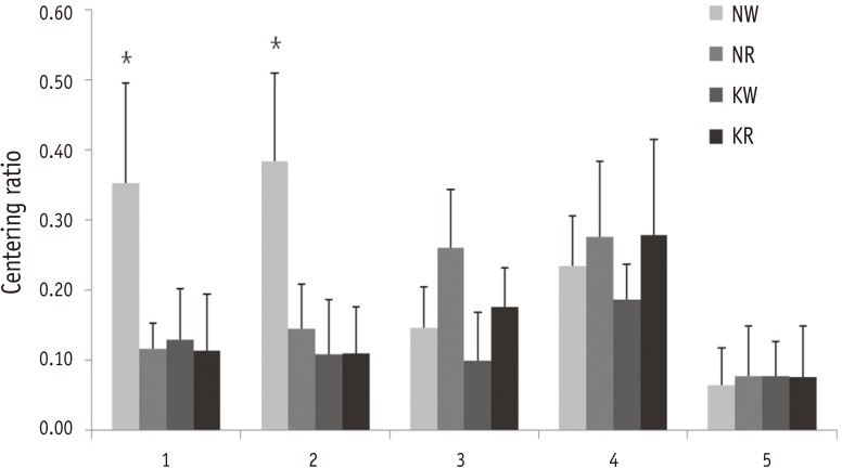
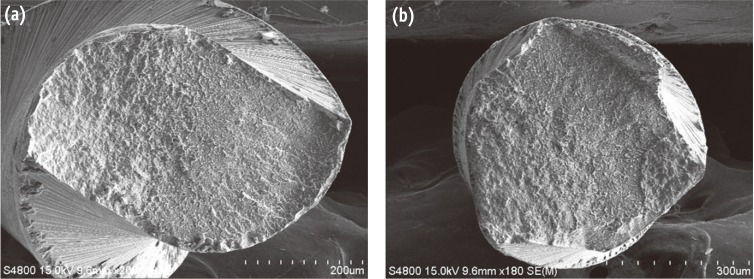
Figure 1 (a) The picture indicates the points at which the canal width was measured after superimposition of pre- and post-operative images; (b) X1 represents the maximum extent of canal movement in one direction and X2 is the movement in the opposite direction. Y is the diameter of the final canal preparation.
Figure 2 Centering ratio of canals at different apical levels. Values are mean ± SD. *A significant difference was determined at p < 0.05. NW, no glide-path / Wave·One; NR, no glide-path / Reciproc; KW, glidepath with K-file / Wave·One; KR, glidepath with K-file / Reciproc.
Figure 3 Scanning electron micrographs of fracture surface of separated fragments. (a) Reciproc (×200); (b) Wave·One (×180).
Figure 1
Figure 2
Figure 3
Comparison of the centering ability of Wave·One and Reciproc nickel-titanium instruments in simulated curved canals

