Articles
- Page Path
- HOME > Restor Dent Endod > Volume 30(1); 2005 > Article
-
Original Article
Bacteriologic
in vitro coronal leakage study of before and after post space preparation - Hyo-An Lee, Eui-Seong Kim
-
2005;30(1):-21.
DOI: https://doi.org/10.5395/JKACD.2005.30.1.016
Published online: January 31, 2005
Department of Conservative Dentistry, College of Dentistry, Yonsei University, Korea.
- Corresponding author: Eui-Seong Kim. Department of Conservative Dentistry, College of Dentistry, Yonsei University, 134 Shinchon-Dong, Seodaemoon Gu, Seoul, 120-752, Korea. Tel: 82-2-361-8700, Fax: 82-2-313-7575, andyendo@yumc.yonsei.ac.kr
• Received: July 16, 2004 • Revised: November 12, 2004 • Accepted: November 23, 2004
Copyright © 2005 Korean Academy of Conservative Dentistry
- 725 Views
- 1 Download
Abstract
-
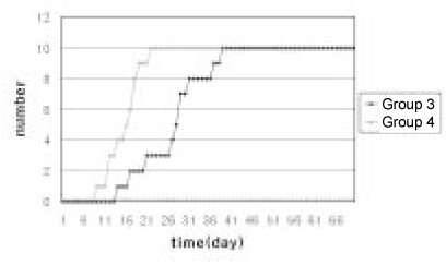
The purpose of present study was to compare the speed of coronal leakage before and after post space preparation using Streptococcus mutans.Forty straight extracted human teeth were selected. The crowns were removed to a uniform remaining root length 14 mm. Canals were enlarged by 06 taper Profiles® to a size #40 as a master apical file. And these were filled with gutta percha point and Tubuliseal® sealer, using continuous wave technique. Groupings are as follows.Group 1 - These teeth were obturated without sealer.Group 2 - These teeth were obturated and covered the surface of the root completely with sticky wax.Group 3 - These teeth were obturated.Group 4 - These teeth were obturated and prepared for post space remaining 5 mm of gutta percha.The teeth were suspended in plastic tubes. The upper chamber received the bacterial suspension everyday to simulate clinical situation. The lower chamber consisted of BHI added Andrade's indicator.All roots in the positive control group (Group 1) turned yellow within 24 h and those of negative control group (Group 2) remained red throughout the experimental period (70 days). The samples of group 3 were contaminated within an average of 27.2 days. The samples of group 4 were contaminated within an average of 15.7 days, ranging from 9 to 22 days.There was significant difference between group 3 and group 4 statistically (p < 0.05).
- 1. Wu MK, Wesselink PR, Boersma J. A 1-year follow-up study on leakage of four root canal sealers at different thickness. Int Endod J. 1995;28(4):185-189.PubMed
- 2. Torabinejad M, Ung B, Kettering JD. In vitro bacterial penetration of coronally unsealed endodontically treated teeth. J Endod. 1990;16(12):566-569.ArticlePubMed
- 3. Swanson K, Madison S. An evaluation of coronal microleakage in endodontically treated teeth. PartI.Time periods. J Endod. 1987;13(2):56-59.ArticlePubMed
- 4. Chailertvanitkul P, Saunders WP, MacKenzie D, Weetman DA. An in vitro study of the coronal leakage of two root canal sealers using an obligate anaerobic microbial marker. Int Endod J. 1996;29(4):249-255.PubMed
- 5. Barrieshi KM, Walton RE, Johnson WT, Drake DR. Coronal leakage of mixed anaerobic bacteria after obturation and post space preparation. Oral Surg Oral Med Oral Pathol Oral Radiol Endod. 1997;84(3):310-314.ArticlePubMed
- 6. Alves J, Walton RE, Drake DR. Coronal leakage: Endotoxin penetration from mixed bacterial communities through obturated post-prepared root canals. J Endod. 1998;24(9):587-591.ArticlePubMed
- 7. Trope M, Chow E, Nissan R. In vitro endotoxin penetration of coronally unsealed endodontically treated teeth. Endod Dent Traumatol. 1995;11(2):90-94.ArticlePubMed
- 8. Metzger Z, Abramovitz R, Abramovitz I, Tagger M. Correlation between remaining length of root canal filings after immediate post space preparation and coronal leakage. J Endod. 2000;26(12):724-728.PubMed
- 9. Mattison GD, Delivanis PD, Thacker RW, Hassell KJ. Effect of post preparation on the apical seal. J Prosthet Dent. 1984;51(6):785-789.ArticlePubMed
- 10. Portell FR, Bernier WE, Lorton L. The effect of immediate versus delayed dowel space preparation on the integrity of apical seal. J Endod. 1982;8(4):154-160.PubMed
- 11. Neagley RL. The effect of dowel preparation on the apical seal of endodontically treated teeth. Oral Surg Oral Med Oral Pathol. 1969;28(5):739-744.ArticlePubMed
- 12. Abramovitz I, Tagger M, Tamse A, Metzger Z. The effect of immediate vs. delayed post space preparation on the apical seal of a root canal filling. A study in an increased-sensitivity pressure-driven system. J Endod. 2000;26(8):435-439.ArticlePubMed
- 13. Khayat A, Lee SJ, Torabinejad M. Human saliva penetration of coronally unsealed obturated root canals. J Endod. 1993;19(9):458-461.ArticlePubMed
- 14. Madison S, Zakariasen KL. Linear and volumetric analysis of apical leakage in teeth prepared for posts. J Endod. 1984;10(9):422-427.ArticlePubMed
- 15. Dickey DJ, Harris GZ, Lemon RR, Luebke RG. Effect of post preparation of apical seal using solvent techniques and peeso reamers. J Endod. 1982;8(8):351-354.PubMed
- 16. Seltzer S. Endodontology. 1989;2nd ed. Philadelphia: Lea and Febiger; 359.
- 17. Saunders EM, Sunders WP. Long term coronal leakage of JS Quickfill root fillings with sealpex and apexit sealers. Endod Dent Traumatol. 1995;11(4):181-185.PubMed
- 18. Ray HA, Trope M. Periapical status of endodontically treated teeth in relation to the technical quality of the root filling and the coronal restoration. Int Endod J. 1995;28(1):12-18.PubMed
- 19. Tronstad L, Asbjornsen K, Doving L, Pedersen I, Eriksen HM. Influence of coronal restorations on the periapical health of endodontically treated teeth. Endod Dent Traumatol. 2000;16(5):218-221.ArticlePubMed
- 20. Fuss Z, Rickoff BD, Mazza LS, Wikarczuk M, Leon SA. Comparative sealing quality of gutta percha following the use of McSpadden compactor and engined plugger. J Endod. 1985;11(3):117-121.PubMed
- 21. Rhome BH, Solomon EA, Rabinowitz JL. Isotopic evaluation of the sealing properties of lateral condensation, vertical condensation and Hydron. J Endod. 1981;7(10):458-461.ArticlePubMed
- 22. Williams S, Goldman M. Penetrability of the smeared layer by strain of Proteus vulgaris. J Endod. 1985;11(9):385-388.PubMed
- 23. McComb D, Smith DC. A preliminary scanning electron microscopic study of root canals after endodontic procedures. J Endod. 1975;1(7):238-242.ArticlePubMed
- 24. Meryon SD, Jakeman KJ, Browne RM. Investigation of the cytotoxicity of bacterial species implicated in pulpal inflammation. Int Endod J. 1986;19(1):11-20.PubMed
- 25. Czonstkowsky M, Wilson EG, Holstein FA. The smear layer in endodontic. Dent Clin North Am. 1990;34(1):13-25.PubMed
- 26. Saunders WP, Sunders EM. The effect of smear layer upon the coronal leakage of gutta-percha root fillings and a glass ionomer sealer. Int Endod J. 1992;25(5):245-249.ArticlePubMed
- 27. Haddix JE, Mattison GD, Shulman CA, Pink FE. Post preparation techniques and their effect on the apical seal. J Prosthet Dent. 1990;64(5):515-519.ArticlePubMed
REFERENCES
Tables & Figures
REFERENCES
Citations
Citations to this article as recorded by 

Bacteriologic in vitro coronal leakage study of before and after post space preparation


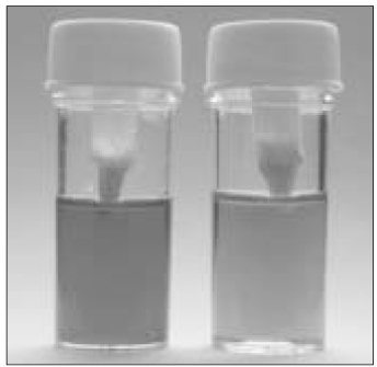
Figure 1
Color change of model system at before(left) and after(right) coronal leakage
Figure 2
Comparison of time periods at which color change showed in lower chamber of model system
Figure 1
Figure 2
Bacteriologic in vitro coronal leakage study of before and after post space preparation

 KACD
KACD


 ePub Link
 Cite
Cite

