
 KACD

Articles
- Page Path
- HOME > Restor Dent Endod > Ahead-of print articles > Article
- Research Article Antimicrobial activity of cobalt oxide nanoparticles against Fusobacterium nucleatum: an in vitro study
-
Srilekha Jayakumar,*
 , Harishma S, Janani Karunakaran, Sreeram Rayar, Jwaalaa Rajkumar, Sreelakshmi PS
-
Restor Dent Endod [Epub ahead of print]
DOI: https://doi.org/10.5395/rde.2026.51.e47
Published online: November 17, 2026
Department of Conservative Dentistry and Endodontics, Chettinad Dental College and Research Institute, Kelambakkam, India
- *Correspondence to Srilekha Jayakumar, MDS Department of Conservative Dentistry and Endodontics, Chettinad Dental College and Research Institute, Kelambakkam, Chengalpattu District, Tamil Nadu 603103, India Email: srilekhajayakumar@gmail.com
• Received: December 12, 2025 • Revised: July 13, 2026 • Accepted: July 13, 2026
© 2026 The Korean Academy of Conservative Dentistry
This is an Open-Access article distributed under the terms of the Creative Commons Attribution Non-Commercial License (http://creativecommons.org/licenses/by-nc/4.0) which permits unrestricted non-commercial use, distribution, and reproduction in any medium, provided the original work is properly cited.
- 48 Views
- 6 Download


Abstract
-
Objectives Endodontic infections are biofilm-mediated diseases. Since biofilms are more resistant to antimicrobial agents, nanoparticles have emerged as promising candidates for endodontic disinfection due to their ability to disrupt microbial growth and biofilm formation. The aim of this in vitro investigation is to analyze the antimicrobial, antibiofilm, and cytocompatibility potential of cobalt oxide nanoparticles (Co3O4).
-
Methods Co3O4 nanoparticles were synthesized via the precipitation method and characterized using scanning electron microscopy, energy-dispersive X-ray spectroscopy, and X-ray diffraction. Antimicrobial activity against Fusobacterium nucleatum was assessed using the broth microdilution method to determine the minimum inhibitory concentration; an antibiofilm assay and bacterial growth inhibition kinetics assay were also carried out. Acridine orange/ethidium bromide fluorescence staining was employed to differentiate between live and dead microbial cells, while the MTT assay was done to evaluate the cytocompatibility of Co3O4 nanoparticles. The following groups were evaluated: Group 1, 3% sodium hypochlorite; Group 2, cobalt oxide nanoparticles; and Group 3, saline.
-
Results Data were analyzed using one-way analysis of variance. Biofilm inhibition by Co3O4 nanoparticles exceeded 80% at concentrations of 500 and 250 µg/mL and showed moderate biofilm inhibition of more than 60% at concentrations of 62.5 µg/mL and 31.25 µg/mL. Bacterial growth inhibition kinetics assay demonstrated rapid bacterial reduction within 15 minutes with Co3O4 nanoparticles. The MTT assay indicated acceptable cytocompatibility at concentrations ≤31.25 µg/mL, maintaining over 80% cell viability.
-
Conclusions Co3O4 nanoparticles in this investigation revealed strong antimicrobial action with effective antibiofilm activity comparable to or higher than 3% NaOCl at certain concentrations (500, 250, and 125 µg/mL). Co3O4 nanoparticles showed ≥80% cytocompatibility at concentrations ≤31.25 µg/mL and demonstrated moderate antibiofilm activity.
INTRODUCTION
The success of the root canal therapy depends mainly on complete debridement and elimination of microorganisms from the root canal system. Anatomic complexities of the root canals such as isthmuses, lateral canals, and ramifications challenge disinfection strategies because these sites harbor microbes [1]. Therefore, endodontic disinfection relies on thorough chemo-mechanical debridement that integrates instrumentation with antimicrobial irrigants for optimal disinfection, thereby enhancing treatment outcomes [2].
Endodontic infections are polymicrobial in nature, with gram-negative organisms predominating in primary infections, while gram-positive organisms are found in secondary and persistent endodontic infections. Fusobacterium nucleatum, a gram-negative obligate anaerobe, plays a pivotal role in endodontic biofilm formation, serving as a “key bridging” species between the primary and secondary colonizers [2]. This organism possesses multiple virulence factors that play vital roles, including cell invasion, proteolytic activity, and biofilm formation via coaggregation. The ability of this organism to survive within the dentinal tubules challenges conventional disinfection strategies, thereby affecting treatment outcomes [3].
Widely used conventional endodontic irrigants, such as sodium hypochlorite (NaOCl) and chlorhexidine, have limitations, including reduced effectiveness against complex biofilm structures [1,4]. NaOCl, though considered the gold-standard irrigant because of its good antimicrobial activity and tissue-dissolving capability, is associated with a few drawbacks, including cytotoxic effects when extruded beyond the root canal, deleterious effects on the radicular dentin matrix, and reduced antimicrobial efficacy at lower concentrations [1,5].
Owing to the above disadvantages, the search for a newer disinfection strategy in endodontics has gained significant attention. In recent years, nanotechnology has emerged as a promising domain for endodontic disinfection. Nanoparticles (1–100 nm) possess unique physical and chemical properties that are entirely different from their bulk counterparts [6]. The antimicrobial activity of the nanoparticles is greatly influenced by their smaller size, shape, and high surface area-to-volume ratio. The surface characteristics of nanoparticles, such as facets and porosities, enable them to penetrate into biofilms, disrupt bacterial cell membranes, and aid in the production of reactive oxygen species (ROS), ultimately leading to bacterial cell death. Several metal oxide nanoparticles, such as zinc oxide, magnesium oxide, copper oxide, and titanium oxide, have been investigated for their antimicrobial potential [7].
Among metal oxide nanoparticles, cobalt oxide nanoparticles (Co3O4) have gained significant interest in recent years because of their unique magnetic properties, large surface area, high porosity, and tunable surface chemistry for drug loading, making them effective for biomedical applications [8–10]. Prior research on Co3O4 nanoparticles showed significant evidence of their use as an antimicrobial agent against a wide range of microorganisms, including both gram-positive and gram-negative species [11–13]. This provides a strong rationale for investigating Co3O4 nanoparticles as a next-generation antimicrobial agent in endodontic disinfection.
Despite the expanding interest in the field of nanotechnology, literature evidence supporting the efficacy of Co3O4 nanoparticles against root canal pathogens remains scarce. To the best of our knowledge, this is the first study to systematically evaluate the antibacterial, antibiofilm properties, and cytocompatibility profile of cobalt oxide nanoparticles against F. nucleatum. Using 3% NaOCl as the reference standard, the study tested the hypothesis that Co3O4 nanoparticles could exhibit a concentration-dependent antimicrobial activity comparable to that of NaOCl.
METHODS
The study protocol was approved by the Institutional Human Ethics Committee of Chettinad Dental College and Research Institute, Kelambakkam, India (No. IHEC-CDCRI/2025/FAC-0089).
Co3O4 nanoparticles were synthesized by the precipitation method. A solution containing 1.422 g of Na2CO3 (Sisco Research Laboratories Pvt. Ltd., Chennai, India) in 10 mL of water was stirred for 10 minutes. Then 0.889 g of CoCl2•6H2O (Sisco Research Laboratories Pvt. Ltd.) was added to the above solution and sonicated for 20 minutes, followed by 10 minutes of stirring. Then the resultant mixture was placed in the hot air oven at 80°C for 16 hours to evaporate the water. The obtained solid powder was calcined in the tubular furnace for 4 hours at 320°C with a ramping rate of 3°C/min. The black powder obtained after calcination was washed with water and ethanol three times at 6,000 revolutions per minute (rpm) for 5 minutes, then dried at 40°C for 12 hours in a vacuum oven [14].
The surface morphology and elemental composition of the synthesized Co3O4 nanoparticles were analyzed using scanning electron microscopy (SEM; Hitachi N3400, Singapore) coupled with energy-dispersive X-ray spectroscopy (EDS; Horiba Instruments, Kyoto, Japan). Crystalline characteristics were determined using X-ray diffraction (XRD; Thermo Fisher Scientific India Pvt. Ltd., Mumbai, India). The diffraction peaks were observed.
1. Groups evaluated
The following groups were evaluated: Group 1, 3% NaOCl as the positive control; Group 2, cobalt oxide nanoparticles (Co3O4); and Group 3, saline as the negative control.
2. Broth microdilution assay
The minimum inhibitory concentration (MIC) of nanoparticles against F. nucleatum (ATCC 23726; HiMedia Laboratories Pvt. Ltd., Mumbai, India) was determined using the broth microdilution method. A 0.5 McFarland standard bacterial suspension was prepared in pre-reduced brain heart infusion (BHI) broth (HiMedia Laboratories Pvt. Ltd.) supplemented with 5% defibrinated sheep blood and 1 µg/mL vitamin K, followed by dilution (1:100) to achieve a final inoculum of approximately 5 × 10⁵ colony-forming units/mL. The nanoparticle solution was prepared by dispersing the nanoparticles in 1% dimethyl sulfoxide (DMSO). Equal volumes (50 µL) of bacterial suspension and nanoparticle solutions at serial concentrations (500, 250, 125, 62.5, 31.25, 15.6, 7.8, 3.9, 1.9, and 0.9 μg/mL) were added to sterile 96-well microtiter plates (MTP; Tarsons Products Ltd., Kolkata, India). Plates were incubated at 37°C for 24 hours under anaerobic conditions. Optical density was measured at 600 nm using a microplate reader (Thermo Fisher Scientific, Waltham, MA, USA). All experiments were performed in triplicate.
3. Antibiofilm assay
The antibiofilm activity was assessed using a MTP assay following the method described by Christensen et al. [15]. A sterile 96-well plate was inoculated with 180 µL of BHI broth and 10 µL of overnight bacterial culture, followed by incubation at 37°C for 24 hours to allow biofilm formation under strict anaerobic conditions. Subsequently, 10 µL of Co3O4 nanoparticle solutions at varying concentrations (500, 250, 125, 62.5, 31.25, 15.6, 7.8, 3.9, 1.9, and 0.9 μg/mL) were added and incubated further. Wells were washed with phosphate-buffered saline to remove planktonic cells. The adherent biofilm was fixed with 2% sodium acetate and stained with 0.1% crystal violet (HiMedia Laboratories Pvt. Ltd.). Excess stain was removed, and plates were air-dried. Optical density was measured at 600 nm using a microplate reader. The percentage of biofilm inhibition was calculated as follows: % Inhibition = [(control optical density − test optical density) / control optical density] × 100.
4. Selection of MIC₅₀ concentration for acridine orange/ethidium bromide staining and bacterial growth inhibition kinetics assay
For the live/dead cell assay using acridine orange/ethidium bromide stain and bacterial growth inhibition kinetics assay, the MIC50 concentration was chosen because it is a biologically acceptable intermediate dose at which about 50% inhibition occurs. This concentration is biologically relevant, enabling the assessment of dynamic microbial killing.
5. Acridine orange/ethidium bromide staining assay
Cell viability was assessed using acridine orange and ethidium bromide dual staining (Sigma-Aldrich, Bengaluru, India). Briefly, 5 × 10⁶ cells/mL of F. nucleatum were plated on a coverslip placed inside a 24-well plate and treated with broth containing a saline sample, 3% NaOCl, and the MIC50 of the Co3O4 sample. After an incubation period of 48 hours at 37°C, 1 mg/mL of acridine orange/ethidium bromide stain was added and subjected to centrifugation (800 rpm) for 2 minutes. Following this, the samples were observed under a fluorescent microscope (ZOE; Bio-Rad, Hercules, CA, USA) to evaluate bacterial cell viability.
6. Bacterial growth inhibition kinetics assay
The bacterial kinetics of Co3O4 nanoparticles were evaluated using a bacterial growth inhibition kinetics assay. Bacterial suspension (150 µL) was combined with 50 µL of nanoparticle solution (MIC₅₀) in 96-well plates. Positive and negative controls included NaOCl and saline, respectively. Plates were incubated at 37°C, and optical density measurements were recorded at 600 nm at 15-minute intervals over 4 hours.
Human gingival fibroblasts for cytotoxicity analysis were purchased from Saveetha Dental College and Hospital, Chennai, India. Cells were cultured in Dulbecco’s Modified Eagle’s Medium (DMEM F12 medium; Sigma-Aldrich) supplemented with 20% fetal bovine serum and antibiotics (penicillin-streptomycin) under standard conditions (37°C, 5% CO2). Cells were seeded in 96-well plates at a density of 1 × 10⁵ cells/mL (200 μL/well) and incubated for 24–48 hours for adherence. Cells were then exposed to varying concentrations of Co3O4 nanoparticles (500–0.9 μg/mL) in serum-free DMEM to eliminate serum protein interference; untreated cells served as a control. After 24 hours, MTT reagent (10 μL of 5 mg/mL) was added and incubated for 2–4 hours. Formazan crystals were dissolved in DMSO, and absorbance was measured at 570 nm using a microplate reader. Cell viability (%) was calculated using GraphPad Prism ver. 6.0 (GraphPad Software, Boston, MA, USA).
The microbiological assay data were collected and subjected to statistical analysis using IBM SPSS for Windows (ver. 26.0; IBM Corp., Armonk, NY, USA). Normality was assessed and confirmed (p > 0.05), allowing the use of parametric statistics such as one-way analysis of variance (ANOVA). Group comparisons were performed using one-way ANOVA. All results are reported using appropriate graphs and tables. A p-value <0.05 was considered statistically significant.
RESULTS
The SEM images showed particles in the form of nanosheets with sizes ranging from 20 to 60 nm (Figure 1). EDS spectra confirmed the presence of cobalt and oxygen with the elemental composition of about 56.43% and 43.57% (Figure 1). XRD analysis showed diffraction peaks at 31º, 36º, 45º, 59º, and 65º corresponding to (220), (311), (400), (511), and (440) planes, confirming the presence of Co3O4 (Figure 2). The increase in the background intensity between 15º and 30º indicates that the material is partially amorphous in nature.
The MIC of cobalt oxide nanoparticles was 0.97 µg/mL and MIC50 was 26.20 µg/mL.
One-way ANOVA showed significant variations in % inhibition (F = 1,586.7, p < 0.001) and OD values (F = 2,258.2, p < 0.001) among all groups. Saline exhibited the highest OD and the least amount of inhibition (3.1 %), whereas 3% NaOCl showed significantly lower OD and strong antibiofilm action, with an inhibition percentage of 71.29%. A definite and increasing dose-dependent pattern was seen across the concentrations of Co3O4 nanoparticles. Higher concentrations of Co3O4 nanoparticles, 500, 250, and 125 µg/mL, showed higher inhibition percentages of 83.38%, 81.13%, and 75.76%, respectively, which were comparable to or higher than those of 3% NaOCl. The concentration of 62.5 µg/mL showed 69.79% inhibition, which was slightly lower than that of 3% NaOCl. The inhibition percentage reduced proportionately to 65.07% at 31.25 µg/mL. The lowest inhibition percentages, ranging from 60.77% to 43.34%, were observed at the lowest concentrations of Co3O4 nanoparticles, 15.6–0.9 µg/mL (Table 1). These integrated findings confirm that the cobalt oxide nanoparticles exhibit a robust, concentration-dependent antimicrobial effect.
Fluorescence microscopy with acridine orange/ethidium bromide staining was interpreted based on apoptotic cell death models [16] as follows: (1) live cells have normal nuclear staining, which presents green chromatin with organized structures; (2) apoptotic cells contain condensed or fragmented chromatin, green or orange; and (3) necrotic cells have similar normal nuclear staining as live cells, except the chromatin is orange instead of green. Results of acridine orange/ethidium bromide staining in this study revealed that in the 3% NaOCl group, bacterial cells appeared green-yellow, yellow in color indicating dying cells with early apoptotic changes. In the Co3O4 nanoparticles group, bacterial cells appeared more orange, indicating a lot of dead cells. Saline group bacterial cells appeared green, indicating more live bacteria (Figure 3).
The bacterial growth inhibition kinetics experiment was monitored for 4 hours at intervals of 15 minutes. OD readings increased continually over the incubation period, indicating a steady rise in bacterial growth in saline-treated wells. In contrast, the bacterial growth was gradually reduced after treatment with Co3O4 nanoparticles and 3% NaOCl. Within 15 minutes, there was a modest decrease in OD and a rise in the percentage of inhibition; the inhibition then gradually increased. Overall, the 15-minute interval monitoring demonstrated how quickly Co3O4 nanoparticles and NaOCl work within that time frame. There was no inhibition or activity seen in saline-treated wells (Figure 4).
The results of the MTT experiment showed that human gingival fibroblast cells treated with varying concentrations of nanoparticles (0.9 µg/mL to 500 µg/mL) had a strong dose-dependent cytotoxic profile. The concentration of Co3O4 nanoparticles ≤31.25 µg/mL maintained cell viability over 80%, consistent with ISO 10993-5:2009 guidelines [17]. However, concentrations ≥62.5 µg/mL showed a gradual decrease in cell viability. The maximum concentration tested in this investigation, 500 µg/mL, exhibited a sharp decline in cell viability to 17.56%, suggesting a strong cytotoxic response (Table 2). These findings provide a clear therapeutic window ranging from 1.95 µg/mL to 31.25 µg/mL for Co3O4 nanoparticles, beyond which cytotoxic effects become apparent (Figure 5).
DISCUSSION
Microbial dynamics of endodontic infections require a deeper understanding for employing optimal disinfection strategies. F. nucleatum is commonly isolated from symptomatic cases with purulent discharge and acute exacerbations [18,19]. Research by Gomes et al. [20,21] identified F. nucleatum as a predominant organism in inflamed root canals. This organism has the ability to coaggregate with species like Prevotella intermedia, Porphyromonas gingivalis, and Peptostreptococcus micros. This particular characteristic feature of this organism enables us to understand its role as a “keystone species” in biofilm formation, enhancing stability, virulence, and resistance of the endodontic biofilm, thereby justifying its selection in the present study [20,21]. However, traditional irrigants have limited efficacy against these resistant biofilms. Therefore, this study evaluated Co3O4 nanoparticles synthesized via the precipitation method for their efficacy against F. nucleatum, which could pave the way for a novel nanotherapeutic agent for root canal disinfection.
Broth microdilution assay was used for the determination of the MIC of Co3O4 nanoparticles, which revealed a MIC of 0.97 µg/mL and MIC₅₀ of 26.20 µg/mL. This substantially low MIC of the nanoparticles denotes the strong inhibitory potential against F. nucleatum. A study reported by Alsharaeh et al. [22] revealed that cobalt oxide-based nanomaterials are effective in reducing bacterial cell integrity at low doses. Similar research by Alahmadi et al. [12] evaluated cobalt-cellulose magnetic nanocomposites and found them effective against Staphylococcus aureus and Escherichia coli, emphasizing the broad-spectrum bacterial action of cobalt oxide nanoparticles. The consistencies across these studies emphasize the expanding evidence to advocate for cobalt oxide nanoparticles as a promising agent for root canal disinfection. Adding to this, Co3O4 nanoparticles exhibit their antibacterial potential through multiple synergistic mechanisms. The initial electrostatic interaction of the nanoparticles with bacterial cells results in membrane destabilization and penetration into the cell, followed by cobalt ion release into the cytoplasm that disrupts the enzymatic systems by binding to thiol groups of proteins [23]. Co3O4 nanoparticles are also capable of generating ROS such as superoxide anions, leading to oxidative damage to the bacterial cell components. Additionally, Co3O4 nanoparticles have the capacity to interfere with iron-sulfur clusters of the bacteria, impairing respiration and DNA repair [24].
Co3O4 nanoparticles also showed good antibiofilm activity, thereby addressing the crucial challenge in root canal infections. Extracellular polymeric substances (EPS) in root canal biofilms act as a “biological glue,” hindering the penetration of conventional root canal disinfectants and thus lowering their efficacy [25,26]. The MTP assay in the present study showed a clear concentration-dependent antibiofilm activity, with higher concentrations exhibiting good antibiofilm efficacy. Notably, maximum antibiofilm activity was observed at 500, 250, and 125 µg/mL of Co3O4 nanoparticles, which was comparable to or higher than that of 3% NaOCl, the gold-standard endodontic irrigant, suggesting that at specific concentrations Co3O4 nanoparticles match the antibiofilm efficacy of NaOCl. Moderate antibiofilm efficacy was evident at the concentrations of 62.5 and 31.25 µg/mL, aligning with the factor that EPS destabilization is nanoparticle-density dependent, occurring only when the concentration of the nanoparticle surpasses the critical threshold.
This mechanism aligns with findings from the studies by Shkodenko et al. [27] and Fulaz et al. [28], which demonstrated that metal and metal oxide nanoparticles can penetrate even mature biofilms through electrostatic interactions between positively charged nanoparticles and negatively charged EPS matrix components, promoting their adherence and penetration into deeper biofilm layers. This is particularly advantageous, as it overcomes a major limitation of conventional irrigants, which often exhibit restricted penetration into mature biofilms. This phenomenon may explain why, in the present study, Co3O4 nanoparticles demonstrated greater or comparable antibiofilm effects than NaOCl at specific concentrations. Furthermore, Ma et al. [29] also reported significant antibiofilm activity of Co3O4 nanoparticles, including reduced biofilm biomass and decreased viability of multidrug-resistant bacterial biofilms following treatment with cobalt oxide nanostructures. This is primarily attributed to ROS-mediated matrix disruption, increased membrane permeability, DNA damage, and impairment of bacterial energy metabolism. The present findings support the proposed mechanisms.
These findings were further substantiated by qualitative assessment using acridine orange/ethidium bromide staining. The saline group presented predominantly green fluorescence, consistent with viable intact cells. In contrast, NaOCl-treated samples exhibited yellowish-orange fluorescence, indicative of early apoptotic-like changes or transitional stages of membrane compromise. Crucially, the Co3O4 nanoparticles (MIC₅₀ concentration was used) treated group demonstrated intense orange-red fluorescence, reflecting widespread membrane damage and nonviable cells, suggesting irreversible intracellular oxidative injury. These qualitative observations substantiate the hypothesis that cobalt oxide exerts bactericidal effects primarily via membrane permeabilization and ROS-mediated cytotoxicity, consistent with findings of Anele et al. [30].
Co3O4 nanoparticles exhibited good bactericidal activity within 15 minutes of exposure, as observed in the bacterial growth inhibition kinetics assay. This assay was evaluated over a period of 4 hours, during which a progressive increase in inhibition was evident, indicating sustained antibacterial activity. This rapid onset of action with sustained efficacy is attributed to the synergistic mechanism of cobalt oxide nanoparticles. This early onset of action, within 15 minutes, reflects the Co2+/Co3+ redox recycling process that continuously releases ROS, causing oxidative damage to the bacterial cell [30]. In contrast, the oxidative damage caused by NaOCl rapidly declines when organic constituents consume the hypochlorous acid [31,32]. The sustained ROS-generating capability of Co3O4 nanoparticles offers a distinct advantageous action on resistant root canal biofilms.
Biocompatibility remains an important determinant of materials for their clinical applicability. The cytotoxicity of Co3O4 nanoparticles was evaluated using the MTT assay with human gingival fibroblasts; concentrations ≤31.25 µg/mL showed more than 80% cell viability, satisfying the ISO 10993-5:2009 cytocompatibility criteria [17], while exhibiting moderate antibiofilm activity (65% biofilm inhibition). However, at a concentration of 62.5 µg/mL, cell viability decreased to 69% and further markedly declined to 17.56% at a concentration of 500 µg/mL. This underscores the significance of dose optimization for nanotherapeutic antimicrobial agents. Despite the strong antibiofilm action at higher concentrations, a therapeutic window is essential for balancing the safety and antibiofilm efficacy. The therapeutic window observed in the present study ranges between 1.95 µg/mL and 31.25 µg/mL for Co3O4 nanoparticles, revealing a moderate antibiofilm efficacy while remaining within cytocompatibility limits. At this cytocompatible concentration of 31.25 µg/mL, repeated and prolonged irrigation with larger volumes increases the frequency of exposure and contact time between bacterial cells and nanoparticles. This strategy not only enhances the antibacterial potential of nanoparticles but also preserves host cell viability by remaining within the cytocompatibility range.
Though this study showed promising results, there are certain limitations that have to be taken into consideration. This is an in vitro design that does not completely reflect the clinical scenario, where host tissue remnants, immune response, and fluid dynamics affect the antimicrobial performance of the agents. Furthermore, a multispecies biofilm model was not used in this study, thereby limiting direct clinical applicability. Additionally, the MTT assay utilized in the study evaluates only short-term cytotoxicity in gingival fibroblasts and did not evaluate the long-term cytotoxic effects in clinically relevant cells such as periodontal fibroblasts. Few studies also reported that Co3O4 nanoparticle antibacterial properties could be influenced by synthesis parameters, particle aggregation, and crystalline structure, which have to be considered in future studies [33,34]. Despite the above limitations, findings of the present study suggest that Co3O4 nanoparticles could serve as an adjunct in endodontic disinfection due to their potent antibiofilm activity, rapid bactericidal kinetics, and acceptable cytocompatibility range. Possible clinical implications in the near future could be nanoparticle irrigant solutions, incorporation into intracanal medicaments such as cobalt oxide nanoparticle-incorporated hydrogels, and cobalt oxide nanoparticle-coated gutta-percha. Future research should focus on well-designed in vivo investigations that support clinical translation, and studies should also focus on nanoparticle penetration into dentinal tubules as well as their effects on radicular dentin.
CONCLUSIONS
Within the limitations of this study, Co3O4 nanoparticles showed marked antimicrobial activity at higher concentrations exceeding 3% NaOCl while maintaining cytocompatibility at 31.25 µg/mL with moderate antibiofilm activity. So, this concentration provides a favorable balance between safety and antimicrobial activity. These findings suggest that cobalt oxide nanoparticles could be an adjunct for endodontic disinfection. Future in vivo and clinical investigations are needed for clinical translation.
-
CONFLICT OF INTEREST
No potential conflict of interest relevant to this article was reported.
-
FUNDING/SUPPORT
The authors have no financial relationships relevant to this article to disclose.
-
AUTHOR CONTRIBUTIONS
Conceptualization, Resources, Data curation, Formal analysis: Jayakumar S, S H, Karunakaran J. Investigation, Methodology, Project administration, Software, Visualization: Jayakumar S, S H. Supervision, Validation: Karunakaran J, Rayar S, Rajkuma J. Writing - original draft: Rayar S, Rajkumar J, PS S. Writing - review & editing: Jayakumar S, Rajkumar J, PS S. All authors read and approved the final manuscript.
-
DATA SHARING STATEMENT
The datasets are not publicly available but are available from the corresponding author upon reasonable request.
-
DISCLOSURE OF GENERATIVE AI IN SCIENTIFIC WRITING
CHATGPT (GPT-6) developed by OpenAI (San Francisco, CA, USA) was used only for grammar correction and language editing to improve the readability of the manuscript. No AI tools were used for data analysis, interpretation of results, or drawing conclusions. The authors take full responsibility for the content and integrity of the manuscript.
Figure 1.
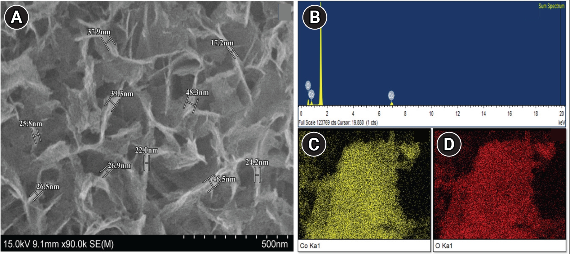
(A) Scanning electron microscopy image of cobalt oxide nanoparticles. (B) Energy-dispersive X-ray spectroscopy (EDS) spectrum. (C, D) EDS mapping of cobalt oxide nanoparticles.
Figure 2.
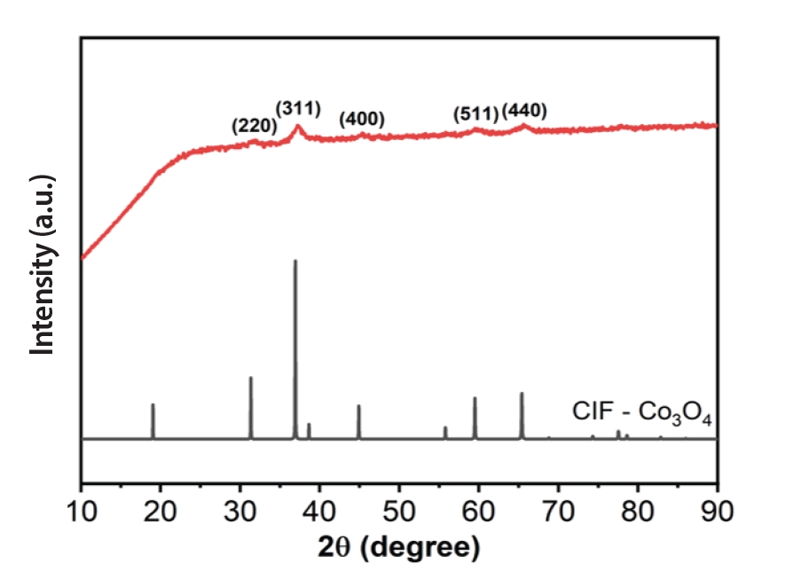
X-ray diffraction analysis of cobalt oxide nanoparticles. a.u., arbitrary units; CIF, crystallographic information file.
Figure 3.

Acridine orange/ethidium bromide staining assay for cell viability and apoptotic cell detection. (A) Saline-treated group. (B) Sodium hypochlorite-treated group. (C, D) Cobalt oxide nanoparticle-treated group.
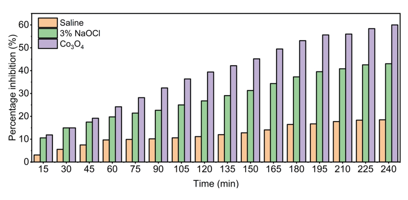
Figure 5.
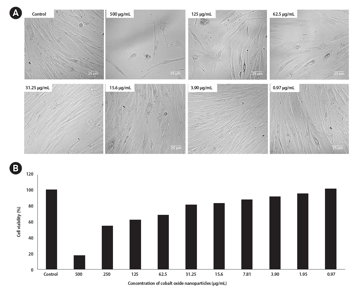
(A) Cell viability of human gingival fibroblasts (HGFs) treated with different concentrations of cobalt oxide nanoparticles (500–0.97 µg/mL). (B) Graphical representation of the cell viability assay. Control indicates untreated cells.
Table 1.
Intergroup analysis of OD and percentage of biofilm inhibition from the microtiter plate assay
Table 2.
Intergroup analysis of OD and percentage of cell viability from MTT assay
- 1. Josic U, Mazzitelli C, Maravic T, Fidler A, Breschi L, Mazzoni A. Biofilm in endodontics: in vitro cultivation possibilities, sonic-, ultrasonic- and laser-assisted removal techniques and evaluation of the cleaning efficacy. Polymers (Basel) 2022;14:1334.ArticlePubMedPMC
- 2. Arias-Moliz MT, Pérez-Carrasco V, Uroz-Torres D, Santana Ramos JD, García-Salcedo JA, Soriano M. Identification of keystone taxa in root canals and periapical lesions of post-treatment endodontic infections: next generation microbiome research. Int Endod J 2024;57:933-942.ArticlePubMed
- 3. Gomes BPFA, Bronzato JD, Almeida-Gomes RF, Pinheiro ET, Sousa ELR, Jacinto RC. Identification of Fusobacterium nucleatum in primary and secondary endodontic infections and its association with clinical features by using two different methods. Clin Oral Investig 2021;25:6249-6258.ArticlePubMedPDF
- 4. Capuano N, Amato A, Dell'Annunziata F, Giordano F, Folliero V, Di Spirito F, et al. Nanoparticles and their antibacterial application in endodontics. Antibiotics (Basel) 2023;12:1690.ArticlePubMedPMC
- 5. Luddin N, Ahmed HM. The antibacterial activity of sodium hypochlorite and chlorhexidine against Enterococcus faecalis: a review on agar diffusion and direct contact methods. J Conserv Dent 2013;16:9-16.ArticlePubMedPMC
- 6. Wong J, Zou T, Lee AHC, Zhang C. The potential translational applications of nanoparticles in endodontics. Int J Nanomedicine 2021;16:2087-2106.ArticlePubMedPMCPDF
- 7. Sharifi R, Vatani A, Sabzi A, Safaei M. A narrative review on application of metal and metal oxide nanoparticles in endodontics. Heliyon 2024;10:e34673.ArticlePubMedPMC
- 8. Ansari SM, Bhor RD, Pai KR, et al. Cobalt nanoparticles for Biomedical Applications: Facile synthesis, physiochemical characterization, cytotoxicity behavior and biocompatibility. Appl Surf Sci 2017;414:171-187.Article
- 9. Annu , Sahu M, Singh S, Prajapati S, Verma DK, Shin DK. From green chemistry to biomedicine: the sustainable symphony of cobalt oxide nanoparticles. RSC Adv 2024;14:32733-32758.ArticlePubMedPMC
- 10. Chang EL, Simmers C, Knight DA. Cobalt complexes as antiviral and antibacterial agents. Pharmaceuticals (Basel) 2010;3:1711-1728.ArticlePubMedPMC
- 11. Parada J, Atria A, Wiese G, Rivas E, Corsini G. Synthesis, characterization and antibacterial activity of cobalt (III) complex with phenanthroline and maltose. J Chil Chem Soc 2014;59:2636-2639.Article
- 12. Alahmadi NS, Betts JW, Cheng F, Francesconi MG, Kelly SM, Kornherr A, et al. Synthesis and antibacterial effects of cobalt-cellulose magnetic nanocomposites. RSC Adv 2017;7:20020-20026.Article
- 13. Alkheraif AA, AlMufareh NA, AlQhtani FA, Asiri W, Abuhadi RI, Hamoud M, et al. Minocycline incorporated cobalt oxide nanoparticles in dental sealants: an in vitro study. Microsc Res Tech 2022;85:2558-2566.ArticlePubMedPDF
- 14. Meng Z, Yao JX, Sun CN, Kang X, Gao R, Li HR, et al. Efficient ammonia production beginning from enhanced air activation. Adv Energy Mater 2022;12:2202105.ArticlePDF
- 15. Christensen GD, Simpson WA, Younger JJ, Baddour LM, Barrett FF, Melton DM, et al. Adherence of coagulase-negative staphylococci to plastic tissue culture plates: a quantitative model for the adherence of staphylococci to medical devices. J Clin Microbiol 1985;22:996-1006.ArticlePubMedPMCPDF
- 16. Byczkowska A, Kunikowska A, Kaźmierczak A. Determination of ACC-induced cell-programmed death in roots of Vicia faba ssp. minor seedlings by acridine orange and ethidium bromide staining. Protoplasma 2013;250:121-128.ArticlePubMedPMC
- 17. International Organization for Standardization (ISO). ISO 10993-5:2009. Biological evaluation of medical devices - Part 5: Tests for in vitro cytotoxicity. Geneva: ISO; 2009.
- 18. Chávez de Paz Villanueva LE. Fusobacterium nucleatum in endodontic flare-ups. Oral Surg Oral Med Oral Pathol Oral Radiol Endod 2002;93:179-183.ArticlePubMed
- 19. Jacinto RC, Montagner F, Signoretti FG, Almeida GC, Gomes BP. Frequency, microbial interactions, and antimicrobial susceptibility of Fusobacterium nucleatum and Fusobacterium necrophorum isolated from primary endodontic infections. J Endod 2008;34:1451-1456.ArticlePubMed
- 20. Gomes BP, Drucker DB, Lilley JD. Associations of specific bacteria with some endodontic signs and symptoms. Int Endod J 1994;27:291-298.PubMed
- 21. Gomes BP, Pinheiro ET, Gadê-Neto CR, Sousa EL, Ferraz CC, Zaia AA, et al. Microbiological examination of infected dental root canals. Oral Microbiol Immunol 2004;19:71-76.ArticlePubMed
- 22. Alsharaeh E, Mussa Y, Ahmed F, Aldawsari Y, Al-Hindawi M, Sing GK. Novel route for the preparation of cobalt oxide nanoparticles/reduced graphene oxide nanocomposites and their antibacterial activities. Ceram Int 2016;42:3407-3410.Article
- 23. Abass AA, Abdulridha WAM, Alaarage WK, Abdulrudha NH, Haider J. Evaluating the antibacterial effect of cobalt nanoparticles against multi-drug resistant pathogens. J Med Life 2021;14:823-833.ArticlePubMedPMC
- 24. Omar IM, Sayed ME, Ibrahim MA, El-Jemni MA. Electrosynthesis and phase control of cobalt-based nanoparticles: antibacterial and antifungal evaluation of Co3O4 formed at varied temperatures. Chemistry 2025;7:87.Article
- 25. Kishen A. Advanced therapeutic options for endodontic biofilms. Endod Top 2010;22:99-123.Article
- 26. Neelakantan P, Romero M, Vera J, Daood U, Khan AU, Yan A, et al. Biofilms in endodontics: current status and future directions. Int J Mol Sci 2017;18:1748.ArticlePubMedPMC
- 27. Shkodenko L, Kassirov I, Koshel E. Metal oxide nanoparticles against bacterial biofilms: perspectives and limitations. Microorganisms 2020;8:1545.ArticlePubMedPMC
- 28. Fulaz S, Vitale S, Quinn L, Casey E. Nanoparticle-biofilm interactions: the role of the EPS matrix. Trends Microbiol 2019;27:915-926.ArticlePubMed
- 29. Ma Y, Lin W, Ruan Y, Lu H, Fan S, Chen D, et al. Advances of cobalt nanomaterials as anti-infection agents, drug carriers, and immunomodulators for potential infectious disease treatment. Pharmaceutics 2022;14:2351.ArticlePubMedPMC
- 30. Anele A, Obare S, Wei J. Recent trends and advances of Co3O4 nanoparticles in environmental remediation of bacteria in wastewater. Nanomaterials (Basel) 2022;12:1129.ArticlePubMedPMC
- 31. Krah-Sinan AA, Adou-Assoumou M, Xavier Djolé S, Diemer F, Gurgel M. The effects of sodium hypochlorite on organic matters: influences of concentration, renewal frequency and contact area. Iran Endod J 2020;15:18-22.PubMedPMC
- 32. Drews DJ, Nguyen AD, Diederich A, Gernhardt CR. The interaction of two widely used endodontic irrigants, chlorhexidine and sodium hypochlorite, and its impact on the disinfection protocol during root canal treatment. Antibiotics (Basel) 2023;12:589.ArticlePubMedPMC
- 33. Fan W, Guo M. Research progress on nanotoxicity and detoxification of cobalt in metal-based implants. Ann Med 2025;57:2532120.ArticlePubMedPMC
- 34. Moradpoor H, Safaei M, Rezaei F, Golshah A, Jamshidy L, Hatam R, et al. Optimisation of cobalt oxide nanoparticles synthesis as bactericidal agents. Open Access Maced J Med Sci 2019;7:2757-2762.ArticlePubMedPMCPDF
REFERENCES
Tables & Figures
REFERENCES
Citations
Citations to this article as recorded by 
 Cite
 XML Download
Antimicrobial activity of cobalt oxide nanoparticles against Fusobacterium nucleatum: an in vitro study
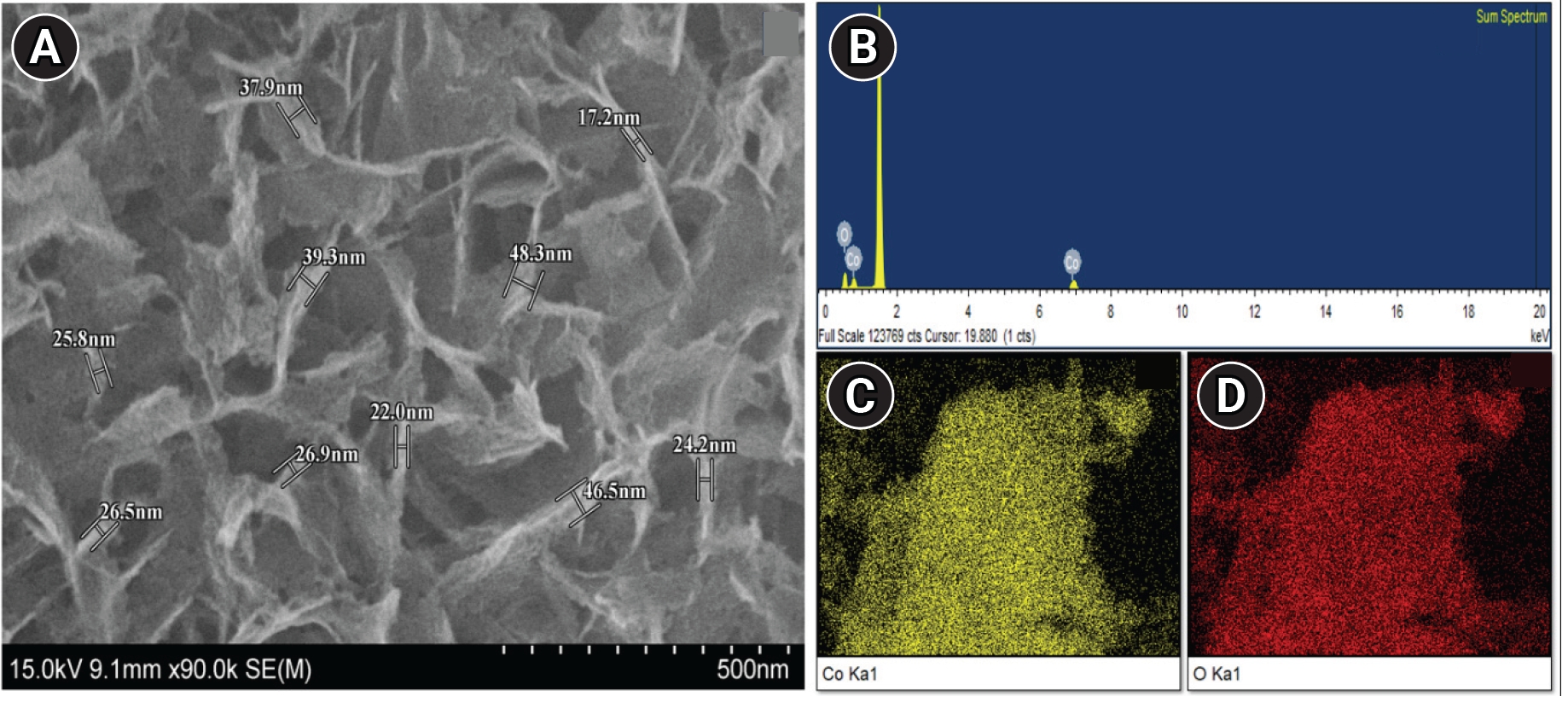
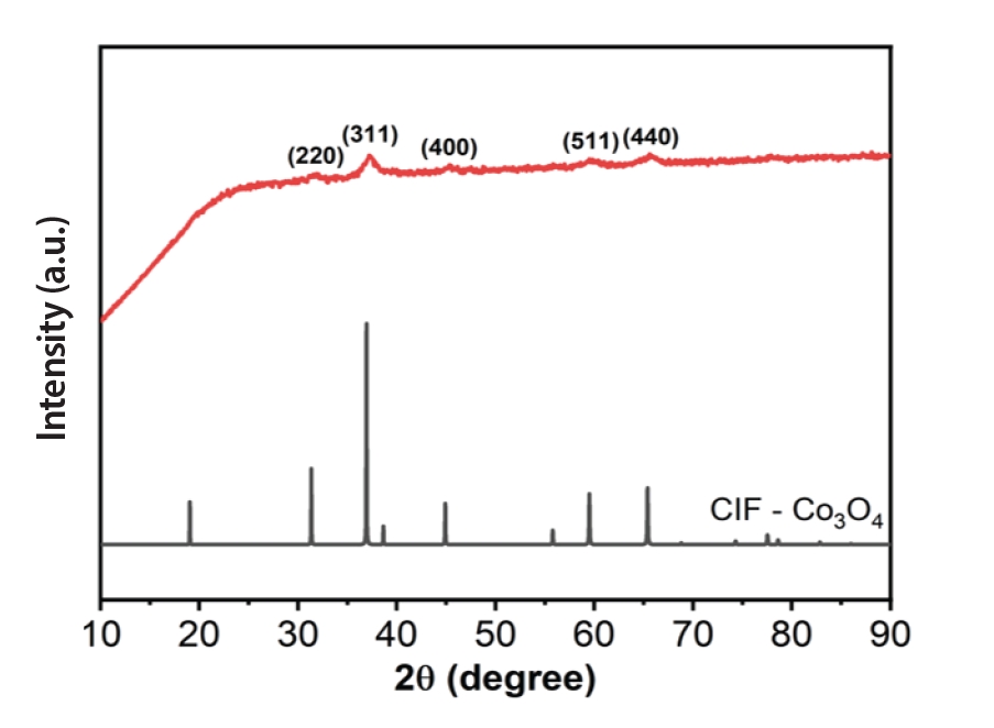

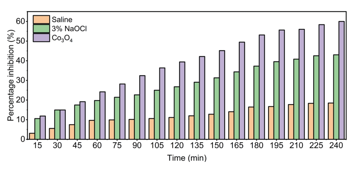
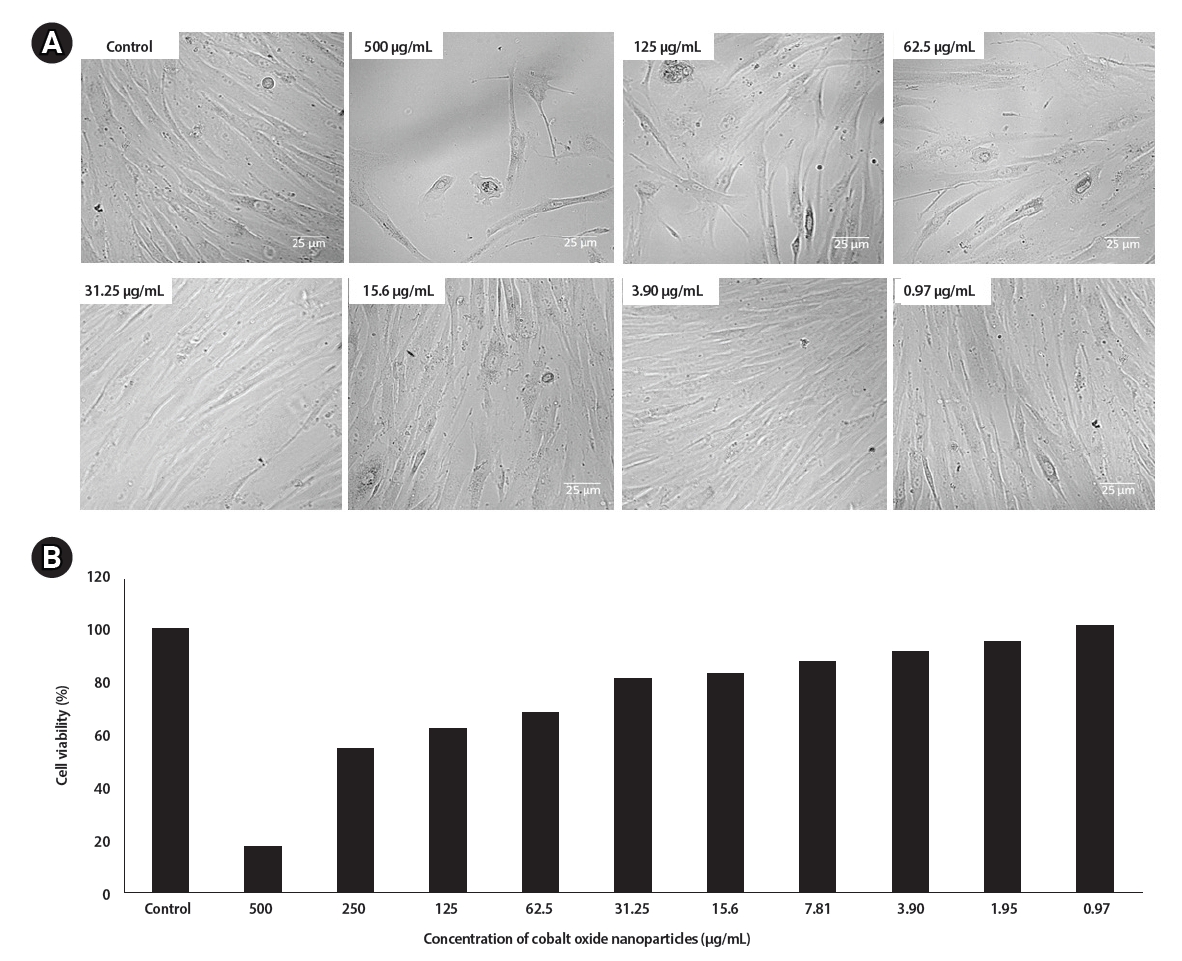
Figure 1. (A) Scanning electron microscopy image of cobalt oxide nanoparticles. (B) Energy-dispersive X-ray spectroscopy (EDS) spectrum. (C, D) EDS mapping of cobalt oxide nanoparticles.
Figure 2. X-ray diffraction analysis of cobalt oxide nanoparticles. a.u., arbitrary units; CIF, crystallographic information file.
Figure 3. Acridine orange/ethidium bromide staining assay for cell viability and apoptotic cell detection. (A) Saline-treated group. (B) Sodium hypochlorite-treated group. (C, D) Cobalt oxide nanoparticle-treated group.
Figure 4. Bacterial growth inhibition kinetics assay.
Figure 5. (A) Cell viability of human gingival fibroblasts (HGFs) treated with different concentrations of cobalt oxide nanoparticles (500–0.97 µg/mL). (B) Graphical representation of the cell viability assay. Control indicates untreated cells.
Figure 1.
Figure 2.
Figure 3.
Figure 4.
Figure 5.
Antimicrobial activity of cobalt oxide nanoparticles against Fusobacterium nucleatum: an in vitro study
| Parameter | Group | Concentration (µg/mL) | Mean ± SD | Mean (95% CI) | F | p-value |
|---|---|---|---|---|---|---|
| OD | Saline | – | 1.3343 ± 0.00929 | 1.3343 (1.3113–1.3574) | 2,258.2 | <0.001 |
| 3% NaOCl | – | 0.3957 ± 0.02040 | 0.3957 (0.3450–0.4464) | |||
| Co3O4 nanoparticles | 500 | 0.2290 ± 0.00854 | 0.2290 (0.2078–0.2502) | |||
| 250 | 0.2600 ± 0.02352 | 0.2600 (0.2016–0.3184) | ||||
| 125 | 0.3340 ± 0.00500 | 0.3340 (0.3216–0.3464) | ||||
| 62.5 | 0.4163 ± 0.01021 | 0.4163 (0.3910–0.4417) | ||||
| 31.25 | 0.4813 ± 0.00681 | 0.4813 (0.4644–0.4982) | ||||
| 15.6 | 0.5407 ± 0.01106 | 0.5407 (0.5132–0.5681) | ||||
| 7.8 | 0.5860 ± 0.01375 | 0.5860 (0.5518–0.6202) | ||||
| 3.9 | 0.6353 ± 0.01290 | 0.6353 (0.6033–0.6674) | ||||
| 1.9 | 0.7170 ± 0.00721 | 0.7170 (0.6991–0.7349) | ||||
| 0.9 | 0.7810 ± 0.01400 | 0.7810 (0.7462–0.8158) | ||||
| Inhibition (%) | Saline | - | 3.1923 ± 0.67412 | 3.1923 (1.5177–4.8669) | 1,586.7 | <0.001 |
| 3% NaOCl | - | 71.2937 ± 1.48038 | 71.2937 (67.6163–74.9712) | |||
| Co3O4 nanoparticles | 500 | 83.3857 ± 0.61989 | 83.3857 (81.8458–84.9256) | |||
| 250 | 81.1366 ± 1.70612 | 81.1366 (76.8984–85.3748) | ||||
| 125 | 75.7678 ± 0.36275 | 75.7678 (74.8666–76.6689) | ||||
| 62.5 | 69.7944 ± 0.74109 | 69.7944 (67.9534–71.6353) | ||||
| 31.25 | 65.0785 ± 0.49386 | 65.0785 (63.8517–66.3053) | ||||
| 15.6 | 60.7738 ± 0.80243 | 60.7738 (58.7804–62.7672) | ||||
| 7.8 | 57.4848 ± 0.99743 | 57.4848 (55.0070–59.9626) | ||||
| 3.9 | 53.9056 ± 0.93571 | 53.9056 (51.5811–56.2300) | ||||
| 1.9 | 47.9805 ± 0.52317 | 47.9805 (46.6809–49.2801) | ||||
| 0.9 | 43.3372 ± 1.01576 | 43.3372 (40.8140–45.8605) |
| Parameter | Group | Concentration (µg/mL) | Mean ± SD | Mean (95% CI) | F | p-value |
|---|---|---|---|---|---|---|
| OD | Control | – | 0.77433 ± 0.002517 | 0.77433 (0.76808–0.78058) | 959.009 | <0.001 |
| Co3O4 nanoparticles | 500 | 0.13600 ± 0.002000 | 0.13600 (0.13103–0.14097) | |||
| 250 | 0.42400 ± 0.012124 | 0.42400 (0.39388–0.45412) | ||||
| 125 | 0.48467 ± 0.003512 | 0.48467 (0.47594–0.49339) | ||||
| 62.5 | 0.53467 ± 0.011060 | 0.53467 (0.50719–0.56214) | ||||
| 31.25 | 0.63067 ± 0.010504 | 0.63067 (0.60457–0.65676) | ||||
| 15.6 | 0.64867 ± 0.010504 | 0.64867 (0.62257–0.67476) | ||||
| 7.8 | 0.68367 ± 0.013317 | 0.68367 (0.65059–0.71675) | ||||
| 3.9 | 0.71367 ± 0.003786 | 0.71367 (0.70426–0.72307) | ||||
| 1.9 | 0.74167 ± 0.023692 | 0.74167 (0.68281–0.80052) | ||||
| 0.9 | 0.78500 ± 0.003606 | 0.78500 (0.77604–0.79396) | ||||
| Cell viability (%) | Control | – | 100.0000 ± 0.000000 | 100.0000 (100.00000–100.00000) | 963.030 | <0.001 |
| Co3O4 nanoparticles | 500 | 17.56417 ± 0.314957 | 17.56417 (16.78177–18.34656) | |||
| 250 | 54.75810 ± 1.624274 | 54.75810 (50.72318–58.79302) | ||||
| 125 | 62.59257 ± 0.606183 | 62.59257 (61.08673–64.09841) | ||||
| 62.5 | 69.05010 ± 1.513506 | 69.05010 (65.29034–72.80986) | ||||
| 31.25 | 81.44810 ± 1.478326 | 81.44810 (77.77574–85.12046) | ||||
| 15.6 | 83.76990 ± 1.219770 | 83.76990 (80.73982–86.79998) | ||||
| 7.8 | 88.28833 ± 1.469071 | 88.28833 (84.63896–91.93771) | ||||
| 3.9 | 92.16593 ± 0.568218 | 92.16593 (90.75440–93.57746) | ||||
| 1.9 | 95.78247 ± 3.097946 | 95.78247 (88.08674–103.47819) | ||||
| 0.9 | 101.3773 ± 0.297377 | 101.3773 (100.63861–102.11606) |
Table 1. Intergroup analysis of OD and percentage of biofilm inhibition from the microtiter plate assay
CI, confidence interval; NaOCl, sodium hypochlorite; OD, optical density; SD, standard deviation. Differences among groups were analyzed using one-way analysis of variance. A
Table 2. Intergroup analysis of OD and percentage of cell viability from MTT assay
CI, confidence interval; MTT, 3-(4,5-dimethylthiazol-2-yl)-2,5-diphenyltetrazolium bromide; OD, optical density; SD, standard deviation. Differences among groups were analyzed using one-way analysis of variance. A


