
 KACD

Articles
- Page Path
- HOME > Restor Dent Endod > Ahead-of print articles > Article
- Research Article Direct cellular responses of a human periodontal ligament stem cell line to root canal sealers and white mineral trioxide aggregate: an in vitro study
-
Atsushi Tomokiyo1
 , Kenta Tsuchiya1,2,*, Keita Ipposhi3, Salvatore Sauro2, Jukka P. Matinlinna4,5, Fumiko Minowa1, Hidehiko Sano1, Hidefumi Maeda3
-
Restor Dent Endod [Epub ahead of print]
DOI: https://doi.org/10.5395/rde.2026.51.e45
Published online: October 23, 2026
1Department of Restorative Dentistry, Faculty of Dental Medicine, Hokkaido University, Sapporo, Japan
2Dental Biomaterials and Minimally Invasive Dentistry, Departamento de Odontología, Facultad de Ciencias de la Salud, Universidad CEU Cardenal Herrera, Valencia, Spain
3Department of Endodontology and Operative Dentistry, Division of Oral Rehabilitation, Faculty of Dental Science, Kyushu University, Fukuoka, Japan
4Faculty of Dentistry, The University of Jordan, Amman, Jordan
5Biomaterials Science, Division of Dentistry, School of Medical Sciences, The University of Manchester, Manchester, UK
- *Correspondence to Kenta Tsuchiya, DDS, PhD Department of Restorative Dentistry, Faculty of Dental Medicine, Hokkaido University, Kita 13, Nishi 7, Kita-ku, Sapporo 060-8586, Japan Email: kentatsuchiya1222@gmail.com
• Received: April 20, 2026 • Revised: June 2, 2026 • Accepted: June 20, 2026
© 2026 The Korean Academy of Conservative Dentistry
This is an Open-Access article distributed under the terms of the Creative Commons Attribution Non-Commercial License (http://creativecommons.org/licenses/by-nc/4.0) which permits unrestricted non-commercial use, distribution, and reproduction in any medium, provided the original work is properly cited.
- 15 Views
- 4 Download


Abstract
-
Objectives Root canal sealers may contact periodontal ligament (PDL) tissues through the apical foramen; therefore, their biological compatibility is critical for periapical healing. This study evaluated the direct cellular responses of human PDL stem cells (PDLSCs) to selected root canal sealers and white mineral trioxide aggregate (WMTA).
-
Methods Disc-shaped specimens of AH Plus (AH; Dentsply Sirona), GuttaFlow 2 (GF; Coltene), GuttaFlow Bioseal (Bio; Coltene), and ProRoot MTA (WMTA) were directly placed onto cultured clonal human PDLSCs (cell line 2–23). Cell viability, gene expression related to proliferation (CCND1, CCNE1), collagen synthesis (COL1, COL3), and mineralization (BMP2, OPN, CEMP1), collagen deposition, mineralized matrix formation, surface characteristics, and Ca2+ release were evaluated using MTT assay, real-time quantitative polymerase chain reaction, histochemical staining, scanning electron microscopy/energy-dispersive X-ray spectroscopy, and ion release analysis.
-
Results AH exhibited marked cytotoxicity, whereas GF, Bio, and WMTA showed higher cytocompatibility. Bio significantly increased proliferation-related gene expression at 7 days. Although early collagen deposition was lower in the Bio and WMTA groups, both materials induced significantly greater mineralized matrix formation after 28 days, accompanied by upregulation of BMP2, OPN, and CEMP1. WMTA showed a rapid initial Ca2+ release, whereas Bio demonstrated a gradual and sustained release profile.
-
Conclusions The evaluated materials exhibited distinct cellular effects on PDLSCs. The bioceramic silicone-based sealer (Bio) demonstrated favorable cytocompatibility and promoted mineralization-related responses comparable to WMTA, suggesting its potential to support periapical tissue healing. Therefore, Bio and WMTA may enhance PDLSC viability and mineralization, supporting improved periapical healing and long-term treatment outcomes.
INTRODUCTION
Root canal treatment aims to eliminate microorganisms and their by-products from the infected root canal system and to prevent reinfection of the canal space, thereby promoting periapical tissue healing through effective microbial control [1,2]. Given that, successful root canal treatment requires adequate mechanical shaping, effective irrigation for disinfection, and subsequent three-dimensional obturation of the root canal system [3]. The quality of root canal obturation is a key determinant of endodontic outcomes [4,5]. It is noteworthy that obturation is typically achieved using a core material with a sealer, which penetrates canal irregularities to establish an apical seal [6,7].
Calcium silicate-based cements (CSCs), including mineral trioxide aggregate (MTA), are widely used in endodontics due to their excellent sealing ability, biocompatibility, and antibacterial potential. Chemically, these materials hydrate in the presence of water and can induce cementum-like tissue formation [8,9]. However, CSCs have certain drawbacks, including their difficulty in obturating curved canals, challenges in removal during retreatment, and relatively long setting times [10]. To overcome these limitations, bioceramic-based root canal sealers with improved handling properties have been developed. These materials may promote remineralization and exhibit lower cytotoxicity than conventional resin-based sealers such as AH Plus (AH; Dentsply Sirona, Tulsa, OK, USA) [11–13].
Silicone-based sealers incorporating gutta-percha particles have also been observed to improve flowability, dimensional stability, and adaptation to canal irregularities [14,15]. Current examples include GuttaFlow (Coltene, Altstätten, Switzerland) and its improved formulation GuttaFlow 2 (GF; Coltene), which consist of gutta-percha powder dispersed in a polydimethylsiloxane matrix. They have demonstrated favorable biocompatibility with periodontal ligament (PDL) and gingival fibroblasts [16,17]. More recently, GuttaFlow Bioseal (Bio; Coltene) was launched as a modified silicone-based sealer containing calcium silicate. Such a bioceramic formulation combines polydimethylsiloxane with gutta-percha particles, nanosilver, and calcium silicate in a glassy matrix has also been reported to induce surface biomineralization and provide favorable sealing ability [18–20].
Root canal sealers come into contact with dentin and PDL tissues at the root apex. The PDL contains diverse cell types, including human PDL stem cells (PDLSCs), which possess self-renewal and multilineage differentiation abilities and clinically contribute to periapical healing [21,22]. Although numerous studies have investigated the biological properties of root canal sealers, most have relied on extract-based assays, which may not fully reflect the direct interactions between set sealers and periapical tissues under clinically relevant conditions. However, laboratory studies evaluating PDLSC responses to direct contact with sealers remain limited, partly because PDLSCs represent a small population and their proliferative capacity declines with age [23,24]. To address these limitations, we previously established a clonal human PDLSC line (cell line 2–23) that exhibits unlimited proliferation and characteristics similar to those of naïve PDLSCs [25]. To the best of our knowledge, no previous laboratory study has comprehensively evaluated the direct cellular responses of a clonal human PDLSC line to contemporary root canal sealers under direct-contact conditions. Therefore, the present laboratory study aimed to evaluate the direct cellular responses of PDLSCs (cell line 2–23) to three contemporary root canal sealers and ProRoot MTA (WMTA; Dentsply Sirona), focusing on cell viability, proliferation-related gene expression, collagen synthesis, mineralized matrix formation, and also mineralization-related gene expression.
METHODS
Sealer disc specimens were prepared according to ISO 10993-5 as previously described [26]. Rubber ring molds (5 mm diameter, 2 mm height) were placed on glass plates and filled with freshly mixed cement materials. An epoxy resin-based sealer (AH), a silicone-based sealer (GF), and a bioceramic silicone-based sealer (Bio) were prepared according to the manufacturers’ instructions under sterile conditions by one operator only. WMTA powder was mixed with distilled water at a powder-to-liquid ratio of 3:1 (w/w), according to the manufacturer's instructions. WMTA was included as a reference bioactive cement material because of its well-established cytocompatibility and mineralization-inducing properties in endodontic applications.
Specimens were then incubated at 37°C in a humidified atmosphere of 5% CO2 for 24 hours to allow complete setting. After removal from the molds under sterile conditions, discs were rinsed with α-minimum essential medium (α-MEM; Gibco-BRL, Grand Island, NY, USA). To evaluate direct cellular responses under contact conditions, the discs were placed directly onto cultured cells rather than using material extracts. The disc morphology is shown in Supplementary Figure 1A–D, and material details are provided in Table 1.
A clonal human PDL stem cell line, the line 2–23 [25], was cultured in α-MEM supplemented with 10% fetal bovine serum and penicillin-streptomycin (50 U/mL penicillin and 50 μg/mL streptomycin) at 37°C in a humidified atmosphere of 5% CO2 and 95% air. After reaching subconfluency, the cells were seeded into 6-well plates at a density of 5 × 10⁴ cells per well with 2 mL medium and incubated for 24 hours. A single material disc was then gently placed onto the cultured cells (n = 4), and the cells were further cultured for 3 or 7 days. Control cells were cultured without discs.
This laboratory study was awarded ethical approval by the Kyushu University Certified Institutional Review Board for Clinical Trials (approval No. 2-115).
Cell morphology was examined using an inverted microscope (BX41; Olympus, Tokyo, Japan) at 3 and 7 days. Images were obtained from areas proximal and distal to the discs with magnifications of ×100 (Supplementary Figure 1E) using a CMOS camera (Bio Tools Inc., Gunma, Japan) and ToupView imaging software.
Cell viability was assessed by the MTT assay in accordance with ISO 10993-5. After 3 and 7 days of culture, 200 μL of MTT solution (Nacalai Tesque, Kyoto, Japan) was added to each well and incubated for 2 hours at 37°C. Solubilization solution (2.0 mL) was then added to dissolve the formazan crystals. Absorbance was measured at 590 nm (reference 690 nm) using a microplate reader (iMARK; Bio-Rad Laboratories, Hercules, CA, USA). Cell viability was normalized to the control group and expressed as relative values, with the control set to 1.0.
Total RNA was extracted from cells cultured for 3 and 7 days using the TRIzol reagent (Invitrogen, Carlsbad, CA, USA), and cDNA was synthesized from 1.0 μg of total RNA using an ExScript RT reagent kit (Takara Bio Inc., Shiga, Japan). Real-time quantitative polymerase chain reaction (RT-qPCR) was performed on a Thermal Cycler Dice Real Time System (Takara Bio Inc.) under standard cycling conditions, with β-actin as the internal control. Relative expression levels of CCND1, CCNE1, COL1, COL3, BMP2, OPN, and CEMP1 were calculated using the ΔΔCt method. Primer sequences are shown in Table 2.
Collagen synthesis was evaluated by Picro-Sirius Red staining after 3 and 7 days of culture. Cells were fixed with 4% paraformaldehyde, stained for 1 hour, and rinsed with 1% acetic acid. Mineralized matrix formation was assessed after 28 days by Alizarin Red S staining. Cells were fixed with 10% formalin and stained with 2% Alizarin Red S (pH 4.1–4.3). Images were obtained using a BZ-9000 microscope (Keyence, Osaka, Japan) with magnification ×100, and stained areas were quantified in four randomly selected fields per sample using BZ-X Analyzer software.
Endodontic cement material discs were incubated in culture medium for 3 days, mounted on aluminum stubs, and sputter-coated with gold. Surface morphology was examined by scanning electron microscopy (SEM; S-3400N, Hitachi High-Technologies, Tokyo, Japan) at 15 kV at low (×200) and high (×1,000) magnifications. Elemental composition was analyzed by energy-dispersive X-ray spectroscopy (EDS; EDAX Genesis XM4, Hitachi High-Technologies). Elemental mapping for O, Ca, Si, Bi, Zr, and Al was obtained.
Material discs were incubated in α-MEM (2.0 mL) at 37°C (n = 4). The medium samples were collected on days 1, 3, 7, 14, and 28. Ca2+ concentrations were analyzed using a QuantiChrom calcium assay kit (BioAssay Systems, Hayward, CA, USA) according to the manufacturer’s instructions.
Four independent specimens were prepared for each experimental group (n = 4). All values are expressed as mean ± standard deviation. Data normality was assessed using the Shapiro-Wilk test. Statistical analysis was performed using one-way analysis of variance followed by the Bonferroni post hoc test for multiple comparisons. Statistical significance was defined as p < 0.05. To confirm reproducibility, all experiments were independently repeated at least three times and showed consistent trends.
RESULTS
Microscopic observation showed an abundant line 2–23 in the control group (without the discs) after 3 and 7 days of culture (Figure 1A and B). In contrast, extensive cell death was observed in the AH group, both proximal and distal to the discs, at both time points. In the GF, Bio, and WMTA groups, most cells remained viable and exhibited morphology comparable to that of the control group. MTT assay results confirmed these observations (Figure 1C). Cell viability in the AH group was extremely low at both 3 and 7 days. The GF and WMTA groups showed lower viability than the control group, whereas the Bio group showed viability comparable to the control. RT-qPCR analysis of proliferation-related genes was performed for the GF, Bio, and WMTA groups because insufficient cells were obtained from the AH group. Expression of CCND1 was significantly higher in the Bio group than in the control, GF, and WMTA groups at both time points (Figure 1D). CCNE1 expression was higher in the WMTA group at 3 days, whereas the Bio group exhibited higher expression at 7 days (Figure 1E).
Picro-Sirius Red staining revealed positive collagen deposition in the control group at both 3 and 7 days, whereas little staining was observed in the AH group because of extensive cell death (Figure 2A and B). The control and GF groups exhibited stronger staining than the Bio and WMTA groups at 3 days (Figure 2A). At 7 days, collagen deposition increased in all groups except AH (Figure 2B). Quantitative analysis suggested that the collagen-positive areas were larger in the control and GF groups than in the Bio and WMTA groups at 3 days, and larger in the control, GF, and WMTA groups than in the Bio group at 7 days (Figure 2C). Gene expression analysis showed that COL1 expression was significantly lower in the WMTA group than in the control and GF groups at both time points (Figure 2D). COL3 expression was lower in the Bio and WMTA groups than in the control group at 3 days; however, it exhibited no significant differences at 7 days (Figure 2E).
Alizarin Red S staining demonstrated minimal mineralized deposition in the control or GF groups at 28 days, although viable cells were present (Figure 3A and B). In contrast, strong mineralization was observed both adjacent to and distant from the discs in the Bio and WMTA groups (Figure 3A). Nevertheless, quantitative analysis showed that mineralized areas were significantly larger in the Bio and WMTA groups than in the control, AH, and GF groups, with no significant difference between Bio and WMTA (Figure 3B). Expression analysis of mineralization-related genes showed that BMP2 expression was significantly higher in the Bio group than in the other groups at both 3 and 7 days (Figure 3C). OPN expression was higher in the Bio group than in the control and GF groups; the WMTA group showed elevated OPN expression at 3 days (Figure 3D). It was observed that the CEMP1 expression was significantly higher in the Bio and WMTA groups than in the control group (Figure 3E).
SEM analysis showed that GF discs had a relatively smooth surface, whereas Bio discs were generally smooth, with occasional particulate structures visible at higher magnification (×1,000). In contrast, WMTA discs exhibited a more irregular surface morphology, with aggregated particles, including apatite-like precipitation (Figure 4A–C). EDS analysis revealed that GF and Bio contained high levels of Si and Zr, while Ca was mainly detected in Bio (3.02 wt%) and WMTA (33.5 wt%) discs. The Ca content was markedly higher in WMTA discs, whereas Si was more abundant in Bio discs (Table 3). Interestingly, the Ca2+ release differed among the cement materials (Figure 4D). It appeared that WMTA released the highest amount of Ca2+ at day 1, while Bio released significantly more Ca2+ than GF at days 3 and 7, and the cumulative Ca2+ release from Bio gradually increased, reaching levels comparable to WMTA at days 14 and 28.
DISCUSSION
Root canal sealers can contact periapical tissues through the apical foramen, so their biological compatibility with PDL cells is vital for periapical healing [21,22]. Our current laboratory results demonstrated marked differences in cytocompatibility among the tested root canal cement materials. For example, the epoxy resin-based sealer (AH) showed severe cytotoxicity, yielding too few surviving cells for further gene expression analysis. These findings are consistent with some previous reports stating that resin-based sealers may release unreacted components that adversely affect cell viability [12,13]. In contrast, the silicone-based sealer (GF) and the bioceramic silicone-based sealer (Bio) exhibited substantially higher cytocompatibility.
On the other hand, the Bio group showed increased expression of proliferation-related genes including CCND1 and CCNE1 in the line 2–23, suggesting that this material may promote PDLSC proliferation. Nonetheless, CCND1 regulates early G1 phase progression by activating CDK4/6, whereas CCNE1 complexes with CDK2 to control the G1/S transition that initiates DNA replication [27].
Although early collagen production was lower in the Bio and WMTA groups, robust mineralized matrix formation was observed at 28 days. Substantial mineralized deposition was observed in the Bio and WMTA groups, whereas only minimal deposition was observed in the control and GF groups. It was observed that mineralization-related genes including BMP2, OPN, and CEMP1 were upregulated in the Bio and WMTA groups, indicating that calcium silicate-containing materials may stimulate osteogenic and cementogenic differentiation of PDLSCs [28,29]. BMP2 is a key regulator of osteogenic differentiation, OPN is associated with mineralized tissue formation, and CEMP1 is considered a marker of cementogenic differentiation. The increased expression of these genes suggests that CSC-containing materials may stimulate mineralization-related cellular responses and promote osteogenic/cementogenic differentiation pathways in PDLSCs. These results suggest that mineralization induced by CSC materials depends not only on early collagen synthesis but also on ion release-mediated signaling and subsequent matrix maturation [30,31]. The limited mineralized deposition observed in the GF group may be explained by its chemical composition, which mainly consists of polydimethylsiloxane, gutta-percha powder, zirconium dioxide, and silica fillers. Unlike Bio and WMTA, GF does not contain calcium silicate phases or a clear calcium ion-releasing component; therefore, it may show cytocompatibility without strongly inducing mineralization-related responses. However, the interpretation of mineralized matrix formation should be made with caution. Although Alizarin Red S staining demonstrated enhanced mineral deposition in the Bio and WMTA groups, this assay alone cannot distinguish true osteogenic/cementogenic differentiation from nonspecific or dystrophic mineral accumulation. Therefore, the present findings should rather be interpreted together with the concomitant upregulation of mineralization-related genes, including BMP2, OPN, and CEMP1. Moreover, because the cells were cultured in standard growth medium rather than osteogenic induction medium, the observed differences are likely attributable to the biological effects of the tested materials rather than exogenous osteogenic supplements.
Furthermore, surface and compositional analyses revealed material-specific differences. SEM showed relatively smooth surfaces in GF and Bio discs at ×1,000 magnification, whereas WMTA discs had a more irregular morphology, such as apatite-like precipitation [31]. EDS detected calcium in Bio and WMTA discs, with only a trace amount detected in GF discs. Ca2+ release analysis showed that WMTA released a large amount of Ca2+ at early time points, whereas Bio exhibited a gradual increase in Ca2+ release over time. Given the crucial role of Ca2+ in remineralization and cellular signaling, the sustained Ca2+ release from Bio may have contributed to the mineralization observed in this study [30]. The gradual Ca2+ release observed in the Bio group may be related to its silicone-based matrix, which could partially restrict water penetration because of its hydrophobic nature and slow the hydration of the embedded calcium silicate particles. Consequently, Ca2+ may be released in a more sustained manner over time compared with WMTA. Mechanistically, Ca2+ release from calcium silicate-containing materials may activate Ca2+-dependent intracellular signaling pathways, leading to upregulation of osteogenic and cementogenic markers such as BMP2, OPN, and CEMP1. This would, thereby, promote mineralized matrix formation [32]. Furthermore, hydration of calcium silicate phases may generate soluble silicate species, which have been reported to promote mineralization-related cellular responses and apatite formation through the development of a silica-rich surface layer and subsequent nucleation of calcium phosphate phases [33]. In addition, it is understood that silicate ions may synergistically enhance osteogenic differentiation by modulating gene expression profiles associated with mineralization [34]. Recent evidence has also suggested that soluble silicate species released from calcium silicate-based materials may contribute to apatite formation and mineralization-related cellular responses through the development of silica-rich surface layers and subsequent calcium phosphate nucleation [35]. Taken together, these new findings suggest that the combined release of Ca2+ and silicate ions in Bio may contribute to sustained mineralization-related responses and support hard tissue formation over time [36].
This laboratory study used a direct-contact model to evaluate cell-material interactions, which may not fully replicate clinical conditions. In addition, the present study focused exclusively on biological responses and did not evaluate physical or physicochemical properties of the tested materials, such as the sealing ability, solubility, dimensional stability, or mechanical performance, which may also significantly influence the clinical outcomes. We also employed a clonal human PDLSC line with high proliferative capacity to ensure experimental reproducibility; however, responses in primary cells may differ. Furthermore, only a single PDLSC line was investigated, and future studies incorporating primary cells and additional cell types relevant to the periapical tissues are warranted. In the present study, specimens were allowed to set for 24 hours before cell culture in order to evaluate the biological responses to set materials and to minimize the influence of transient cytotoxic effects associated with the initial setting reaction. Freshly mixed sealers may exhibit different cellular responses because of the release of unreacted components during the setting process. Nevertheless, the direct-contact model enabled assessment of immediate cell-material interactions that extract-based methods cannot fully capture. Unlike extract-based assays, which primarily evaluate the biological effects of soluble components released from materials, the direct-contact model allows cells to interact directly with the cement material surface and its surrounding microenvironment. This approach enables evaluation of cellular responses to the surface characteristics, localized ion release, and interfacial mineralization processes that may occur in periapical tissues following clinical cement application. Similar three-dimensional direct-contact models have been proposed to better simulate clinically relevant material–tissue interactions [37]. To our knowledge and understanding, this current research provides novel evidence of the direct cellular responses of a clonal human PDLSC line to a bioceramic silicone-based root canal sealer, highlighting its potential bioactivity relative to conventional materials.
CONCLUSIONS
Within the described limitations of this in vitro study and model, the bioceramic silicone-based sealer GuttaFlow Bioseal exhibited favorable cytocompatibility and promoted mineralization-related responses in PDLSCs comparable to those observed with ProRoot MTA. These findings suggest that bioactive silicone-based sealers may support periapical tissue healing. Further studies using primary cells and in vivo models are needed to confirm clinical relevance.
-
CONFLICT OF INTEREST
No potential conflict of interest relevant to this article was reported.
-
FUNDING/SUPPORT
This work was supported by JSPS KAKENHI (Grant Number JP25K24051) and the Tokuyama Science Foundation (KT).
-
AUTHOR CONTRIBUTIONS
Conceptualization, Methodology: all authors. Data curation, Formal analysis, Investigation: Tomokiyo A, Ipposhi K. Supervision: Maeda H. Writing - original draft: Tomokiyo A, Tsuchiya K. Writing - review & editing: Maeda H, Sauro S, Matinlinna JP, Sano H. All authors read and approved the final manuscript.
-
DATA SHARING STATEMENT
The datasets are not publicly available but are available from the corresponding author upon reasonable request.
-
DISCLOSURE OF GENERATIVE AI IN SCIENTIFIC WRITING
No generative AI tools were used in the conduct of this study or in the preparation of this manuscript.
SUPPLEMENTARY MATERIALS
Supplementary Fig. 1 can be found via https://doi.org/10.5395/rde.2026.51.e45.
Supplementary Figure 1.
Disc picture of experimental materials and morphological analysis.
Figure 1.
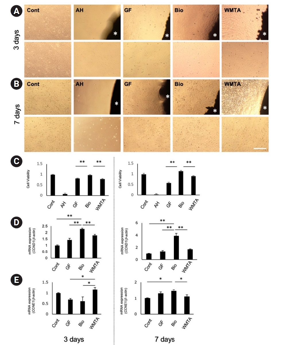
Effects of root canal sealers and mineral trioxide aggregate on the viability and proliferation of line 2–23. (A) Representative microscopic images of line 2–23 cultured with or without material discs (control [Cont], AH, GF, Bio, and WMTA) after 3 days. (B) Representative microscopic images after 7 days of culture. (C) Cell viability was determined by the MTT assay at 3 and 7 days. Cell viability was normalized to the control group and expressed as relative values, with the control set to 1.0. (D) Relative messenger RNA (mRNA) expression of the proliferation-related gene CCND1 in cells cultured with GF, Bio, and WMTA discs at 3 and 7 days. (E) Relative mRNA expression of CCNE1 in cells cultured with GF, Bio, and WMTA discs at 3 and 7 days. Gene expression levels were normalized to β-actin and calculated using the ΔΔCt method. Data are presented as mean ± standard deviation (n = 4). Statistical analysis was performed using one-way analysis of variance followed by the Bonferroni test (*p < 0.05, **p < 0.01). White asterisks in panels A and B indicate the location of the material discs. For the full product names and manufacturers of AH, Bio, GF, and WMTA, see Table 1.
Figure 2.
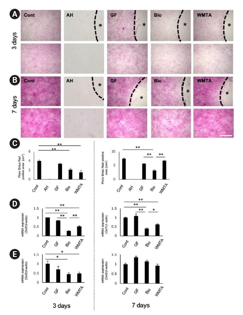
Collagen production of line 2–23 cultured with root canal sealers and white mineral trioxide aggregate discs. (A) Representative images of Picro-Sirius Red staining showing collagen deposition after 3 days of culture with or without material discs (control [Cont], AH, GF, Bio, and WMTA). (B) Representative images after 7 days of culture. (C) Quantitative analysis of collagen-positive areas at 3 and 7 days. (D) Relative messenger RNA (mRNA) expression of COL1 in cells cultured with GF, Bio, and WMTA discs at 3 and 7 days determined by real-time quantitative polymerase chain reaction. (E) Relative mRNA expression of COL3 in cells cultured with GF, Bio, and WMTA discs at 3 and 7 days. The gene expression levels were normalized to β-actin and calculated using the ΔΔCt method. Data are presented as mean ± standard deviation (n = 4). Statistical analysis was performed using one-way analysis of variance followed by the Bonferroni test (*p < 0.05; **p < 0.01). Black asterisks in panels A and B indicate the location of the material discs. For the full product names and manufacturers of AH, Bio, GF, and WMTA, see Table 1.
Figure 3.
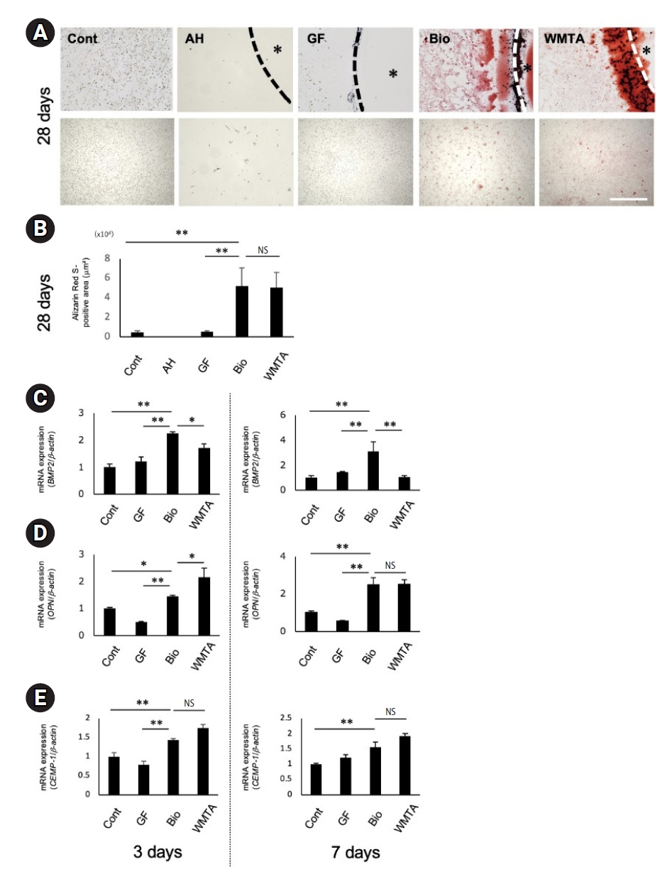
Mineralized matrix formation of line 2–23 cultured with root canal sealers and white mineral trioxide aggregate discs. (A) Representative images of Alizarin Red S staining showing mineralized matrix deposition after 28 days of culture with or without material discs (control [Cont], AH, GF, Bio, and WMTA). (B) Quantitative analysis of Alizarin Red S-positive areas after 28 days of culture. (C) Relative messenger RNA (mRNA) expression of BMP2 in cells cultured with GF, Bio, and WMTA discs at 3 and 7 days determined by RT-qPCR. (D) Relative mRNA expression of OPN in cells cultured with GF, Bio, and WMTA discs at 3 and 7 days. (E) Relative mRNA expression of CEMP1 in cells cultured with GF, Bio, and WMTA discs at 3 and 7 days. Gene expression levels were normalized to β-actin and calculated using the ΔΔCt method. Data are presented as mean ± standard deviation (n = 4). Statistical analysis was performed using one-way analysis of variance followed by the Bonferroni test (*p < 0.05, **p < 0.01). NS, not significant. Black asterisks in panel A indicate the location of the material discs. For the full product names and manufacturers of AH, Bio, GF, and WMTA, see Table 1.
Figure 4.
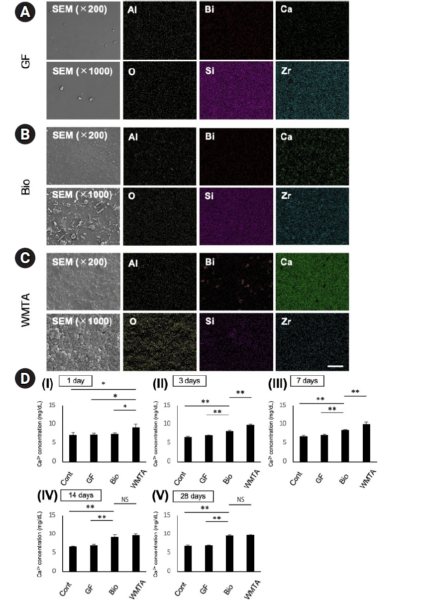
Surface characteristics and calcium ion release of the analyzed materials. (A–C) Representative scanning electron microscopy (SEM) images showing the surface morphology of (A) GF, (B) Bio, and (C) WMTA discs after incubation in culture medium. SEM images were obtained at low (×200) and high (×1,000) magnifications. Elemental distributions on the disc surfaces were visualized by energy-dispersive X-ray spectroscopy (EDS) mapping for Al, O, Bi, Si, Zr, and Ca. (D) Calcium ion release from GF, Bio, and WMTA discs. Discs were immersed in alpha-minimum essential medium (α-MEM), and Ca2+ concentrations in the medium were measured after immersion for (Ⅰ) 1 day, (Ⅱ) 3 days, (Ⅲ) 7 days, (Ⅳ) 14 days, and (Ⅴ) 28 days. Data are presented as mean ± standard deviation (n = 4). Statistical analysis was performed using one-way analysis of variance followed by the Bonferroni test (*p < 0.05, **p < 0.01). NS, not significant. For the full product names and manufacturers of Bio, GF, and WMTA, see Table 1.
Table 1.
Root canal sealers and MTA used in this study and their main compositions
Table 2.
Primer sequences used for real-time quantitative polymerase chain reaction analysis
Table 3.
Elemental composition of the tested materials determined by energy-dispersive X-ray spectroscopy
| Materiala) | Al | Bi | Ca | Na | O | Si | Zr | Total (wt%) |
|---|---|---|---|---|---|---|---|---|
| GF | 0.54 | 0.00 | 0.19 | 0.00 | 24.56 | 51.39 | 23.32 | 100 |
| Bio | 0.45 | 0.00 | 3.02 | 5.29 | 30.60 | 42.70 | 17.94 | 100 |
| WMTA | 0.86 | 0.64 | 33.5 | 1.39 | 60.04 | 2.14 | 1.43 | 100 |
GF and Bio showed high Si and Zr contents, whereas Ca was predominantly detected in Bio and WMTA, with the highest Ca level observed in WMTA.
a)For the full product names and manufacturers of GF, Bio, and WMTA, see Table 1.
- 1. Siqueira JF Jr, Rôças IN. Clinical implications and microbiology of bacterial persistence after treatment procedures. J Endod 2008;34:1291-1301.e3.ArticlePubMed
- 2. Islam R, Islam MRR, Tanaka T, Alam MK, Ahmed HMA, Sano H. Direct pulp capping procedures: evidence and practice. Jpn Dent Sci Rev 2023;59:48-61.ArticlePubMedPMC
- 3. Bürklein S, Arias A. Effectiveness of root canal instrumentation for the treatment of apical periodontitis: a systematic review and meta-analysis. Int Endod J 2023;56 Suppl 3:395-421.ArticlePubMed
- 4. Moura MS, Guedes OA, De Alencar AH, Azevedo BC, Estrela C. Influence of length of root canal obturation on apical periodontitis detected by periapical radiography and cone beam computed tomography. J Endod 2009;35:805-809.ArticlePubMed
- 5. Ribeiro DM, Réus JC, Felippe WT, Pacheco-Pereira C, Dutra KL, Santos JN, et al. Technical quality of root canal treatment performed by undergraduate students using hand instrumentation: a meta-analysis. Int Endod J 2018;51:269-283.ArticlePubMedPDF
- 6. Li GH, Niu LN, Zhang W, Olsen M, De-Deus G, Eid AA, et al. Ability of new obturation materials to improve the seal of the root canal system: a review. Acta Biomater 2014;10:1050-1063.ArticlePubMed
- 7. Tait C, Camilleri J, Blundell K. Non-surgical endodontics: obturation. Br Dent J 2025;238:487-496.ArticlePubMedPMCPDF
- 8. Tsuchiya K, Sauro S, Sano H, Matinlinna JP, Yamauti M, Hoshika S, et al. Clinical applications and classification of calcium silicate-based cements based on their history and evolution: a narrative review. Clin Oral Investig 2025;29:187.ArticlePubMedPDF
- 9. Tsuchiya K, Sauro S, Matinlinna JP, Sano H, Yamauti M, Mehta D, et al. Sealing ability of mineral trioxide aggregate: a scoping review of laboratory assessment methods. Eur J Oral Sci 2025;133:e70044.ArticlePubMedPDF
- 10. Torabinejad M, Parirokh M. Mineral trioxide aggregate: a comprehensive literature review: part II: leakage and biocompatibility investigations. J Endod 2010;36:190-202.ArticlePubMed
- 11. Torabinejad M, Parirokh M, Dummer PMH. Mineral trioxide aggregate and other bioactive endodontic cements: an updated overview: part II: other clinical applications and complications. Int Endod J 2018;51:284-317.ArticlePubMedPDF
- 12. Camilleri J, Atmeh A, Li X, Meschi N. Present status and future directions: hydraulic materials for endodontic use. Int Endod J 2022;55(Suppl 3):710-777.ArticlePubMedPMCPDF
- 13. Almeida MM, Rodrigues CT, Matos AA, Carvalho KK, Silva EJ, Duarte MA, et al. Analysis of the physicochemical properties, cytotoxicity and volumetric changes of AH Plus, MTA Fillapex and TotalFill BC Sealer. J Clin Exp Dent 2020;12:e1058-e1065.ArticlePubMedPMC
- 14. Wu D, Tang Z, Zhang G, Liu W. The sealing ability of a new silicone-based root canal filling material (GuttaFlow): an in vitro study using the percentage of gutta-percha-filled area. Dent Mater J 2011;30:569-575.ArticlePubMed
- 15. Lyu WJ, Bai W, Wang XY, Liang YH. Physicochemical properties of a novel bioceramic silicone-based root canal sealer. J Dent Sci 2022;17:831-835.ArticlePubMed
- 16. Accardo C, Himel VT, Lallier TE. A novel GuttaFlow sealer supports cell survival and attachment. J Endod 2014;40:231-234.ArticlePubMed
- 17. Mandal P, Zhao J, Sah SK, Huang Y, Liu J. In vitro cytotoxicity of guttaflow 2 on human gingival fibroblasts. J Endod 2014;40:1156-1159.ArticlePubMed
- 18. Hoikkala NJ, Wang X, Hupa L, Smått JH, Peltonen J, Vallittu PK. Dissolution and mineralization characterization of bioactive glass ceramic containing endodontic sealer Guttaflow Bioseal. Dent Mater J 2018;37:988-994.ArticlePubMed
- 19. Lee SH, Oh S, Al-Ghamdi AS, Mandorah AO, Kum KY, Chang SW. Sealing ability of AH Plus and GuttaFlow Bioseal. Bioinorg Chem Appl 2020;2020:8892561.ArticlePubMedPMCPDF
- 20. Gandolfi MG, Siboni F, Prati C. Properties of a novel polysiloxane-guttapercha calcium silicate-bioglass-containing root canal sealer. Dent Mater 2016;32:e113-e126.ArticlePubMed
- 21. Seo BM, Miura M, Gronthos S, Bartold PM, Batouli S, Brahim J, et al. Investigation of multipotent postnatal stem cells from human periodontal ligament. Lancet 2004;364:149-155.ArticlePubMed
- 22. Zhu W, Liang M. Periodontal ligament stem cells: current status, concerns, and future prospects. Stem Cells Int 2015;2015:972313.ArticlePubMedPMCPDF
- 23. Zheng W, Wang S, Ma D, Tang L, Duan Y, Jin Y. Loss of proliferation and differentiation capacity of aged human periodontal ligament stem cells and rejuvenation by exposure to the young extrinsic environment. Tissue Eng Part A 2009;15:2363-2371.ArticlePubMed
- 24. Zhang J, An Y, Gao LN, Zhang YJ, Jin Y, Chen FM. The effect of aging on the pluripotential capacity and regenerative potential of human periodontal ligament stem cells. Biomaterials 2012;33:6974-6986.ArticlePubMed
- 25. Hasegawa D, Hasegawa K, Kaneko H, Yoshida S, Mitarai H, Arima M, et al. MEST regulates the stemness of human periodontal ligament stem cells. Stem Cells Int 2020;2020:9672673.ArticlePubMedPMCPDF
- 26. Tomokiyo A, Hasegawa D, Ono T, Nagano R, Ipposhi K, Yamashita K, et al. Characterization of a clonal human periodontal ligament stem cell line exposed to methacrylate resin-, bioactive glass-, or silicon-based root canal sealers. Odontology 2022;110:127-137.ArticlePubMedPDF
- 27. Hwang HC, Clurman BE. Cyclin E in normal and neoplastic cell cycles. Oncogene 2005;24:2776-2786.ArticlePubMed
- 28. López-García S, Sánchez-Bautista S, García-Bernal D, Lozano A, Forner L, Sanz JL, et al. Premixed calcium silicate-based ceramic sealers promote osteogenic/cementogenic differentiation of human periodontal ligament stem cells: a microscopy study. Microsc Res Tech 2024;87:1584-1597.ArticlePubMed
- 29. Tomokiyo A, Wada N, Maeda H. Periodontal ligament stem cells: regenerative potency in periodontium. Stem Cells Dev 2019;28:974-985.ArticlePubMed
- 30. Prati C, Gandolfi MG. Calcium silicate bioactive cements: biological perspectives and clinical applications. Dent Mater 2015;31:351-370.ArticlePubMed
- 31. Tsuchiya K, Sauro S, Sano H, Matinlinna JP, Yamauti M, Hoshika S, et al. Physicochemical properties and apatite precipitation behavior of experimental calcium silicate-based cements doped with phosphate compounds. Dent Mater 2025;41:1058-1066.ArticlePubMed
- 32. Gandolfi MG, Shah SN, Feng R, Prati C, Akintoye SO. Biomimetic calcium-silicate cements support differentiation of human orofacial mesenchymal stem cells. J Endod 2011;37:1102-1108.ArticlePubMedPMC
- 33. Simila HO, Karpukhina N, Hill RG. Bioactivity and fluoride release of strontium and fluoride modified Biodentine. Dent Mater 2018;34:e1-e7.ArticlePubMed
- 34. Uribe P, Johansson A, Jugdaohsingh R, Powell JJ, Magnusson C, Davila M, et al. Soluble silica stimulates osteogenic differentiation and gap junction communication in human dental follicle cells. Sci Rep 2020;10:9923.ArticlePubMedPMCPDF
- 35. Tsuchiya K, Sauro S, Matinlinna JP, Yamauti M, Sano H, Min KS, et al. Trends and future perspectives in additive modifications of calcium silicate-based cements: a scoping review (2015-2024). Aust Endod J 2026;52:606-618.ArticlePubMedPMCPDF
- 36. Xynos ID, Edgar AJ, Buttery LD, Hench LL, Polak JM. Gene-expression profiling of human osteoblasts following treatment with the ionic products of Bioglass 45S5 dissolution. J Biomed Mater Res 2001;55:151-157.ArticlePubMed
- 37. Choi MJ, Kang ES, Kim MK, Tsuchiya K, Matinlinna JP, Lee KW, et al. Biological effects of a premixed calcium silicate pulp-capping material containing dimethyl sulphoxide as a vehicle: In vitro and in vivo study. Eur J Oral Sci 2026;134:e70096.Article
REFERENCES
Tables & Figures
REFERENCES
Citations
Citations to this article as recorded by 
 Cite
 XML Download
Direct cellular responses of a human periodontal ligament stem cell line to root canal sealers and white mineral trioxide aggregate: an in vitro study
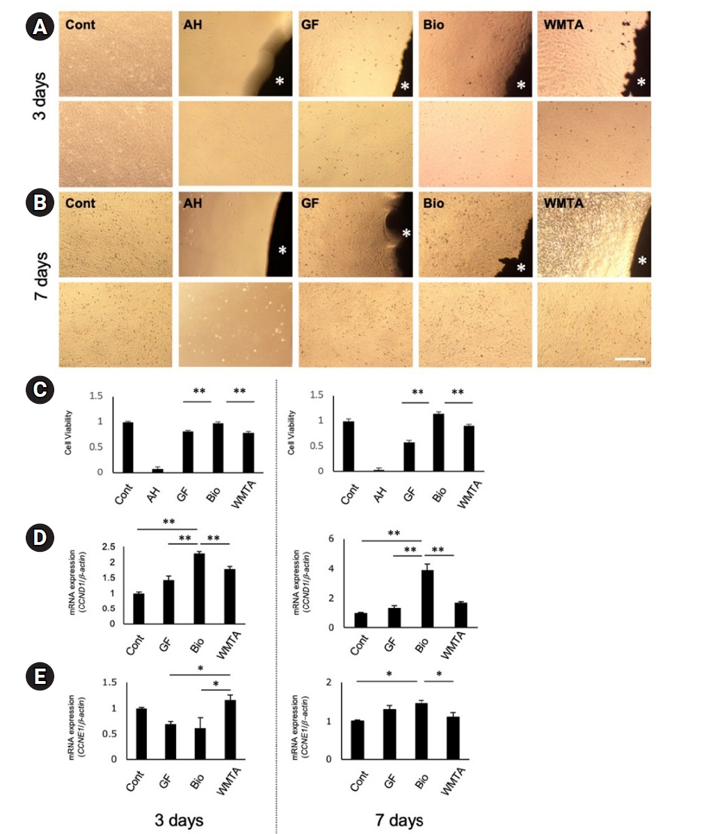
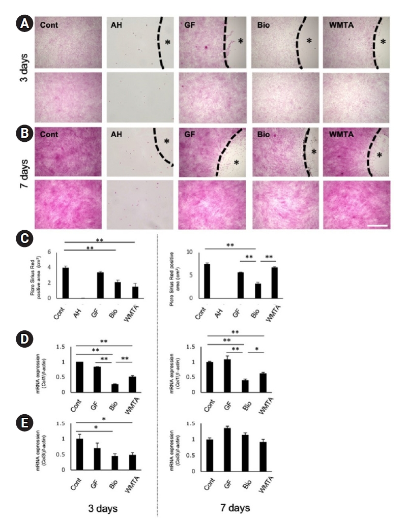
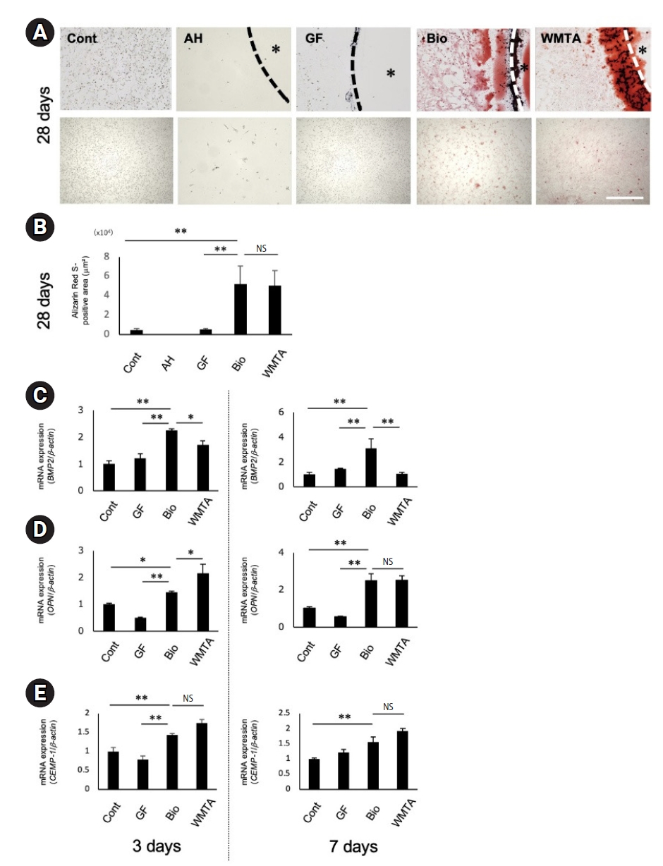
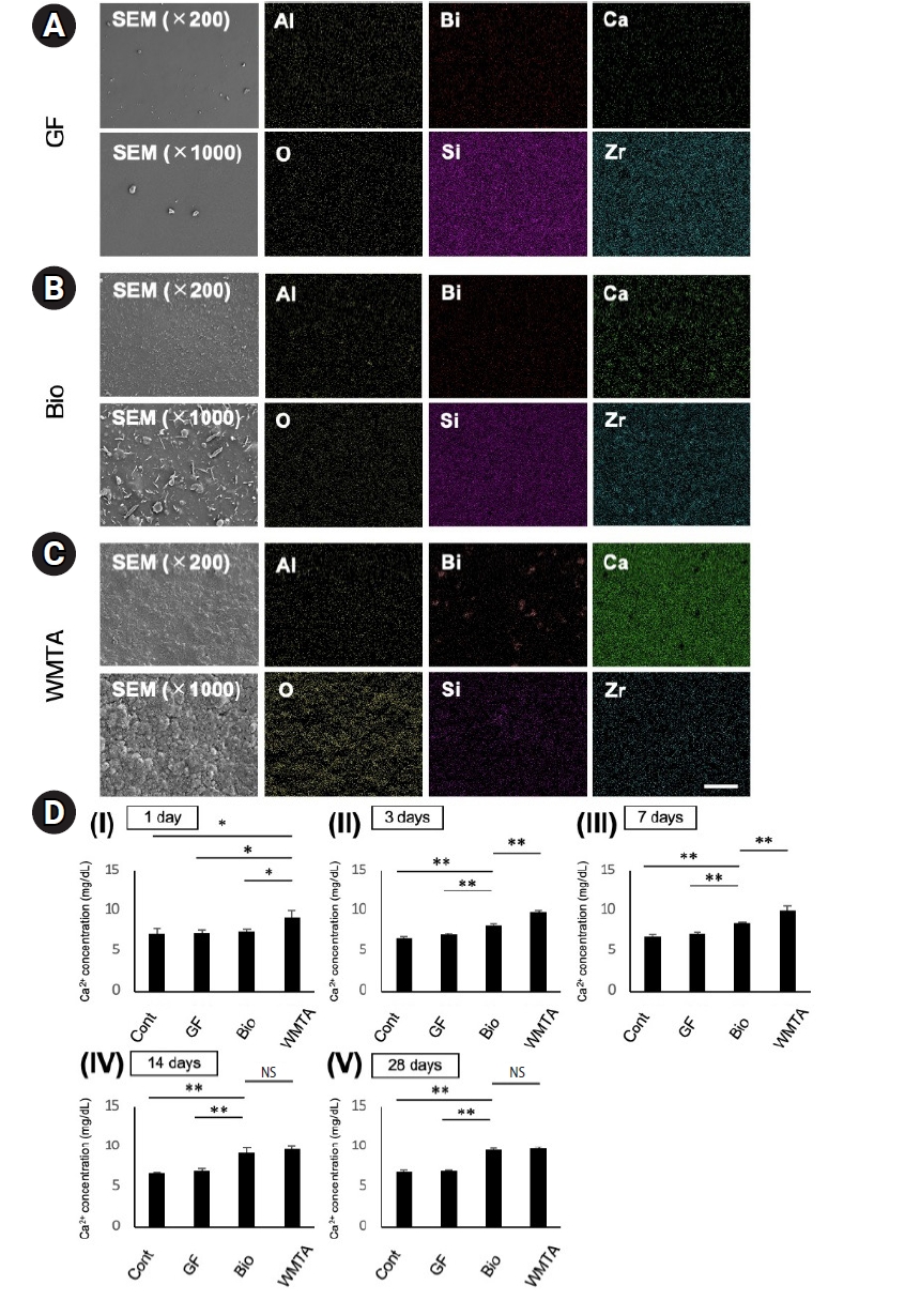
Figure 1. Effects of root canal sealers and mineral trioxide aggregate on the viability and proliferation of line 2–23. (A) Representative microscopic images of line 2–23 cultured with or without material discs (control [Cont], AH, GF, Bio, and WMTA) after 3 days. (B) Representative microscopic images after 7 days of culture. (C) Cell viability was determined by the MTT assay at 3 and 7 days. Cell viability was normalized to the control group and expressed as relative values, with the control set to 1.0. (D) Relative messenger RNA (mRNA) expression of the proliferation-related gene CCND1 in cells cultured with GF, Bio, and WMTA discs at 3 and 7 days. (E) Relative mRNA expression of CCNE1 in cells cultured with GF, Bio, and WMTA discs at 3 and 7 days. Gene expression levels were normalized to β-actin and calculated using the ΔΔCt method. Data are presented as mean ± standard deviation (n = 4). Statistical analysis was performed using one-way analysis of variance followed by the Bonferroni test (*p < 0.05, **p < 0.01). White asterisks in panels A and B indicate the location of the material discs. For the full product names and manufacturers of AH, Bio, GF, and WMTA, see Table 1.
Figure 2. Collagen production of line 2–23 cultured with root canal sealers and white mineral trioxide aggregate discs. (A) Representative images of Picro-Sirius Red staining showing collagen deposition after 3 days of culture with or without material discs (control [Cont], AH, GF, Bio, and WMTA). (B) Representative images after 7 days of culture. (C) Quantitative analysis of collagen-positive areas at 3 and 7 days. (D) Relative messenger RNA (mRNA) expression of COL1 in cells cultured with GF, Bio, and WMTA discs at 3 and 7 days determined by real-time quantitative polymerase chain reaction. (E) Relative mRNA expression of COL3 in cells cultured with GF, Bio, and WMTA discs at 3 and 7 days. The gene expression levels were normalized to β-actin and calculated using the ΔΔCt method. Data are presented as mean ± standard deviation (n = 4). Statistical analysis was performed using one-way analysis of variance followed by the Bonferroni test (*p < 0.05; **p < 0.01). Black asterisks in panels A and B indicate the location of the material discs. For the full product names and manufacturers of AH, Bio, GF, and WMTA, see Table 1.
Figure 3. Mineralized matrix formation of line 2–23 cultured with root canal sealers and white mineral trioxide aggregate discs. (A) Representative images of Alizarin Red S staining showing mineralized matrix deposition after 28 days of culture with or without material discs (control [Cont], AH, GF, Bio, and WMTA). (B) Quantitative analysis of Alizarin Red S-positive areas after 28 days of culture. (C) Relative messenger RNA (mRNA) expression of BMP2 in cells cultured with GF, Bio, and WMTA discs at 3 and 7 days determined by RT-qPCR. (D) Relative mRNA expression of OPN in cells cultured with GF, Bio, and WMTA discs at 3 and 7 days. (E) Relative mRNA expression of CEMP1 in cells cultured with GF, Bio, and WMTA discs at 3 and 7 days. Gene expression levels were normalized to β-actin and calculated using the ΔΔCt method. Data are presented as mean ± standard deviation (n = 4). Statistical analysis was performed using one-way analysis of variance followed by the Bonferroni test (*p < 0.05, **p < 0.01). NS, not significant. Black asterisks in panel A indicate the location of the material discs. For the full product names and manufacturers of AH, Bio, GF, and WMTA, see Table 1.
Figure 4. Surface characteristics and calcium ion release of the analyzed materials. (A–C) Representative scanning electron microscopy (SEM) images showing the surface morphology of (A) GF, (B) Bio, and (C) WMTA discs after incubation in culture medium. SEM images were obtained at low (×200) and high (×1,000) magnifications. Elemental distributions on the disc surfaces were visualized by energy-dispersive X-ray spectroscopy (EDS) mapping for Al, O, Bi, Si, Zr, and Ca. (D) Calcium ion release from GF, Bio, and WMTA discs. Discs were immersed in alpha-minimum essential medium (α-MEM), and Ca2+ concentrations in the medium were measured after immersion for (Ⅰ) 1 day, (Ⅱ) 3 days, (Ⅲ) 7 days, (Ⅳ) 14 days, and (Ⅴ) 28 days. Data are presented as mean ± standard deviation (n = 4). Statistical analysis was performed using one-way analysis of variance followed by the Bonferroni test (*p < 0.05, **p < 0.01). NS, not significant. For the full product names and manufacturers of Bio, GF, and WMTA, see Table 1.
Figure 1.
Figure 2.
Figure 3.
Figure 4.
Direct cellular responses of a human periodontal ligament stem cell line to root canal sealers and white mineral trioxide aggregate: an in vitro study
| Material | Type | Main composition | Manufacturer |
|---|---|---|---|
| AH Plus (AH) | Epoxy resin-based root canal sealer | Bisphenol-A epoxy resin, amine hardeners, zirconium oxide, calcium tungstate, silica fillers, iron oxide pigments | Dentsply Sirona, Tulsa, OK, USA |
| GuttaFlow 2 (GF) | Silicone-based root canal sealer | Polydimethylsiloxane, gutta-percha powder, zirconium dioxide, silica fillers | Coltene, Altstätten, Switzerland |
| GuttaFlow Bioseal (Bio) | Bioceramics-modified silicone-based root canal sealer | Polydimethylsiloxane, gutta-percha particles, calcium silicate bioactive glass, nanosilver, zirconium dioxide | Coltene, Altstätten, Switzerland |
| ProRoot MTA (WMTA) | Hydraulic calcium silicate-based cement | Tricalcium silicate, dicalcium silicate, tricalcium aluminate, calcium sulfate, bismuth oxide | Dentsply Sirona, Tulsa, OK, USA |
| Target gene (abbreviation) | Forward primer (top) and reverse (bottom) primer sequences | Size of amplified products (bp) | Annealing temperature (°C) | No. of cycles |
|---|---|---|---|---|
| CCND1 | AGGAGAACAAACAGATCA/ | 223 | 60 | 40 |
| TAGGACAGGAAGTTGTTG | ||||
| CCNE1 | GCCAGCCTTGGGACAATAATG/ | 104 | 60 | 40 |
| CTTGCACGTTGAGTTTGGGT | ||||
| COL1 | CCCGGGTTTCAGAGACAACTTC/ | 148 | 60 | 40 |
| TCCACATGCTTTATTCCAGCAATC | ||||
| COL3 | GCAAATTCACCTACACAGTTCTGGA/ | 147 | 60 | 40 |
| CTTGATCAGGACCACCAATGTCATA | ||||
| BMP2 | TCCACTAATCATGCCATTGTTCAGA/ | 74 | 60 | 40 |
| GGGACACAGCATGCCTTAGGA | ||||
| OPN | ACACATATGATGGCCGAGGTGA/ | 115 | 60 | 40 |
| TGTGAGGTGATGTCCTCGTCTGT | ||||
| CEMP1 | CTGGCACAGGGTATACAGGGTTAG/ | 142 | 60 | 40 |
| ACTGGTGCCGTTTATGCCTTG | ||||
| β-actin | ATTGCCGACAGGATGCAGA/ | 89 | 60 | 40 |
| GAGTACTTGCGCTCAGGAGGA |
| Material |
Al | Bi | Ca | Na | O | Si | Zr | Total (wt%) |
|---|---|---|---|---|---|---|---|---|
| GF | 0.54 | 0.00 | 0.19 | 0.00 | 24.56 | 51.39 | 23.32 | 100 |
| Bio | 0.45 | 0.00 | 3.02 | 5.29 | 30.60 | 42.70 | 17.94 | 100 |
| WMTA | 0.86 | 0.64 | 33.5 | 1.39 | 60.04 | 2.14 | 1.43 | 100 |
Table 1. Root canal sealers and MTA used in this study and their main compositions
Table 2. Primer sequences used for real-time quantitative polymerase chain reaction analysis
Table 3. Elemental composition of the tested materials determined by energy-dispersive X-ray spectroscopy
GF and Bio showed high Si and Zr contents, whereas Ca was predominantly detected in Bio and WMTA, with the highest Ca level observed in WMTA. For the full product names and manufacturers of GF, Bio, and WMTA, see


