
 KACD

Articles
- Page Path
- HOME > Restor Dent Endod > Ahead-of print articles > Article
- Research Article Monomer conversion, flexural strength, fluoride release, and color stability of experimental resin composites containing bioactive glass nanoparticles: an in vitro study
-
Tongjai Chotitanmapong1
 , Sawaskorn Temkitthaworn1, Linlinee Arnanthigo1, Papawarin Amornchaiyakij1, Pakkapol Assawaarsu1, Phantira Vatthanachairoj1, Wuttipong Chontanasawat1, Nitchakarn Leenutaphong1, Parichart Naruphontjirakul2, Chawal Padunglappisit1, Piyaphong Panpisut1,3,*
-
Restor Dent Endod [Epub ahead of print]
DOI: https://doi.org/10.5395/rde.2026.51.e46
Published online: November 17, 2026
1Faculty of Dentistry, Thammasat University, Pathum Thani, Thailand
2Biological Engineering Program, Faculty of Engineering, King Mongkut’s University of Technology Thonburi, Bangkok, Thailand
3Thammasat University Research Unit in Advanced Dental Biomaterials for Sustainable Oral Health Care, Thammasat University, Pathum Thani, Thailand
- *Correspondence to Piyaphong Panpisut, DDS, MSc, PhD Division of Restorative Dentistry, Faculty of Dentistry, Thammasat University, 99 M. 18, Klong Nueng, Klong Laung, Pathum Thani 12120, Thailand Email: panpisut@tu.ac.th
• Received: January 7, 2026 • Revised: March 9, 2026 • Accepted: June 24, 2026
© 2026 The Korean Academy of Conservative Dentistry
This is an Open-Access article distributed under the terms of the Creative Commons Attribution Non-Commercial License (http://creativecommons.org/licenses/by-nc/4.0) which permits unrestricted non-commercial use, distribution, and reproduction in any medium, provided the original work is properly cited.
- 45 Views
- 4 Download


Abstract
-
Objectives This study evaluated the degree of conversion (DC), mechanical properties, fluoride release, and color stability of a newly developed ion-releasing resin composite (Thamfil) containing calcium phosphate and bioactive glass compared to commercial materials.
-
Methods DC was measured using attenuated total reflectance Fourier-transform infrared spectroscopy (n = 5). Biaxial flexural strength (BFS) and modulus (BFM) were tested after 24 hours of immersion in deionized water at 37°C (n = 5). The cumulative fluoride release in deionized water for 4 weeks was also determined (n = 4). Color stability (∆E*00) was assessed after continuous immersion in deionized water or red wine at 37°C for 2 weeks (n = 5 per solution) using an intraoral spectrophotometer. Comparisons included FiltekBulk (3M Oral Care), BeautifilBulk (Shofu), and CentionN (Ivoclar Vivadent).
-
Results Thamfil showed significantly higher DC (74%) than other materials (46%–54%, p < 0.05). BFS was highest for FiltekBulk (232 MPa), while Thamfil (133 MPa) was comparable to CentionN (160 MPa) and BeautifilBulk (126 MPa) (p > 0.05). BFM of Thamfil (2.6 GPa) was similar to that of BeautifilBulk (2.3 GPa) and CentionN (3.3 GPa) (p > 0.05). The cumulative fluoride release at 28 days for Thamfil (3.3 ppm) was comparable to BeautifilBulk (3.4 ppm) but was lower than CentionN (92.3 ppm). Thamfil showed higher ∆E*00 in water (3.6) than other materials (0.5–1.3) (p < 0.05), but a similar color change in red wine (39.2) to that of FiltekBulk (p > 0.05).
-
Conclusions Thamfil exhibited high polymerization but low color stability. However, the material exhibited comparable strength and fluoride release to those of a commercial ion-releasing material (BeautifilBulk).
INTRODUCTION
Untreated caries is the most common preventable chronic disease. The global burden of caries was estimated to be 2.24 billion cases in permanent teeth and 0.52 billion cases in primary teeth [1]. In 2050, the number of prevalent cases is predicted to reach 2.26 billion [1]. Additionally, Asia is projected to account for approximately half of all new global cases [1]. The direct treatment costs for dental diseases were estimated to be approximately 298 billion US dollars (USD) annually, while the indirect costs due to productivity loss were about 144 billion USD [2]. When caries progresses and forms a cavitated lesion, restorative treatment may be recommended to restore form, function, and esthetics for the patient.
Resin composite is the most commonly selected restorative material due to its excellent esthetics. However, the most common failure of the restoration is often attributed to secondary caries, which can be up to 15% [3]. This may drive the need for replacement and can subsequently compromise tooth integrity through the restorative cycle. Additionally, repeated replacement negatively impacts environmental sustainability through the generation of microparticulate waste, increased chemical release, leachate formation in landfills, and an increased carbon footprint associated with patient travel and material manufacturing [4].
The development of ion-releasing resin-based materials, such as alkasite, is intended to simplify the complex placement procedures associated with traditional resin composites, such as dual curing and bulk placement [5]. The material released multiple ions, such as calcium, fluoride, phosphate, strontium, and hydroxide. These ions are essential to encourage an alkaline environment suitable for acid neutralization and ionic concentration for apatite precipitation [6]. These features are expected to reduce demineralization and potentially reduce the risk of secondary caries. However, a systematic review found no statistically significant difference in the occurrence of secondary caries or marginal adaptation between ion-releasing materials and resin composites [7].
It is hypothesized that the ion-releasing actions are primarily due to ion-releasing fillers, such as bioactive glass particles (BAGs) and alkaline fillers [8]. The dissolution of bioactive glass fillers facilitates the release of multiple ions that could promote remineralization and also reduce bacterial growth [9]. Additionally, the incorporation of calcium phosphates, such as monocalcium phosphate monohydrate (MCPM), also encourages mineral precipitation, which could potentially reduce demineralization [10]. The acidic nature of MCPM also enhances the self-etching effects that may promote adhesion of the material to the tooth structure [11]. The addition of these additives may, however, compromise physical and mechanical properties [12]. The incorporation of different glass fillers may introduce a refractive index mismatch, which affects light penetration and polymerization of the resin composite [13]. Additionally, the increase in water sorption due to hydrophilic components may affect the color stability of the materials [14].
The objective of the current study was to prepare ion-releasing resin composites containing MCPM and BAGs and to evaluate monomer conversion, biaxial flexural strength (BFS), fluoride release, and color stability compared with commercial materials. The null hypotheses were that there would be no significant differences among materials in (i) degree of conversion (DC), (ii) BFS, and biaxial flexural modulus (BFM) after 24 hours of water immersion, (iii) cumulative fluoride release over 4 weeks, and (iv) color change (ΔE*00) after immersion in deionized water or red wine (2 weeks).
METHODS
As this in vitro study did not involve human subjects or animal models, Institutional Review Board approval was not required. The experimental design of the current study is provided in Figure 1.
BAGs were prepared via a sol-gel approach with subsequent functionalization and thermal treatment, following protocols described previously [15]. The synthesis involved two main stages: the initial formation of silica nanoparticles, followed by the incorporation of calcium, strontium, and fluoride. An aqueous-ethanol mixture containing deionized water, ammonium hydroxide, and ethanol was stirred at 600 revolutions/min (rpm) for 30 minutes in an Erlenmeyer flask. After adding tetraethyl orthosilicate, the solution was stirred overnight at ambient temperature to facilitate silica network formation. Centrifugation (5,000 rpm for 30 minutes) was used to isolate the SiO2 nanoparticles, which were subsequently washed and redispersed in water. Elemental doping was achieved by introducing Ca(NO3)2•4H2O, Sr(NO3)2, and NaF to obtain a nominal (precursor-based) SiO2:CaO:SrO:NaF molar composition of 1.0:0.33:1.0:0.5. Following overnight drying, the particles underwent calcination at 680°C to eliminate nitrate residues, then received two ethanol wash cycles. Particle size analysis revealed a mean diameter of approximately 200 nm [15]. Elemental composition was examined using scanning electron microscopy coupled with energy-dispersive X-ray spectroscopy using a JSM-7800F scanning electron microscope (JEOL, Tokyo, Japan) and an X-Max 20 EDX detector (Oxford Instruments, Abingdon, UK) (Figure 2).
The experimental resin composites were prepared using a protocol similar to that in the previous study [16]. The composition of the experimental material is provided in Table 1. The liquid phase consisted of urethane dimethacrylate (Sigma-Aldrich, St. Louis, MO, USA), triethylene glycol dimethacrylate (Sigma-Aldrich), 2-hydroxyethyl methacrylate (HEMA; Sigma-Aldrich), 10-methacryloyloxydecyl dihydrogen phosphate (Watson International, Jiangsu, China), and trimethylbenzoyldiphenylphosphine oxide (TPO; Sigma-Aldrich). Furthermore, benzoyl peroxide (Sigma-Aldrich) and N,N-dimethyl-p-toluidine (DMPT; Sigma-Aldrich, St. Louis, MO, USA) were used as an initiator and activator, respectively. The powder phase of the experimental composite contained silanated barium aluminosilicate glass (diameter of 0.18–7 µm; Esstech, Essington, PA, USA), BAGs (diameter of approximately 200 nm), and MCPM (diameter of approximately 10 µm; Himed, Old Bethpage, NY, USA).
The liquid and powder phases were mixed at a 2:1 powder-to-liquid mass ratio for 20 seconds until a homogeneous appearance was obtained. The mixed initiator and activator pastes were separately loaded into double-barrel syringes (medmix Switzerland AG, Haag, Switzerland). The initiator and activator pastes of experimental composites were then mixed using the syringe mixing tip and dispenser (medmix Switzerland AG, Haag, Switzerland). The commercial comparisons (Table 1) were Filtek One Bulk Fill Restorative (3M Oral Care, St. Paul, MN, USA), Cention N (Ivoclar Vivadent, Schaan, Liechtenstein), and Beautifil-Bulk Restorative (Shofu, Kyoto, Japan).
This study conducted a prior power analysis using a BFS as the main outcome. The power analysis was performed in IBM SPSS ver. 31 (IBM Corp, Armonk, NY, USA) using the strength results from a similar previous study [17] that examined the physical and mechanical properties of an ion-releasing resin composite. The power was set to 0.95, and the calculated effect size, based on the root-mean-square standardized effect, was 3.894. The result suggests that a sample size of n = 5 per group should provide power greater than 0.95 for one-way analysis of variance (ANOVA) at α = 0.05.
The degree of monomer conversion (DC, n = 5) for each material was determined using attenuated total reflectance Fourier-transform infrared spectroscopy (ATR-FTIR; Nicolet iS5 Fourier Transform Infrared Spectrometer, Thermo Fisher Scientific, Waltham, MA, USA). Each material was placed on a metal circlip (1 mm thick, 10 mm in diameter) positioned over the ATR diamond. Initial spectra were obtained, and FTIR spectra in the range of 400–4,000 cm–1 were recorded from the bottom of the specimen with eight repetitions at a resolution of 4 cm–1. The specimens were then light-cured from the top for 40 seconds using a light-emitting diode light-curing unit (irradiance of approximately 1,500 mW/cm2; Eighteeth Curing Pen, Eighteeth, Shenzhen, China). The FTIR spectra were then immediately obtained after light-curing. The DC (%) was then calculated using Equation 1.
where ∆A0 and ∆At represent the peak’s height at the C-O of the methacrylate group (1,320 cm–1) above the background level at 1,335 cm–1 before curing and after initiating curing at 40 seconds (t), respectively.
The mechanical properties of the materials were evaluated using a ball-on-ring testing jig to determine their BFS and BFM. To prepare disc specimens (n = 5), each material was placed within a metal circlip (1 mm thick and 10 mm in diameter) and pressed with acetate sheets and glass slabs on both the upper and lower surfaces. The specimens were light-cured for 40 seconds on each side, then stored at 25°C for an additional 24 hours. Subsequently, the samples were removed from the molds, any excess material was trimmed, and the specimens were immersed in 5 mL of deionized water maintained at 37°C for 24 hours. Before testing, each specimen’s thickness was measured with a digital vernier caliper. The specimens were then placed on a ball-on-ring testing jig in a mechanical testing frame (AGSX, Shimadzu, Kyoto, Japan). Testing was conducted at a crosshead speed of 1 mm/min using a 500 N load cell. The failure load (N) was recorded. Then, BFS (Pa) and BFM (Pa) values were determined using Equations 2 and 3.
F indicates the failure load measured in newtons (N), d denotes the thickness of the disc specimens in meters (m), r refers to the radius of the circular support of the ball-on-ring testing jig (m), and v represents Poisson’s ratio, set at 0.3. Additionally, ΔH/ΔWc is the gradient of the elastic region of the force versus central deflection curve (N/m), and βc is the center deflection function (0.5024).
Fluoride release in deionized water was measured using a fluoride-specific electrode (Orion Versastar Pro, Thermo Fisher Scientific). Disc specimens (10 mm diameter, 1 mm thickness; n = 4) were made and immersed in 5 mL of deionized water. Samples were incubated at 37°C for up to 4 weeks. Calibration was performed using fluoride concentrations of 1, 10, 100, and 1,000 ppm. The solution was mixed with TISAB III (1:10 vol ratio), and the fluoride concentration was measured.
The color stability (ΔE*00) of the materials after immersion in deionized water (n = 5) or red wine (n = 5) was evaluated using an intraoral dental spectrophotometer (Easyshade V, VITA Zahnfabrik, Bad Säckingen, Germany). Although the Easyshade V is primarily designed for clinical shade matching, it has been used in a previous in vitro study to obtain objective CIE L*a*b* coordinates under standardized measurement conditions [18]. Disc specimens (1 mm in thickness and 10 mm in diameter) were prepared and placed over a standard gray background (Kodak R-27 Neutral Test Card, Eastman Kodak Company, Rochester, NY, USA) in an illumination box with approximately 1,000 lux illumination, measured by a light meter (LX1330B Digital Illuminance/Light Meter, Dr. Meter, Shenzhen, China). Baseline CIELAB coordinates (CIE L, a, b, C*, and h°) were recorded for each specimen. The specimens were then immersed in 5 mL of deionized water or red wine and incubated at 37°C for up to 2 weeks. Color measurements were repeated after 2 weeks of immersion without changing the storage solution. L* represents lightness, a* and b* represent values along the red-green and yellow–blue axes, respectively, and C* and h° correspond to chroma and hue angle. Color differences (∆E*00) upon immersion were calculated using the CIEDE2000 formula in Equation 4.
where ∆L', ∆C', and ∆H' represent the differences in lightness, chroma, and hue, respectively, before and after immersion. RT represents a rotation function that accounts for the interaction between chroma and hue differences in the blue region. Additionally, SL, SC, and SH are weighting functions, and KL, KC, and KH are parametric factors for adjusting experimental conditions. The complete formula and associated equations were obtained from the previous study [19].
Data are shown as means with 95% confidence intervals. Statistical analysis was performed using Prism version 10.0 for macOS (GraphPad Software, San Diego, CA, USA). The normality of the data was assessed using the Shapiro-Wilk test. For normally distributed results (DC, BFS/BFM, F release, and ΔE*00), comparisons were made using one-way ANOVA followed by Tukey honestly significant difference post hoc test. For non-normally distributed results (ΔL, Δa, Δb), the Kruskal-Wallis test was used, followed by Dunn test for multiple comparisons. The significance level was set at p < 0.05.
RESULTS
The highest DC after light-curing for 40 seconds was detected with Thamfil (74.0% ± 2.6%), and the lowest value was obtained from BeautifilBulk (45.6% ± 7.5%) (Figure 3). The DC of Thamfil was significantly higher than that of CentionN (53.5% ± 5.3%), FiltekBulk (48.8% ± 2.6%), and BeautifilBulk. The DC values of CentionN, FiltekBulk, and BeautifilBulk were comparable (p > 0.05).
The highest BFS was detected with FiltekBulk (232 ± 19 MPa), while the lowest value was detected with BeautifilBulk (126 ± 8 MPa) (Figure 4). The BFS of FiltekBulk was significantly higher than that of CentionN (160 ± 18 MPa, p < 0.01), BeautifilBulk (p < 0.01), and Thamfil (133 ± 14 MPa, p < 0.01). The BFS of Thamfil, however, was comparable to that of CentionN (p = 0.1026) and BeautifilBulk (p = 0.9295).
FiltekBulk also exhibited the highest BFM (4.3 ± 0.7 GPa), while the lowest value was detected with BeautifilBulk (2.3 ± 0.1 GPa). The BFM of FiltekBulk was significantly higher than BeautifilBulk (p = 0.0003) and Thamfil (p = 0.0016). The BFM of Thamfil was comparable to that of CentionN (3.3 ± 0.7 GPa, p = 0.3205) and BeautifilBulk (p = 0.7966).
The highest cumulative fluoride release at 4 weeks was detected with CentionN (92.3 ± 15.0 ppm) (Figure 5). The value was significantly higher than that of BeautifilBulk (3.4 ± 1.1 ppm) (p < 0.01) and Thamfil (3.3 ± 1.0 ppm) (p < 0.01). However, the values of BeautifilBulk and Thamfil were comparable (p > 0.05). The fluoride release from FiltekBulk was below the detection limit.
After water immersion (Table 2), all materials exhibited minimal changes in lightness (∆L). FiltekBulk remained nearly unchanged, while CentionN (0.42 ± 0.37) and BeautifilBulk (0.24 ± 0.68) became slightly lighter. Thamfil showed the highest lightness change (3.08 ± 0.79), significantly different from FiltekBulk (p = 0.0275). For the red-green axis (∆a), FiltekBulk (–0.36 ± 0.16) and CentionN (0.28 ± 0.04) showed minimal changes, while Thamfil (–2.24 ± 0.18) demonstrated the most pronounced green shift, significantly different from CentionN (p = 0.0003). On the yellow–blue axis (∆b), CentionN (0.48 ± 0.17) shifted slightly toward yellow, while Thamfil (–1.94 ± 0.70) showed the greatest blue shift, significantly different from CentionN (p = 0.0011). Thamfil exhibited the highest overall color change (∆E*00 = 3.57 ± 0.65), significantly higher than FiltekBulk (1.33 ± 0.88, p = 0.0003), BeautifilBulk (0.62 ± 0.28, p < 0.0001), and CentionN (0.55 ± 0.13, p < 0.0001), whereas the three commercial materials did not differ from one another (p>0.05) (Figure 6A).
Following wine immersion, all materials exhibited substantial darkening. CentionN showed the greatest darkening (∆L = –47.22 ± 1.60), significantly different from FiltekBulk (–40.34 ± 1.11, p = 0.0105), while BeautifilBulk (–44.94 ± 3.59) and Thamfil (–42.06 ± 1.46) showed intermediate values. All materials shifted toward red (∆a), with BeautifilBulk (19.16 ± 1.57) significantly higher than FiltekBulk (15.48 ± 0.79) and Thamfil (15.42 ± 1.14). BeautifilBulk demonstrated a dramatic yellow shift (∆b = 28.88 ± 3.14), significantly different from CentionN (–0.76 ± 1.13, p = 0.0028) and Thamfil (–0.36 ± 0.99, p = 0.0088), but not from FiltekBulk (5.88 ± 1.58, p > 0.9999. All materials exhibited severe overall color changes (∆E*00) far exceeding clinical acceptability (Figure 6). CentionN (44.7 ± 1.3) and BeautifilBulk (44.8 ± 4.3) showed the highest values with no significant difference (p > 0.05), while Thamfil (39.2 ± 1.4) and FiltekBulk (39.2 ± 1.6) demonstrated comparable lower values (p > 0.05).
DISCUSSION
This study evaluated the DC, BFS/BFM, fluoride release, and color stability of an experimental composite compared with commercial materials. The null hypothesis for DC was rejected because Thamfil demonstrated a higher DC than the commercial materials. The null hypothesis for BFS/BFM was not fully rejected, as Thamfil exhibited BFS and BFM comparable to the ion-releasing commercial materials (CentionN and BeautifilBulk). The null hypothesis for cumulative fluoride release was not fully rejected as Thamfil showed cumulative fluoride release comparable to BeautifilBulk but substantially lower than CentionN. Finally, the null hypothesis of color stability was rejected, as Thamfil showed higher ΔE*00 in water than in the other materials, whereas immersion in red wine produced severe discoloration in all materials.
The high DC was expected to promote an increase in strength and a reduction in water sorption of the materials. Additionally, the DC of Thamfil was higher than that of the composite, with a relatively similar composition reported in the previous study (DC approximately 60%) [16]. The main change involved replacing camphorquinone (CQ) with TPO. It was reported that TPO, a type I photoinitiator, generates significantly more free radicals and is more reactive than CQ, a type II photoinitiator [20]. However, the DC in the current study was assessed at a material thickness of 1 mm. The materials used in the current study were designed for bulk placement. Thus, future studies should examine the DC at greater depth (e.g., 4–5 mm thick) to ensure that the experimental material can be placed in bulk, similar to other commercial bulk-fill materials.
The strength required for posterior composite restorations is 80 MPa according to BS EN ISO 4049:2019 (Dentistry—Polymer-based restorative materials). Accordingly, the BFS of Thamfil exceeded the ISO requirement. As expected, FiltekBulk exhibited the highest strength. Notably, the BFS of FiltekBulk in the present study was higher than that reported previously (approximately 140 MPa) [21]. This discrepancy may be attributable to differences in specimen preparation and curing protocol [22], including the higher light intensity (1,500 mW/cm2 vs 1,200 mW/cm2) and the longer curing time (40 seconds vs 20 seconds) compared with the earlier study.
The higher strength of FiltekBulk compared with other materials could be due to the high concentration of inorganic nanosized fillers and the lack of reactive fillers [21]. It was expected that the small inner fillers may increase filler surface area, thereby enhancing stress distribution and reducing stress concentration at the filler–matrix interface [23]. Thamfil showed satisfactory strength, comparable to that of CentionN and BeautifilBulk. This may be due to the fact that these composites contain reactive fillers, such as pre-reacted and alkaline fillers, which may promote water sorption and allow ion release. The water may act as a plasticizer in the resin matrix, leading to reduced network rigidity, degradation, and reduced strength [24]. In the present study, mechanical properties were assessed after 24 hours of water storage, in accordance with ISO 4049. However, long-term water aging and thermocycling may alter the mechanical performance of ion-releasing composites due to water sorption and hydrolytic degradation; therefore, these aging protocols should be included in future studies.
It has been suggested that fluoride concentrations as low as approximately 0.015 ppm may contribute to a detectable reduction in tooth demineralization [25]. In the present study, Thamfil exhibited substantially lower fluoride release than CentionN, which may be explained by its lower initial fluoride content, with fluoride mainly originating from the bioactive glass phase. In addition, fluoride may bind with calcium released from MCPM to form less soluble fluoride salts, thereby reducing the amount of free fluoride ions detected in solution [26]. The highest fluoride release was observed for CentionN, likely due to its high content of alkaline fluorosilicate glass (approximately 24.6 wt%), which is responsible for releasing fluoride as well as other ions (e.g., calcium and hydroxide) [27]. In contrast, the primary fluoride source in giomer materials (BeautifilBulk) is S-PRG filler; thus, fluoride release may be comparatively limited and may depend on the extent of acid-base reactions and water diffusion through the polymer network [28]. However, fluoride release alone may not fully represent the bioactive potential of ion-releasing restoratives for remineralization [29]. Therefore, a limitation of the present work is the absence of analysis of multi-ion release (e.g., Ca, P), pH/acid-neutralization capacity, or direct assessment of remineralization/apatite formation. Hence, these outcomes should be included in future work to provide a more comprehensive evaluation of bioactivity.
Despite the high fluoride release from CentionN, the material still exhibited an acceptable color change in water. This was consistent with a previous study showing superior color stability of CentionN compared with other ion-releasing materials [30]. A possible explanation is that the satisfactory surface roughness of CentionN reduces pigment accumulation [30]. Furthermore, CentionN can undergo chemical-activated polymerization, which may further enhance the rigidity of the polymer network, reduce polymer degradation, and help maintain its optical properties [31].
Although immersion in deionized water is less aggressive than staining media, Thamfil showed a ΔE*00 that exceeded the commonly reported clinical acceptability threshold, suggesting that the current formulation may be at risk of perceptible and clinically relevant color change even in the absence of strong pigments. This finding is clinically important because color instability in water typically may reflect intrinsic changes in the resin matrix and/or filler–matrix interface rather than purely extrinsic staining. The greater discoloration of Thamfil may be related to its more hydrophilic constituents (e.g., HEMA and MCPM) [32]. This may subsequently increase water sorption and promote plasticization and hydrolytic degradation [33], leading to microstructural changes and altered optical properties [34]. Furthermore, water absorption may alter the refractive index mismatch between the matrix and fillers, thus affecting the optical properties of the materials [35]. Future studies should determine water sorption and solubility in accordance with ISO 4049.
Another possible contributor to the reduced color stability of Thamfil is the use of relatively large filler particles, such as MCPM (approximately 10 µm) and barium aluminosilicate glass (approximately 7 µm). The use of mixed filler sizes was intended to maximize filler packing and overall filler loading in the experimental resin composite. However, larger particles may increase surface irregularities. For example, they can be more prone to plucking during water immersion, leaving a rougher surface that can affect optical properties and increase susceptibility to discoloration [36]. In addition, surface roughness and standardization of finishing/polishing were not evaluated in this study. Surface topography can markedly influence light scattering and stain adsorption [37]. Hence, future work should standardize polishing procedures and measure surface roughness alongside water sorption/solubility to better isolate intrinsic material effects on color stability.
The unacceptable color change upon immersion of the materials in red wine was observed due to its high staining potential, resulting in a large negative ∆L value. This could be due to the generally low pH of wine and its high tannin content [38]. Acidic environments may erode composite surfaces, leading to filler–matrix degradation and increased staining through interactions with organic pigments [39]. This may subsequently result in a substantial change in the color of the resin composite. A limitation of the present study is that the pH of the immersion solution was not measured; therefore, it may not reflect the pH of the specific batch used.
The ∆E*00 values obtained from the current study were within the range (∆E*00 range from 28 to 51) of commercial resin composites immersed in red wine for 4 weeks [40]. The observed color change is also influenced by the surface smoothness following polishing, which may represent a limitation of this study. This factor should be further investigated in future studies. A benchtop spectrophotometer may provide accurate control of geometry and illumination for in vitro color measurement. However, the Easyshade was selected for screening optical properties and used under standardized conditions; nevertheless, confirmation using a benchtop system should be planned for future work.
Several limitations of this study should be acknowledged. This study was conducted in vitro; therefore, caution is required when extrapolating these findings to clinical performance. Several clinically relevant factors were beyond the scope of the present work. In particular, water sorption/solubility (ISO 4049), depth of cure at clinically relevant thicknesses (4–5 mm), bonding performance to tooth structure, or long-term durability (e.g., extended water aging, thermocycling, and/or fatigue loading) may be needed. These evaluations should be addressed in future studies to better characterize the clinical applicability of the experimental composite.
CONCLUSIONS
Thamfil demonstrated superior conversion and satisfactory mechanical properties, as well as fluoride release comparable to commercial ion-releasing materials, particularly BeautifilBulk. However, color stability was compromised, which may limit its application in esthetically critical areas. Further optimization is needed for broader clinical application.
-
CONFLICT OF INTEREST
No potential conflict of interest relevant to this article was reported.
-
FUNDING/SUPPORT
This work was supported by the Office of the Permanent Secretary, Ministry of Higher Education, Science, Research, and Innovation (OPS MHESI), Thailand Science Research and Innovation (TSRI), and Thammasat University (grant No. RGNS 64–123).
-
AUTHOR CONTRIBUTIONS
Conceptualization, Funding acquisition, Project administration: Chotitanmapong T, Panpisut P. Data curation, Formal analysis, Investigation: Temkitthaworn S, Arnanthigo L, Amornchaiyakij P, Assawaarsu P, Vatthanachairoj P, Chontanasawat W. Methodology: Temkitthaworn S, Arnanthigo L, Amornchaiyakij P, Assawaarsu P, Vatthanachairoj P, Chontanasawat W, Leenutaphong N, Naruphontjirakul P, Padunglappisit C, Panpisut P. Resources, Software, Visualization: Panpisut P. Supervision: Chotitanmapong T, Leenutaphong N, Padunglappisit C, Panpisut P. Validation: Leenutaphong N, Naruphontjirakul P, Padunglappisit C, Panpisut P. Writing - original draft: Panpisut P. Writing - review & editing: Panpisut P. All authors read and approved the final manuscript.
-
DATA SHARING STATEMENT
The datasets are not publicly available but are available from the corresponding author upon reasonable request.
-
DISCLOSURE OF GENERATIVE AI IN SCIENTIFIC WRITING
Grammarly (version 1.1999.0.0, Grammarly Inc., San Francisco, CA, USA) was used for language editing and grammar correction during the preparation of this manuscript.
Figure 1.
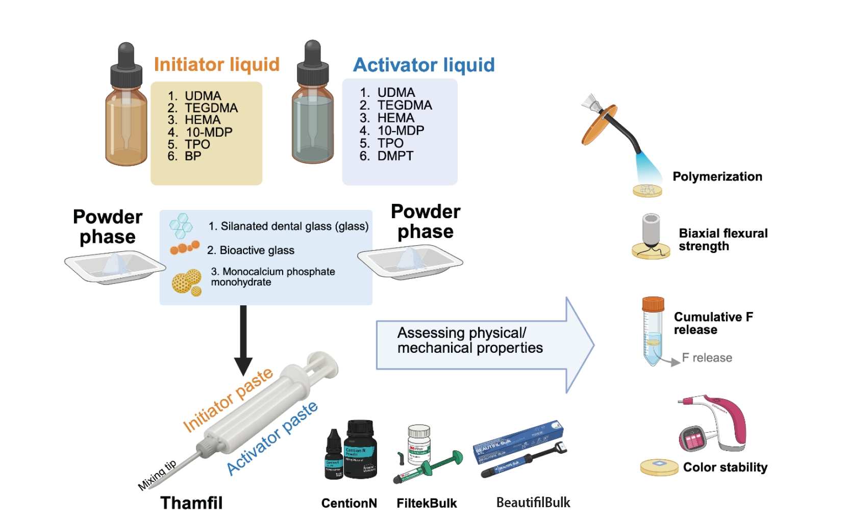
The experimental design of the current study. 10-MDP, 10-methacryloyloxydecyl dihydrogen phosphate; BP, benzoyl peroxide; DMPT, N,N-dimethyl-p-toluidine; HEMA, 2-hydroxyethyl methacrylate; TEGDMA, triethylene glycol dimethacrylate; TPO, diphenyl(2,4,6-trimethylbenzoyl)phosphine oxide; UDMA, urethane dimethacrylate. Created in BioRender (Panpisut P, 2026; https://BioRender.com/q26d152). For manufacturer information on the products listed in this figure, see Table 1.
Figure 2.
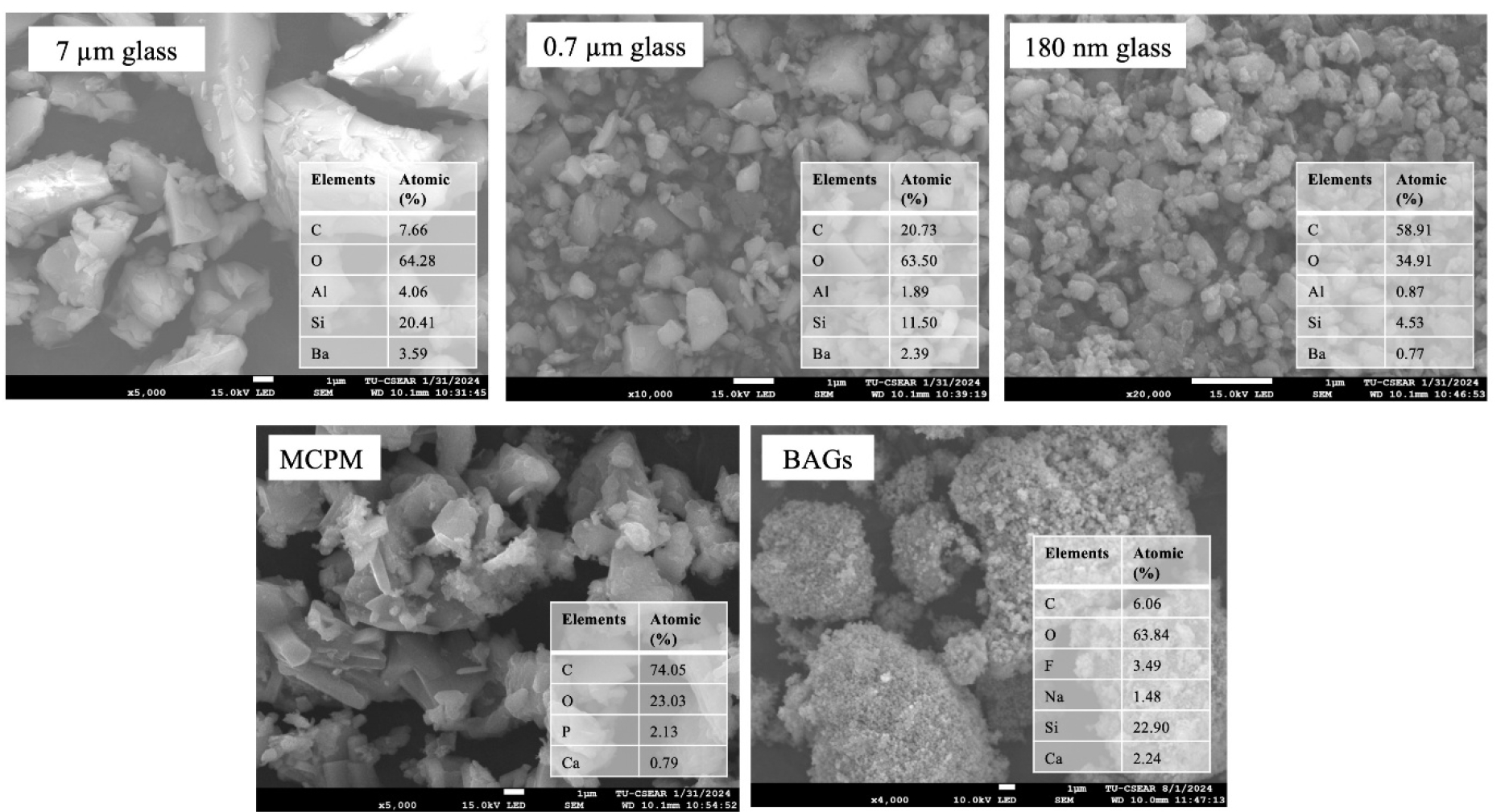
The scanning electron microscopy images and elemental composition of each filler phase in the experimental resin composite. EDX was performed as point analysis; the values are semi-quantitative and may not represent the bulk composition of the particles. BAG, bioactive glass particles; MCPM, monocalcium phosphate monohydrate.
Figure 3.
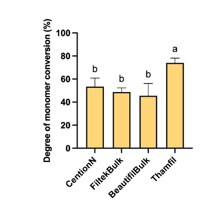
The DC of materials after light-curing for 40 seconds. The same letters indicate p > 0.05. Error bars are 95% confidence intervals (n = 5). For manufacturer information on the products listed in this figure, see Table 1.
Figure 4.
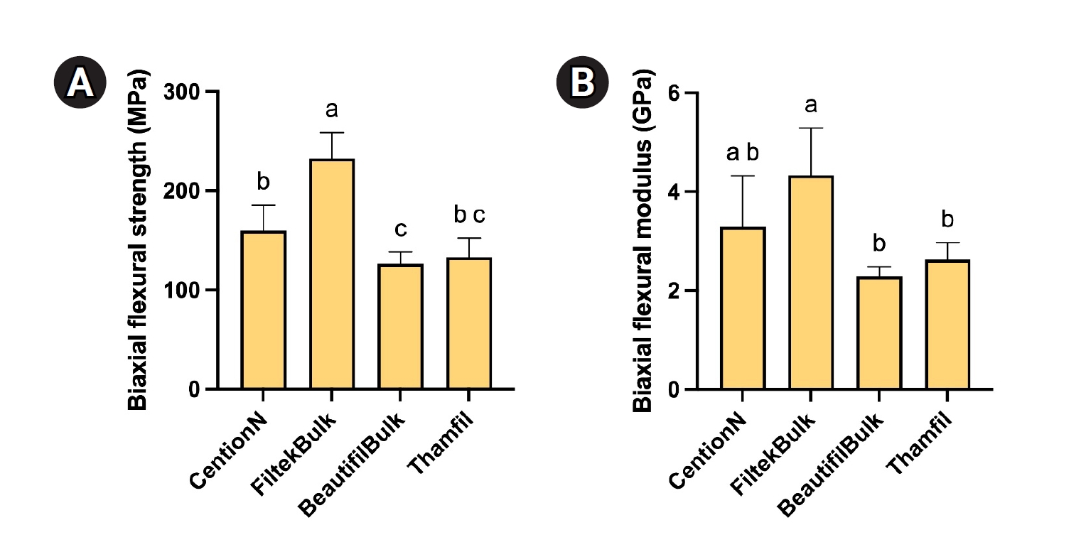
(A) Biaxial flexural strength and (B) biaxial flexural modulus of materials after immersion in deionized water for 24 hours. The same letters indicate p > 0.05. Error bars are 95% confidence intervals (n = 5). For manufacturer information on the products listed in this figure, see Table 1.
Figure 5.
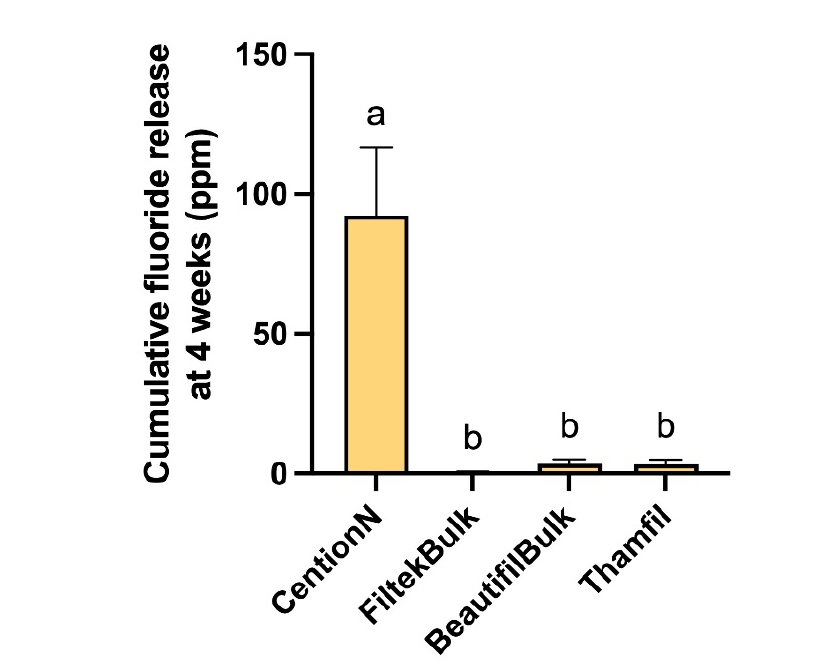
Cumulative fluoride release after 4 weeks of immersion in deionized water. The same letters indicate p > 0.05. Error bars are 95% confidence intervals (n = 4). For manufacturer information on the products listed in this figure, see Table 1.
Figure 6.
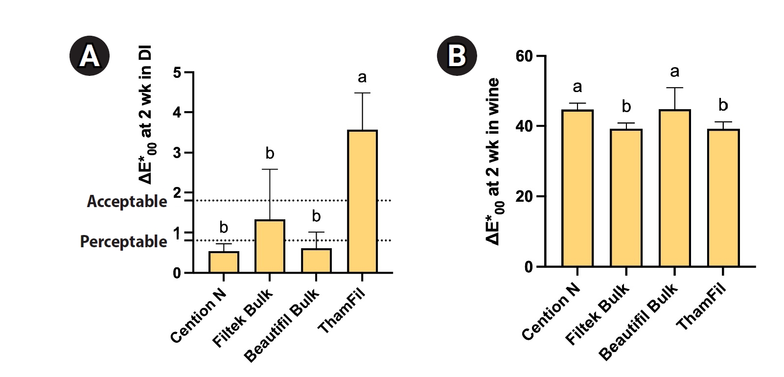
The color change (ΔE*00) of each material after immersion in water (A) and red wine (B) for 2 weeks. The same letters indicate p > 0.05. Error bars are 95% confidence intervals (n = 5). For manufacturer information on the products listed in this figure, see Table 1.
Table 1.
The composition of materials used in the current study. The actual composition of the commercial product was not provided by the manufacturer
Table 2.
The average coordinate of color changes after immersion in deionized water or red wine for 2 weeks
Identical lower- and upper-case letters in the same row indicate p > 0.05 for deionized water (DI) and Wine immersion, respectively.
For manufacturer information on the products listed in this table, see Table 1.
- 1. Li X, Li R, Wang H, Yang Z, Liu Y, Li X, et al. Global burden of dental caries from 1990 to 2021 and future projections. Int Dent J 2025;75:100904.ArticlePubMedPMC
- 2. Peres MA, Macpherson LMD, Weyant RJ, Daly B, Venturelli R, Mathur MR, et al. Oral diseases: a global public health challenge. Lancet 2019;394:249-260.ArticlePubMed
- 3. Sauro S, Carvalho RM, Ferracane J. The rise of advanced bioactive restorative materials: are they redefining operative dentistry? Dent Mater 2025;41:1411-1429.ArticlePubMed
- 4. Mulligan S, Hatton PV, Martin N. Resin-based composite materials: elution and pollution. Br Dent J 2022;232:644-652.ArticlePubMedPMCPDF
- 5. Ilie N. Comparative effect of self- or dual-curing on polymerization kinetics and mechanical properties in a novel, dental-resin-based composite with alkaline filler. Materials (Basel) 2018;11:108.ArticlePubMedPMC
- 6. Theerarath T, Sriarj W. An alkasite restorative material effectively remineralized artificial interproximal enamel caries in vitro. Clin Oral Investig 2022;26:4437-4445.ArticlePubMedPDF
- 7. de Carvalho LF, Gimenes E Silva M, Barboza ADS, Badaró MM, Stolf SC, Cuevas-Suárez CE, et al. Effectiveness of bioactive resin materials in preventing secondary caries and retention loss in direct posterior restorations: a systematic review and meta-analysis. J Dent 2025;152:105460.ArticlePubMed
- 8. Khalid H, Aleesa N, Grosjean M, Hill R, Wong F. Characterisation of a bioactive SiO2-CaO-CaF2-Na2O glass used in composites. Dent Mater 2021;37:1-9.ArticlePubMed
- 9. Simila HO, Boccaccini AR. Sol-gel bioactive glass containing biomaterials for restorative dentistry: a review. Dent Mater 2022;38:725-747.ArticlePubMed
- 10. Alkhouri N, Xia W, Ashley P, Young A. The effect of varying monocalcium phosphate and polylysine levels on dental composite properties. J Mech Behav Biomed Mater 2023;145:106039.ArticlePubMedPMC
- 11. Alkhouri N, Xia W, Ashley PF, Young AM. Renewal MI dental composite etch and seal properties. Materials (Basel) 2022;15:5438.ArticlePubMedPMC
- 12. Aljabo A, Xia W, Liaqat S, Khan MA, Knowles JC, Ashley P, et al. Conversion, shrinkage, water sorption, flexural strength and modulus of re-mineralizing dental composites. Dent Mater 2015;31:1279-1289.ArticlePubMed
- 13. Oivanen M, Keulemans F, Garoushi S, Vallittu PK, Lassila L. The effect of refractive index of fillers and polymer matrix on translucency and color matching of dental resin composite. Biomater Investig Dent 2021;8:48-53.ArticlePubMedPMC
- 14. Go HB, Lee MJ, Seo JY, Byun SY, Kwon JS. Mechanical properties and sustainable bacterial resistance effect of strontium-modified phosphate-based glass microfiller in dental composite resins. Sci Rep 2023;13:17763.ArticlePubMedPMCPDF
- 15. Gesprasert C, Kettratad M, Nimmano N, Wittayanuwat S, Pischom N, Naruphontjirakul P, et al. Effect of a 1.1% NaF toothpaste containing Sr/F-doped bioactive glass on irradiated demineralized dentin: an in vitro study. BMC Oral Health 2024;24:1399.ArticlePubMedPMCPDF
- 16. Thanyasiri S, Naruphontjirakul P, Padunglappisit C, Mirchandani B, Young AM, Panpisut P. Assessment of physical/mechanical properties and cytotoxicity of dual-cured resin cements containing Sr-bioactive glass nanoparticles and calcium phosphate. Dent Mater J 2023;42:806-817.ArticlePubMed
- 17. Pariwatanasak M, Chadarat S, Potiprapanpong W, Kyopun S, Young AM, Panpisut P. The in vitro assessment of the degree of monomer conversion, biaxial flexural strength, and mineral precipitation on demineralised dentine of novel resin composite containing monocalcium phosphate monohydrate and polylysine. Biomater Investig Dent 2025;12:44551.ArticlePubMedPMCPDF
- 18. Hamdy TM, Zaki ZM, Abdelraouf RM. Assessment of toothbrushing, bleaching pen and bleaching mouthwash in removing stains from tooth structure and single-shade resin composite. Sci Rep 2026;16:7948.ArticlePubMedPMCPDF
- 19. Ardu S, Duc O, Di Bella E, Krejci I. Color stability of recent composite resins. Odontology 2017;105:29-35.ArticlePubMedPDF
- 20. Kowalska A, Sokołowski J, Szynkowska-Jóźwik MI, Gozdek T, Kopacz K, Bociong K. Can TPO as photoinitiator replace “golden mean” camphorquinone and tertiary amines in dental composites?: testing experimental composites containing different concentration of diphenyl(2,4,6-trimethylbenzoyl)phosphine Oxide. Int J Mol Sci 2022;23:11594.ArticlePubMedPMC
- 21. Alzahrani B, Alshabib A, Awliya W. Surface hardness and flexural strength of dual-cured bulk-fill restorative materials after solvent storage. BMC Oral Health 2023;23:306.ArticlePubMedPMCPDF
- 22. Alkhudhairy FI. The effect of curing intensity on mechanical properties of different bulk-fill composite resins. Clin Cosmet Investig Dent 2017;9:1-6.ArticlePubMedPMCPDF
- 23. Fu SY, Feng XQ, Lauke B, Mai YW. Effects of particle size, particle/matrix interface adhesion and particle loading on mechanical properties of particulate-polymer composites. Compos B Eng 2008;39:933-961.Article
- 24. Shimizu S, Kotake H, Takagaki T, Shinno K, Miyata S, Burrow MF, et al. Evaluation of bonding performance and multi-ion release of S-PRG fillercontaining self-adhesive resin composite. Dent Mater J 2021;40:1257-1263.ArticlePubMed
- 25. Nicholson JW, Sidhu SK, Czarnecka B. Fluoride exchange by glass-ionomer dental cements and its clinical effects: a review. Biomater Investig Dent 2023;10:2244982.ArticlePubMedPMC
- 26. Billington RW, Williams JA, Dorban A, Pearson GJ. Glass ionomer cement: evidence pointing to fluorine release in the form of monofluorophosphate in addition to fluoride ion. Biomaterials 2004;25:3399-3402.ArticlePubMed
- 27. Kelić K, Par M, Peroš K, Šutej I, Tarle Z. Fluoride-releasing restorative materials: the effect of a resinous coat on ion release. Acta Stomatol Croat 2020;54:371-381.ArticlePubMedPMC
- 28. Soe T, Sunami A, Kyaw O, Hatano K, Kanazawa M, Inokoshi M. Bioactivity and potential applications of surface pre-reacted glass-ionomer (S-PRG) filler-containing dental materials for geriatric oral health: a scoping review. Jpn Dent Sci Rev 2025;61:280-291.ArticlePubMedPMC
- 29. Xu J, Shi H, Luo J, Yao H, Wang P, Li Z, et al. Advanced materials for enamel remineralization. Front Bioeng Biotechnol 2022;10:985881.ArticlePubMedPMC
- 30. Zadeh AK, Haghi S, Drikvand M. In vitro comparison of color stability of resin-modified glass ionomer and Cention N bioactive restorative material. Dent Res J (Isfahan) 2025;22:24.ArticlePubMedPMC
- 31. Gomes de Araújo-Neto V, Sebold M, Fernandes de Castro E, Feitosa VP, Giannini M. Evaluation of physico-mechanical properties and filler particles characterization of conventional, bulk-fill, and bioactive resin-based composites. J Mech Behav Biomed Mater 2021;115:104288.ArticlePubMed
- 32. Mehdawi IM, Pratten J, Spratt DA, Knowles JC, Young AM. High strength re-mineralizing, antibacterial dental composites with reactive calcium phosphates. Dent Mater 2013;29:473-484.ArticlePubMed
- 33. Ferracane JL. Hygroscopic and hydrolytic effects in dental polymer networks. Dent Mater 2006;22:211-222.ArticlePubMed
- 34. Cinelli F, Scaminaci Russo D, Nieri M, Giachetti L. Stain susceptibility of composite resins: pigment penetration analysis. Materials (Basel) 2022;15:4874.ArticlePubMedPMC
- 35. Almasabi W, Tichy A, Abdou A, Hosaka K, Nakajima M, Tagami J. Effect of water storage and thermocycling on light transmission properties, translucency and refractive index of nanofilled flowable composites. Dent Mater J 2021;40:599-605.ArticlePubMed
- 36. Kangwankai K, Sani S, Panpisut P, Xia W, Ashley P, Petridis H, et al. Monomer conversion, dimensional stability, strength, modulus, surface apatite precipitation and wear of novel, reactive calcium phosphate and polylysine-containing dental composites. PLoS One 2017;12:e0187757.ArticlePubMedPMC
- 37. Kareem ASA, Abdel-Fattah WM, El Gayar MIL. Evaluation of color stability and surface roughness of smart monochromatic resin composite in comparison to universal resin composites after immersion in staining solutions. BMC Oral Health 2025;25:1211.ArticlePubMedPMCPDF
- 38. Tanthanuch S, Kukiattrakoon B, Peerasukprasert T, Chanmanee N, Chaisomboonphun P, Rodklai A. The effect of red and white wine on color changes of nanofilled and nanohybrid resin composites. Restor Dent Endod 2016;41:130-136.ArticlePubMedPMCPDF
- 39. Şişmanoğlu S, Sengez G. Effects of acidic beverages on color stability of bulk-fill composites with different viscosities. Odovtos Int J Dent Sci 2022;24:90-99.ArticlePDF
- 40. Ardu S, Duc O, Di Bella E, Krejci I, Daher R. Color stability of different composite resins after polishing. Odontology 2018;106:328-333.ArticlePubMedPDF
REFERENCES
Tables & Figures
REFERENCES
Citations
Citations to this article as recorded by 
 Cite
 XML Download
Monomer conversion, flexural strength, fluoride release, and color stability of experimental resin composites containing bioactive glass nanoparticles: an in vitro study
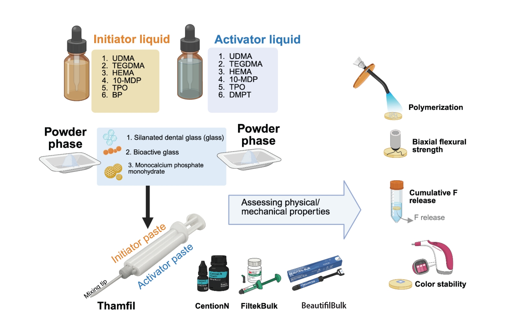
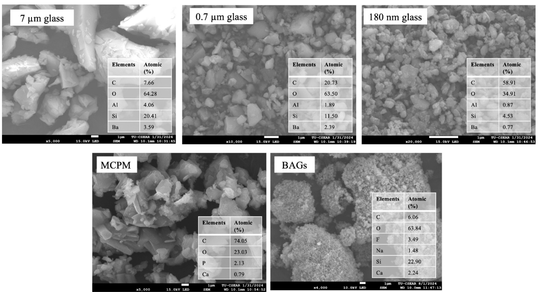
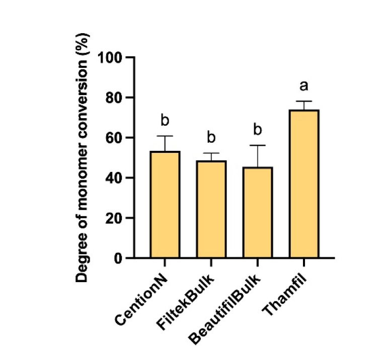
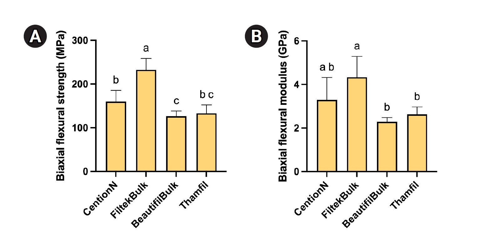
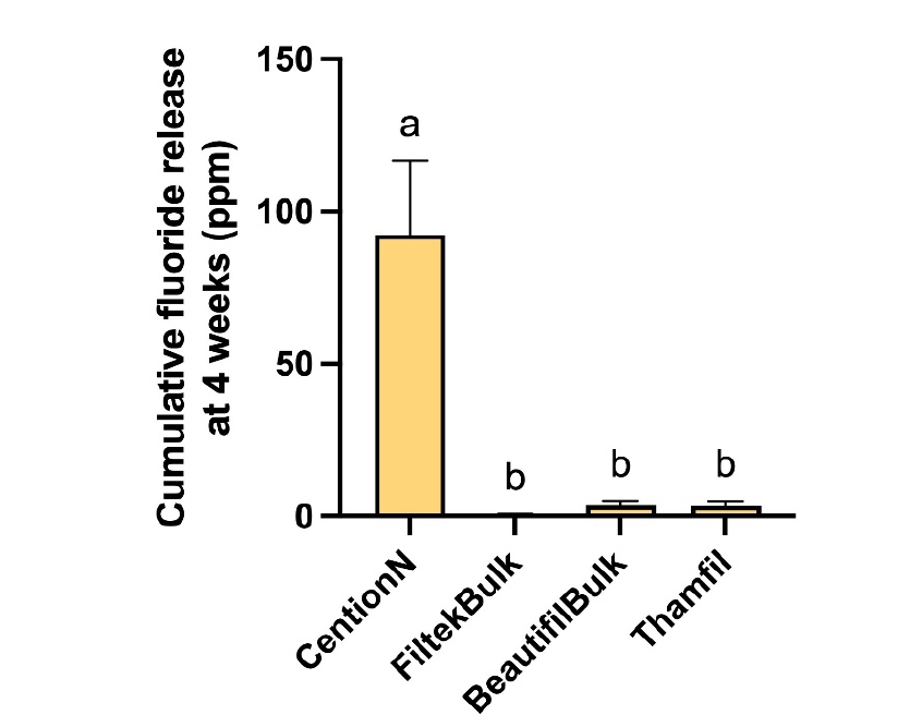
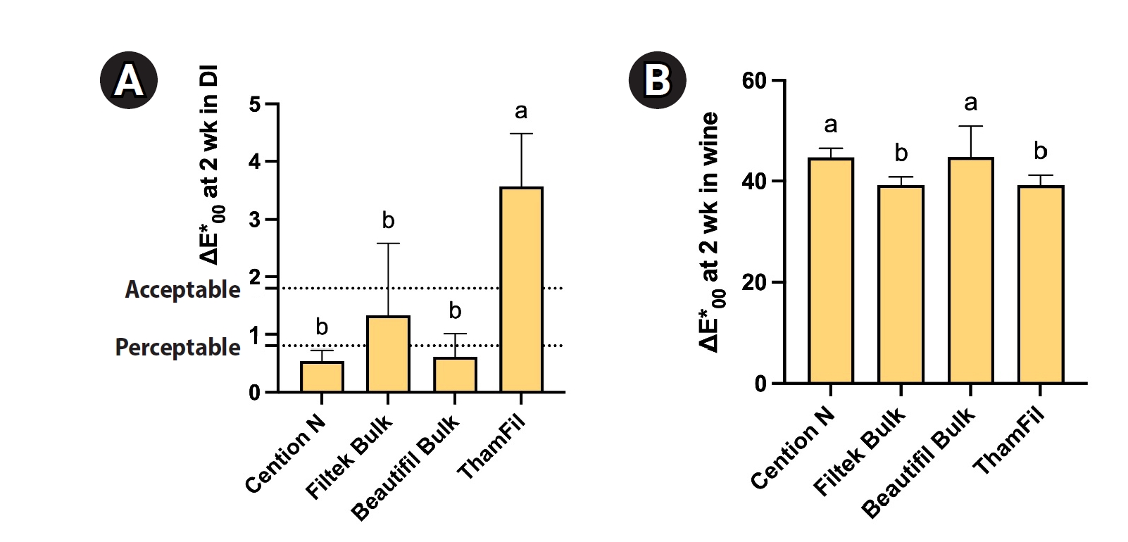
Figure 1. The experimental design of the current study. 10-MDP, 10-methacryloyloxydecyl dihydrogen phosphate; BP, benzoyl peroxide; DMPT, N,N-dimethyl-p-toluidine; HEMA, 2-hydroxyethyl methacrylate; TEGDMA, triethylene glycol dimethacrylate; TPO, diphenyl(2,4,6-trimethylbenzoyl)phosphine oxide; UDMA, urethane dimethacrylate. Created in BioRender (Panpisut P, 2026; https://BioRender.com/q26d152). For manufacturer information on the products listed in this figure, see Table 1.
Figure 2. The scanning electron microscopy images and elemental composition of each filler phase in the experimental resin composite. EDX was performed as point analysis; the values are semi-quantitative and may not represent the bulk composition of the particles. BAG, bioactive glass particles; MCPM, monocalcium phosphate monohydrate.
Figure 3. The DC of materials after light-curing for 40 seconds. The same letters indicate p > 0.05. Error bars are 95% confidence intervals (n = 5). For manufacturer information on the products listed in this figure, see Table 1.
Figure 4. (A) Biaxial flexural strength and (B) biaxial flexural modulus of materials after immersion in deionized water for 24 hours. The same letters indicate p > 0.05. Error bars are 95% confidence intervals (n = 5). For manufacturer information on the products listed in this figure, see Table 1.
Figure 5. Cumulative fluoride release after 4 weeks of immersion in deionized water. The same letters indicate p > 0.05. Error bars are 95% confidence intervals (n = 4). For manufacturer information on the products listed in this figure, see Table 1.
Figure 6. The color change (ΔE*00) of each material after immersion in water (A) and red wine (B) for 2 weeks. The same letters indicate p > 0.05. Error bars are 95% confidence intervals (n = 5). For manufacturer information on the products listed in this figure, see Table 1.
Figure 1.
Figure 2.
Figure 3.
Figure 4.
Figure 5.
Figure 6.
Monomer conversion, flexural strength, fluoride release, and color stability of experimental resin composites containing bioactive glass nanoparticles: an in vitro study
| Material (abbreviation) | Type | Composition | Supplier |
|---|---|---|---|
| Filtek One Bulk Fill Restorative (FiltekBulk) | Bulk-fill resin composite | Silane-treated ceramic (60–70 wt%), aromatic urethane dimethacrylate (10-20 wt%), silane treated silica (1–10 wt%), ytterbium fluoride (YbF3) (1-10 wt%), water (<5 wt%), silane treated zirconia (<5 wt%), 1,12-dodecane dimethacrylate (<2.5 wt%), ethyl-4-dimethylaminobenzoate (<0.3 wt%) | 3M, St. Paul, MN, USA |
| Cention N (CentionN) | Alkasite | Liquid phase: Dimethacrylate (95–97 wt%), initiators (2–3 wt%), stabilizers (<1 wt%), and additives (1–2 wt%). Powder phase: Calcium fluoro-silicate glass (10–20 wt%), prepolymer, ytterbium trifluoride (5–10 wt%), isofiller (copolymer) (15–25 wt%), initiators (<1 wt%), and pigments (<0.1 wt%) | Ivoclar Vivadent, Schaan, Liechtenstein |
| Beautifil-Bulk Restorative (BeautifilBulk) | Giomer | Aluminofluoro-borosilicate glass (60–70 wt%), bisphenol A-glycidyl methacrylate (5–10 wt%), urethane dimethacrylate (1–5 wt%), bisphenol A polyethoxy methacrylate (1–5 wt%), triethylene glycol dimethacrylate (1–5 wt%), silane coupling agent (1–10 wt%), aluminum oxide (1–5 wt%), silicon dioxide (1–5 wt%), Reactive initiator, others | Shofu, Kyoto, Japan |
| Thamfil | Experimental composite | Initiator paste: Monomers: Urethane dimethacrylate (70 wt%), triethylene glycol dimethacrylate (22.75 wt%), 2-hydroxyethyl methacrylate (3 wt%), 10-methacryloyloxydecyl dihydrogen phosphate (3 wt%), trimethylbenzoyldiphenylphosphine oxide (0.5 wt%), benzoyl peroxide (0.75 wt%). Powder: Barium aluminosilicate glass (87 wt%), MCPM (3 wt%), BAGs (10 wt%) | - |
| Activator paste: Monomers: Urethane dimethacrylate (70 wt%), triethylene glycol dimethacrylate (22.75 wt%), 2-hydroxyethyl methacrylate (3 wt%), 10-methacryloyloxydecyl dihydrogen phosphate (3 wt%), trimethylbenzoyldiphenylphosphine oxide (0.5 wt%), N,N-dimethyl-p-toluidine (0.75 wt%). Powder: Barium aluminosilicate glass (87 wt%), MCPM (3 wt%), BAGs (10 wt%) |
| Solution | CentionN | FiltekBulk | BeautifilBulk | Thamfil | ||||
|---|---|---|---|---|---|---|---|---|
| DI | Wine | DI | Wine | DI | Wine | DI | Wine | |
| ∆L (lightness) | 0.42a,b | –47.22B | –0.02b | –40.34A | 0.24a,b | –44.94A,B | 3.08a | –42.06A,B |
| ∆a (red-green) | 0.28a | 17.24A,B | –0.36a,b | 15.48B | –0.14a,b | 19.16A | –2.24b | 15.42B |
| ∆b (yellow-blue) | 0.48a | –0.76B | –0.08a,b | 5.88A,B | –0.38a,b | 28.88A | –1.94b | –0.36B |
Table 1. The composition of materials used in the current study. The actual composition of the commercial product was not provided by the manufacturer
Table 2. The average coordinate of color changes after immersion in deionized water or red wine for 2 weeks
Identical lower- and upper-case letters in the same row indicate For manufacturer information on the products listed in this table, see


