Search
- Page Path
- HOME > Search
- A case report of multiple bilateral dens invaginatus in maxillary anteriors
- Shin Hye Chung, You-Jeong Hwang, Sung-Yeop You, Young-Hye Hwang, Soram Oh
- Restor Dent Endod 2019;44(4):e39. Published online October 21, 2019
- DOI: https://doi.org/10.5395/rde.2019.44.e39

-
 Abstract
 PDF
PDF PubReader
 ePub
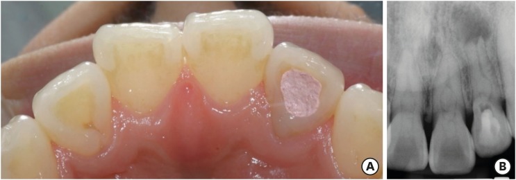
ePub The present report presents a case of dens invaginatus (DI) in a patient with 4 maxillary incisors. A 24-year-old female complained of swelling of the maxillary left anterior region and discoloration of the maxillary left anterior tooth. The maxillary left lateral incisor (tooth #22) showed pulp necrosis and a chronic apical abscess, and a periapical X-ray demonstrated DI on bilateral maxillary central and lateral incisors. All teeth responded to a vitality test, except tooth #22. The anatomic form of tooth #22 was similar to that of tooth #12, and both teeth had lingual pits. In addition, panoramic and periapical X-rays demonstrated root canal calcification, such as pulp stones, in the maxillary canines, first and second premolars, and the mandibular incisors, canines, and first premolars bilaterally. The patient underwent root canal treatment of tooth #22 and non-vital tooth bleaching. After a temporary filling material was removed, the invaginated mass was removed using ultrasonic tips under an operating microscope. The working length was established, and the root canal was enlarged up to #50 apical size and obturated with gutta-percha and AH 26 sealer using the continuous wave of condensation technique. Finally, non-vital bleaching was performed, and the access cavity was filled with composite resin.
-
Citations
Citations to this article as recorded by- The use of three-dimensional-printed guides, static navigation, and bioactive materials to treat bilateral and double dens invaginatus
Parth Patel, Nidhi Bharti, Ankit Arora, C. Nimisha Shah
Saudi Endodontic Journal.2025; 15(2): 207. CrossRef - Endodontic Management of Dens in Dente – A Systematic Review of Case Reports and Case Series
Sanket Dilip Aras, Anamika Chetan Borkar, Sonal Kale, Sayali Maral, Prakriti Jaggi, Shailendra Sonawane
Journal of the International Clinical Dental Research Organization.2024; 16(1): 17. CrossRef - Dens invaginatus of fourteen teeth in a pediatric patient
Momoko Usuda, Tatsuya Akitomo, Mariko Kametani, Satoru Kusaka, Chieko Mitsuhata, Ryota Nomura
Pediatric Dental Journal.2023; 33(3): 240. CrossRef - The Impact of the Preferred Reporting Items for Case Reports in Endodontics (PRICE) 2020 Guidelines on the Reporting of Endodontic Case Reports
Sofian Youssef, Phillip Tomson, Amir Reza Akbari, Natalie Archer, Fayjel Shah, Jasmeet Heran, Sunmeet Kandhari, Sandeep Pai, Shivakar Mehrotra, Joanna M Batt
Cureus.2023;[Epub] CrossRef - Root Maturation of an Immature Dens Invaginatus Despite Unsuccessful Revitalization Procedure: A Case Report and Recommendations for Educational Purposes
Julia Ludwig, Marcel Reymus, Alexander Winkler, Sebastian Soliman, Ralf Krug, Gabriel Krastl
Dentistry Journal.2023; 11(2): 47. CrossRef - Conservative Management of Infraorbital Space Infection Secondary to Type III B Dens Invaginatus: A Case Report
Ashima Goyal, Aditi Kapur, Manoj A Jaiswal, Gauba Krishan, Raja Raghu, Sanjeev K Singh
Journal of Postgraduate Medicine, Education and Research.2022; 56(4): 192. CrossRef
- The use of three-dimensional-printed guides, static navigation, and bioactive materials to treat bilateral and double dens invaginatus
- 2,416 View
- 28 Download
- 6 Crossref

- Clinical study of shade improvement and safety of polymer-based pen type BlancTic Forte whitening agent containing 8.3% Carbamide peroxide
- Jin-Kyung Lee, Sun-Hong Min, Sung-Tae Hong, So-Ram Oh, Shin-Hye Chung, Young-Hye Hwang, Sung-Yeop You, Kwang-Shik Bae, Seung-Ho Baek, Woo-Cheol Lee, Won-Jun Son, Kee-Yeon Kum
- J Korean Acad Conserv Dent 2009;34(2):154-161. Published online March 31, 2009
- DOI: https://doi.org/10.5395/JKACD.2009.34.2.154
-
 Abstract
 PDF
PDF PubReader
 ePub
ePub This clinical study evaluated the whitening effect and safety of polymer based-pen type BlancTis Forte (NIBEC) containing 8.3% carbamide peroxide. Twenty volunteers used the BlancTis Forte whitening agent for 2 hours twice a day for 4 weeks. As a control, Whitening Effect Pen (LG) containing 3% hydrogen peroxide was used by 20 volunteers using the same protocol. The change in shade (ΔE*, color difference) was measured using Shadepilot™ (DeguDent) before, during, and after bleaching (2 weeks, 4 weeks, and post-bleaching 4 weeks). A clinical examination for any side effects (tooth hypersensitivity or soft tissue complications) was also performed at each check-up. The following results were obtained.
1. Both the experimental and control groups displayed a noticeable change in shade (ΔE) of over 2. No significant differences were found between the two groups (p > 0.05), implying that the two agents have a similar whitening effect.
2. The whitening effect was mainly due to changes in a and b values rather than in L value (brightness). The experimental group showed a significantly higher change in b value, thus yellow shade, than the control (p < 0.05).
3. None of the participants complained of tooth hypersensitivity or soft tissue complications, confirming the safety of both whitening agents.
-
Citations
Citations to this article as recorded by- Surface Damage and Bleaching Effect according to the Application Type of Home Tooth Bleaching Applicants
Na-Yeoun Tak, Do-Seon Lim, Hee-Jung Lim, Im-Hee Jung
Journal of Dental Hygiene Science.2020; 20(4): 252. CrossRef - Efficacy of a self - applied paint - on whitening gel combined with wrap
Soo-Yeon Kim, Jae-Hyun Ahn, Ji-Young Kim, Jin-Woo Kim, Se-Hee Park, Kyung-Mo Cho
Journal of Dental Rehabilitation and Applied Science.2018; 34(3): 175. CrossRef
- Surface Damage and Bleaching Effect according to the Application Type of Home Tooth Bleaching Applicants
- 1,224 View
- 2 Download
- 2 Crossref


 KACD
KACD

 First
First Prev
Prev


