
Search
- Page Path
- HOME > Search
Research Article
- Initial attachment, viability, proliferation, and migration of osteoblast-like SaOS-2 cells on two resorbable xenogeneic membranes for guided tissue regeneration: ab in vitro experimental study
- Rafael Fernández-Grisales, Giovanna García-Suárez, Ximena Guerrero-Rodríguez, Carolina Berruecos-Orozco, Marco Calle-Jaramillo, Wilder Javier Rojas, Vanessa Esmeralda Duque, Daniela Serna-Guisao, Néstor Ríos-Osorio
- Restor Dent Endod 2026;51(2):e20. Published online April 13, 2026
- DOI: https://doi.org/10.5395/rde.2026.51.e20
-
 Abstract
 PDF
PDF PubReader
 ePub
ePub - Objectives
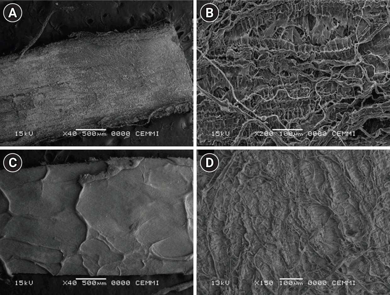
This study evaluated the biocompatibility of a new xenogeneic resorbable membrane derived from porcine esophagus membrane (Quirumatrix, Cells Tech Co.) and compared it with a porcine pericardium membrane (Straumann Jason, Straumann Holding AG.) traditionally used for guided tissue regeneration. The parameters investigated were the viability, migration, and adhesion of SaOS-2 osteoblast-like cells derived from osteosarcoma on both membranes.
Methods
The cells were cultured in 100 mm plates in RPMI 1640 medium (40 mL), supplemented. They were incubated at 37°C in a humidified atmosphere with 95% air and 5% to 10% CO2. Cell morphology and adhesion were evaluated using phase contrast optical microscopy and scanning electron microscope. Cell viability and proliferation were evaluated using a fluorometric resazurin reduction assay, with fluorescence intensity measured at 48, 72, and 96 hours. Cell migration was evaluated using staining with Alexa Fluor 555 Phalloidin (Cell Signaling Technology) and DAPI, with a reference line. Cell migration was analyzed by measuring displacement within the delineated area using an Axio Imager M2 fluorescence microscope (Carl Zeiss). Each membrane was photographed. The statistical analysis was performed using GraphPad Prism ver. 10.2.3 (GraphPad Software). A p-value <0.05 was considered significant between experimental groups.
Results
Both membranes were shown to be biocompatible. The porcine pericardium membrane showed greater cell adhesion and proliferation compared to the porcine esophagus membrane. Cell migration was significantly greater in the Jason membrane.
Conclusions
The results revealed that both evaluated membranes are biocompatible and non-cytotoxic; further research is needed to understand their long-term behavior, interactions with other types of cells, and performance in specific therapeutic situations.
- 1,175 View
- 87 Download

Case Report
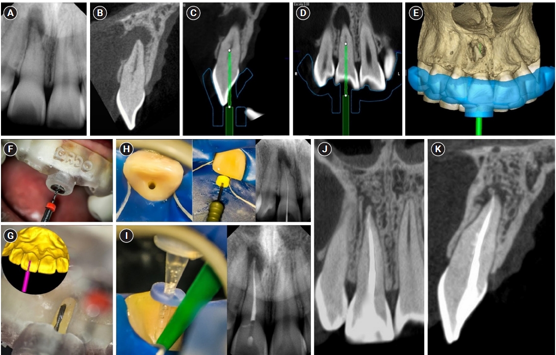
- Guided endodontics, precision and predictability: a case series of mineralized anterior teeth with follow-up cone-beam computed tomography
- Rafael Fernández-Grisales, Wilder Javier Rojas-Gutierrez, Pamela Mejía, Carolina Berruecos-Orozco, Néstor Ríos-Osorio
- Restor Dent Endod 2025;50(1):e4. Published online January 6, 2025
- DOI: https://doi.org/10.5395/rde.2025.50.e4
-
Abstract
PDFPubReaderePub
- Pulp chamber and root canal obliteration (PCO/RCO) presents a challenge for clinicians when nonsurgical endodontic treatment is indicated. Guided endodontics (GE) aims to precisely locate the root canal (RC) system while preserving as much pericervical dentin as possible. GE involves integrating cone-beam computed tomography (CBCT) of the affected tooth with a digital impression of the maxillary/mandibular arch, allowing for careful planning of the drilling path to the RC system through a three-dimensional (3D) static guide. This article reports four cases of teeth with PCO/RCO, accompanied by additional diagnoses of internal and external root resorption and horizontal tooth fracture, all successfully treated with GE. These cases highlight the clinical and radiographic success of GE treatments using CBCT, establishing this technique as a predictable approach for managing mineralized teeth.
-
Citations
Citations to this article as recorded by
- Static Guided Endodontics in Primary Endodontic Treatment of Anterior Teeth: A Narrative Review
Monika Kuczmaja, Wiesława Puchalska, Agata Żółtowska
Dentistry Journal.2026; 14(4): 195. CrossRef - Effect of different rotary instrument designs (protaper ultimate and protaper gold) on postoperative pain and bacterial reduction: a randomized clinical trial
Khaled Hassan Abed, Ahmed Abdel Rahman Hashem, Reem Ahmed Lutfy, Somaia Abdellatif Eissa, Dina Ahmed Morsy
BMC Oral Health.2026;[Epub] CrossRef
- Static Guided Endodontics in Primary Endodontic Treatment of Anterior Teeth: A Narrative Review
- 5,281 View
- 379 Download
- 2 Web of Science
- 2 Crossref
 First
First Prev
Prev

