
Search
- Page Path
- HOME > Search
Research Articles
- Difference in light transmittance and depth of cure of flowable composite depending on tooth thickness: an in vitro experimental study
- Seong-Pyo Bae, Myung-Jin Lee, Kyung-San Min, Mi-Kyung Yu, Kwang-Won Lee
- Restor Dent Endod 2025;50(4):e39. Published online November 28, 2025
- DOI: https://doi.org/10.5395/rde.2025.50.e39
-
 Abstract
 PDF
PDF PubReader
 ePub
ePub - Objectives
This study aimed to quantify light attenuation through varying tooth thicknesses and its impact on the depth of cure of composite resin.
Methods
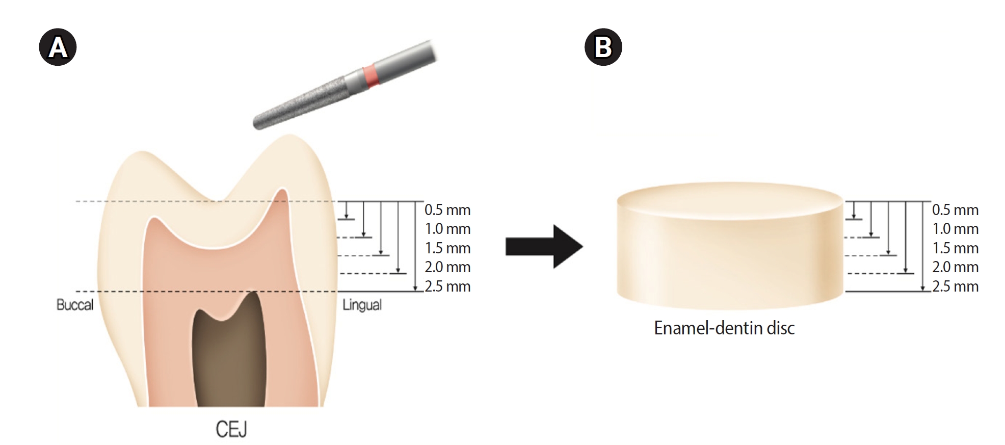
Twenty extracted premolars were used to create enamel-dentin discs that were sanded progressively in 0.5 mm increments from 2.5 mm to 0.5 mm. Light irradiance was measured with and without tooth specimens to evaluate light transmittance. Resin was cured beneath different thicknesses, and the depth of cure was assessed using the Vickers hardness test.
Results
The results demonstrated that light transmittance significantly decreased as tooth thickness increased (p < 0.01), leading to reduced resin polymerization. In the 2.0-mm and 2.5-mm tooth thickness groups, the depth of cure was significantly lower than in the control group without tooth specimens (p < 0.05).
Conclusions
Ultimately, for tooth structures exceeding 2 mm, self-cure or dual-cure resin polymerization is thought to be more efficient than light polymerization.
- 1,842 View
- 141 Download

- Effect of different storage media on elemental analysis and microhardness of cervical cavity margins restored with a bioactive material
- Hoda Saleh Ismail, Brian Ray Morrow, Ashraf Ibrahim Ali, Rabab Elsayed Elaraby Mehesen, Salah Hasab Mahmoud, Franklin Garcia-Godoy
- Restor Dent Endod 2024;49(1):e6. Published online January 17, 2024
- DOI: https://doi.org/10.5395/rde.2024.49.e6
-
Abstract
PDFPubReaderePub
Objectives This study aimed to investigate the elemental analysis and microhardness of a bioactive material (Activa) and marginal tooth structure after storage in different media.
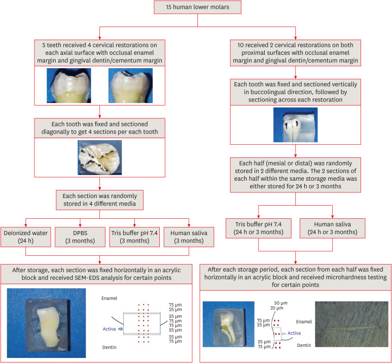
Materials and Methods Fifteen teeth received cervical restorations with occlusal enamel and gingival dentin margins using the tested material bonded with a universal adhesive, 5 of them on the 4 axial surfaces and the other 10 on only the 2 proximal surfaces. The first 5 teeth were sectioned into 4 restorations each, then stored in 4 different media; deionized water, Dulbecco's phosphate buffered saline (DPBS), Tris buffer, and saliva. The storage period for deionized water was 24 hours while it was 3 months for the other media. Each part was analyzed by scanning electron microscopy-energy dispersive spectroscopy (SEM-EDS) analysis for different substrates/distances and the wt% of calcium, phosphorus, silica, and fluoride were calculated. The other 10 teeth were sectioned across the restoration, stored in either Tris buffer or saliva for 24 hours or 3 months, and were evaluated for microhardness of different substrates/areas. Data were analyzed using analysis of variance and Tukey’s
post hoc test.Results Enamel and dentin interfaces in the DPBS group exhibited a significant increase in calcium and phosphorus wt%. Both silica and fluoride significantly increased in tooth structure up to a distance of 75 μm in the 3-month-media groups than the immediate group. Storage media did not affect the microhardness values.
Conclusions SEM-EDS analysis suggests an ion movement between Activa and tooth structure through a universal adhesive while stored in DPBS.
-
Citations
Citations to this article as recorded by
- A two-year randomized clinical trial of bulk-fill and ion-releasing composites with universal adhesives in class V carious lesions
Hoda Saleh Ismail, Hanan Ahmed Nabil Soliman, Ashraf Ibrahim Ali, Ahmed Gamal Raghip, Eman H. Albelasy
Clinical Oral Investigations.2026;[Epub] CrossRef - Elemental and micromorphological analysis of ion releasing restoration/carious dentin interface
Alaa Esmat Abdelsalam, Hoda Saleh Ismail, Hamdi Hosni Hamama
Scientific Reports.2025;[Epub] CrossRef - Influence of curing mode and aging on the bonding performance of universal adhesives in coronal and root dentin
Hoda Saleh Ismail, Ashraf Ibrahim Ali, Mohamed Elshirbeny Elawsya
BMC Oral Health.2024;[Epub] CrossRef
- A two-year randomized clinical trial of bulk-fill and ion-releasing composites with universal adhesives in class V carious lesions
- 2,796 View
- 107 Download
- 3 Web of Science
- 3 Crossref
Original Article
- Surface hardness of the dental composite cured by light that penetrate tooth structure according to thickness of tooth structure, light intensity and curing time
- Soo-Kyung Cho, Dong-Jun Kim, Yun-Chan Hwang, Won-Mann Oh, In-Nam Hwang
- J Korean Acad Conserv Dent 2005;30(2):128-137. Published online March 31, 2005
- DOI: https://doi.org/10.5395/JKACD.2005.30.2.128
-
Abstract
PDFPubReaderePub
In this study we measured the amount of light energy that was projected through the tooth material and analyzed the degree of polymerization by measuring the surface hardness of composites. For polymerization, Optilux 501 (Demetron, USA) with two types of light guide was used: a 12 mm diameter light guide with 840 mW/cm2 light intensity and a 7 mm diameter turbo light guide with 1100 mW/cm2.
Specimens were divided into three groups according to thickness of penetrating tooth (1 mm, 2 mm, 0 mm). Each group was further divided into four subgroups according to type of light guide and curing time (20 seconds, 40 seconds). Vickers'hardness was measured by using a microhardness tester. In 0 mm and 1 mm penetrating tooth group, which were polymerized by a turbo light guide for 40 seconds, showed the highest hardness values. The specimens from 2 mm penetrating tooth group, which were polymerized for 20 seconds, demonstrated the lowest hardness regardless of the types of light guides (p < 0.05).
The results of this study suggest that, when projecting tooth material over a specified thickness, the increase of polymerization will be limited even if light intensity or curing time is increased.
-
Citations
Citations to this article as recorded by- Comparison of Flexural Strength According to Post-Curing Treatment Time of Cast Resin Printed by 3D Printing Method
Jihyun Kim
International Journal of Clinical Preventive Dentistry.2024; 20(4): 170. CrossRef - Comparison of Surface Microhardness of the Flowable Bulk-Fill Resin and the Packable Bulk-Fill Resin according to Light Curing Time and Distance
Hyung-Min Kim, Moon-Jin Jeong, Hee-Jung Lim, Do-Seon Lim
Journal of Dental Hygiene Science.2023; 23(2): 123. CrossRef - Comparison of polymerization by time of light curing for dental 3D printing
Dong-Yeon Kim, Gwang-Young Lee
Journal of Korean Acedemy of Dental Technology.2022; 44(3): 76. CrossRef - Comparative analysis of the flexural strength of provisional restorative resins using a digital light processing printer according to the post-curing method
Young-Dae Park, Wol Kang
Journal of Korean Acedemy of Dental Technology.2020; 42(4): 341. CrossRef
- Comparison of Flexural Strength According to Post-Curing Treatment Time of Cast Resin Printed by 3D Printing Method
- 1,826 View
- 3 Download
- 4 Crossref
 First
First Prev
Prev

