Search
- Page Path
- HOME > Search
- Stress distribution of restorations in external cervical root resorption under occlusal and traumatic loads: a finite element analysis
- Padmapriya Ramanujam, Paul Kevin Abishek Karthikeyan, Vignesh Srinivasan, Selvakarthikeyan Ulaganathan, Velmurugan Natanasabapathy, Nandini Suresh
- Restor Dent Endod 2025;50(2):e21. Published online May 21, 2025
- DOI: https://doi.org/10.5395/rde.2025.50.e21

-
 Abstract
 PDF
PDF PubReader
 ePub
ePub - Objectives
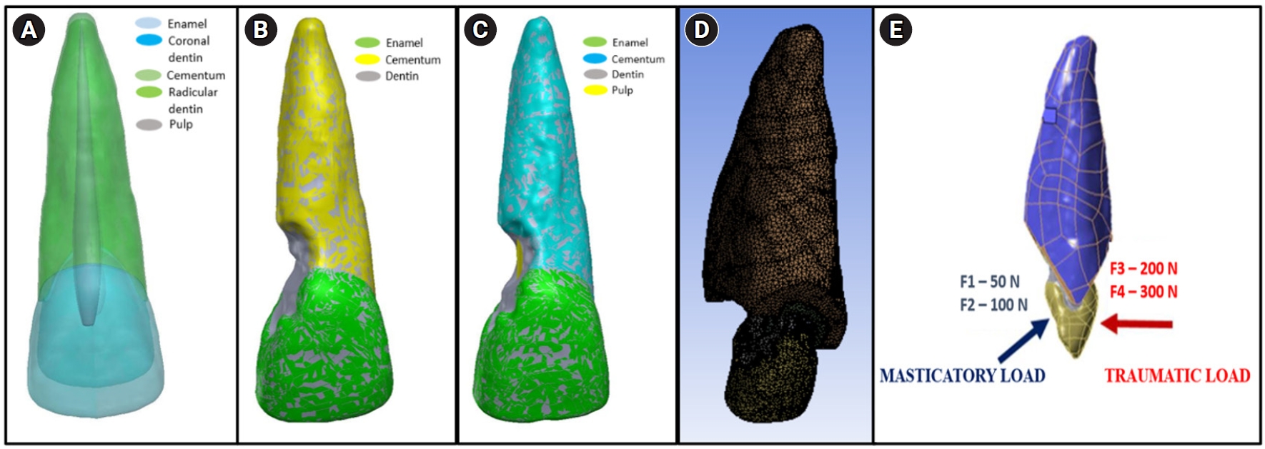
This study analyzed the stress distribution in a maxillary central incisor with external cervical resorptive defect restored with different restorative materials under normal masticatory and traumatic loading conditions using finite element analysis.
Methods
Cone-beam computed tomography of an extracted intact incisor and created resorptive models (Patel’s 3D classification-2Bd and 2Bp) in the maxillary central incisor was performed for finite element models. The 2Bd models were restored either with glass ionomer cement (GIC)/Biodentine (Septodont) or a combination of both with composite resin. 2Bp models were restored externally with a combination technique and internally with root canal treatment. The other model was external restoration with GIC and internal with fiber post. Two masticatory loads were applied at 45˚ to the palatal aspect, and two traumatic loads were applied at 90˚ to the buccal aspect. Maximum von Mises stresses were calculated, and stress distribution patterns were studied.
Results
In 2Bd models, all restorative strategies decreased stress considerably, similar to the control model under all loads. In 2Bp models, the dentin component showed maximum stress at the deepest portion of the resorptive defect, which transfers into the adjacent pulp space. In 2Bp defects, a multilayered restoration externally and root canal treatment internally provides better stress distribution compared to the placement of a fiber post.
Conclusions
Increase in load, proportionally increased von Mises stress, despite the direction or angulation of the load. Multilayered restoration is preferred for 2Bd defects, and using an internal approach of root canal treatment is suggested to restore 2Bp defects.
- 1,856 View
- 128 Download

- Impact of post adhesion on stress distribution: an in silico study
- Kkot-Byeol Bae, Jae-Yoon Choi, Young-Tae Cho, Bin-Na Lee, Hoon-Sang Chang, Yun-Chan Hwang, Won-Mann Oh, In-Nam Hwang
- Restor Dent Endod 2025;50(2):e19. Published online May 21, 2025
- DOI: https://doi.org/10.5395/rde.2025.50.e19

-
 Abstract
 PDF
PDF PubReader
 ePub
ePub - Objectives
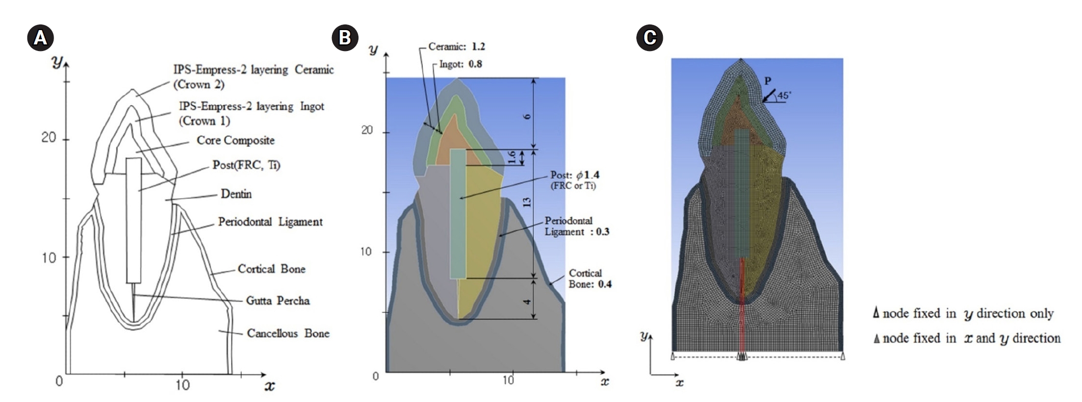
This study aimed to evaluate the stress distribution in teeth restored with different post materials and bonding conditions using finite element analysis (FEA).
Methods
A two-dimensional FEA model of a maxillary central incisor restored with IPS-Empress-2 crown (Ivoclar Vivadent), composite resin core, and posts were created. The model simulated bonded and non-bonded conditions for both fiber-reinforced composite (FRC) and titanium (Ti) posts. Stress distribution was analyzed using ANSYS 14.0 software under a 100-N load applied at a 45° angle to the long axis of the tooth.
Results
The results revealed that stress concentration was significantly higher in non-bonded posts compared to bonded ones. FRC posts exhibited stress values closer to those of dentin, whereas Ti posts demonstrated higher stress concentration, particularly in non-bonded states, increasing the potential risk of damage to surrounding tissues.
Conclusions
FRC posts, with elastic properties similar to dentin and proper adhesion, minimize stress concentration and potential damage to surrounding tissues. Conversely, materials with higher elastic modulus like Ti, can cause unfavorable stress concentrations if not properly bonded, emphasizing the importance of post adhesion in tooth restoration.
- 2,148 View
- 85 Download

- Effect of the restorative technique on load-bearing capacity, cusp deflection, and stress distribution of endodontically-treated premolars with MOD restoration
- Daniel Maranha da Rocha, João Paulo Mendes Tribst, Pietro Ausiello, Amanda Maria de Oliveira Dal Piva, Milena Cerqueira da Rocha, Rebeca Di Nicoló, Alexandre Luiz Souto Borges
- Restor Dent Endod 2019;44(3):e33. Published online August 7, 2019
- DOI: https://doi.org/10.5395/rde.2019.44.e33

-
 Abstract
 PDF
PDF PubReader
 ePub
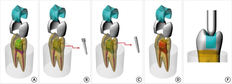
ePub Objectives To evaluate the influence of the restorative technique on the mechanical response of endodontically-treated upper premolars with mesio-occluso-distal (MOD) cavity.
Materials and Methods Forty-eight premolars received MOD preparation (4 groups,
n = 12) with different restorative techniques: glass ionomer cement + composite resin (the GIC group), a metallic post + composite resin (the MP group), a fiberglass post + composite resin (the FGP group), or no endodontic treatment + restoration with composite resin (the CR group). Cusp strain and load-bearing capacity were evaluated. One-way analysis of variance and the Tukey test were used with α = 5%. Finite element analysis (FEA) was used to calculate displacement and tensile stress for the teeth and restorations.Results MP showed the highest cusp (
p = 0.027) deflection (24.28 ± 5.09 µm/µm), followed by FGP (20.61 ± 5.05 µm/µm), CR (17.72 ± 6.32 µm/µm), and GIC (17.62 ± 7.00 µm/µm). For load-bearing, CR (38.89 ± 3.24 N) showed the highest, followed by GIC (37.51 ± 6.69 N), FGP (29.80 ± 10.03 N), and MP (18.41 ± 4.15 N) (p = 0.001) value. FEA showed similar behavior in the restorations in all groups, while MP showed the highest stress concentration in the tooth and post.Conclusions There is no mechanical advantage in using intraradicular posts for endodontically-treated premolars requiring MOD restoration. Filling the pulp chamber with GIC and restoring the tooth with only CR showed the most promising results for cusp deflection, failure load, and stress distribution.
-
Citations
Citations to this article as recorded by- How to adaptively balance ‘classic’ or ‘conservative’ approaches in tooth defect management: a 3D-finite element analysis study
Jiani Xu, Xu Liang, Lili Hu, Chen Sun, Zhipeng Zhang, Jiawei Yang, Jie Wang
BMC Oral Health.2025;[Epub] CrossRef - Inkjet-printed strain gauge sensors: Materials, manufacturing, and emerging applications
Lara Abdel Salam, Samir Mustapha, Alexandra Mikhael, Nisrine Bakri, Sahera Saleh, Massoud L. Khraiche
Sensors and Actuators A: Physical.2025; 394: 116934. CrossRef - Influence of endodontic access cavity design on mechanical properties of a first mandibular premolar tooth: a finite element analysis study
Taha Özyürek, Gülşah Uslu, Burçin Arıcan, Mustafa Gündoğar, Mohammad Hossein Nekoofar, Paul Michael Howell Dummer
Clinical Oral Investigations.2024;[Epub] CrossRef - Comparison of the Effect of Different Cavity Designs and Temporary Restoration Materials on the Fracture Resistance of Upper Premolars, Undergone Re-treatment: An In-Vitro Study
Parnian Alavinejad, Mohammad Yazdizadeh, Ali Mombeinipour, Ebrahim Karimzadeh
Proceedings of the National Academy of Sciences, India Section B: Biological Sciences.2024; 94(3): 677. CrossRef - Fracture resistance and failure mode of endodontically treated premolars reconstructed by different preparation approaches: Cervical margin relocation and crown lengthening with complete and partial ferrule with three different post and core systems
Mehran Falahchai, Naghmeh Musapoor, Soroosh Mokhtari, Yasamin Babaee Hemmati, Hamid Neshandar Asli
Journal of Prosthodontics.2024; 33(8): 774. CrossRef - Comparison of the stress distribution in base materials and thicknesses in composite resin restorations
Min-Kwan Jung, Mi-Jeong Jeon, Jae-Hoon Kim, Sung-Ae Son, Jeong-Kil Park, Deog-Gyu Seo
Heliyon.2024; 10(3): e25040. CrossRef -
Fracture resistance and failure pattern of endodontically treated maxillary premolars restored with transfixed glass fiber post: an
in vitro
and finite element analysis
Saleem Abdulrab, Greta Geerts, Ganesh Thiagarajan
Computer Methods in Biomechanics and Biomedical Engineering.2024; 27(4): 419. CrossRef - Influence of size-anatomy of the maxillary central incisor on the biomechanical performance of post-and-core restoration with different ferrule heights
Domingo Santos Pantaleón, João Paulo Mendes Tribst, Franklin García-Godoy
The Journal of Advanced Prosthodontics.2024; 16(2): 77. CrossRef - Influence of internal angle and shape of the lining on residual stress of Class II molar restorations
Qianqian Zuo, Annan Li, Haidong Teng, Zhan Liu
Computer Methods in Biomechanics and Biomedical Engineering.2024; 27(5): 680. CrossRef - Evaluation of stress distribution in coronal base and restorative materials: A narrative review of finite element analysis studies
Yelda Polat, İzzet Yavuz
Conservative Dentistry Journal.2024; 14(2): 47. CrossRef - The influence of horizontal glass fiber posts on fracture strength and fracture pattern of endodontically treated teeth: A systematic review and meta‐analysis of in vitro studies
Saleem Abdulrab, Greta Geerts, Sadeq Ali Al‐Maweri, Mohammed Nasser Alhajj, Hatem Alhadainy, Raidan Ba‐Hattab
Journal of Prosthodontics.2023; 32(6): 469. CrossRef - Stress distribution of a novel bundle fiber post with curved roots and oval canals
Deniz Yanık, Nurullah Turker
Journal of Esthetic and Restorative Dentistry.2022; 34(3): 550. CrossRef - The Effect of Endodontic Treatment and Thermocycling on Cuspal Deflection of Teeth Restored with Different Direct Resin Composites
Cansu Atalay, Ayse Ruya Yazici, Aynur Sidika Horuztepe, Emre Nagas
Conservative Dentistry and Endodontic Journal.2022; 6(2): 38. CrossRef - The use of different adhesive filling material and mass combinations to restore class II cavities under loading and shrinkage effects: a 3D-FEA
P. Ausiello, S. Ciaramella, A. De Benedictis, A. Lanzotti, J. P. M. Tribst, D. C. Watts
Computer Methods in Biomechanics and Biomedical Engineering.2021; 24(5): 485. CrossRef - Biomechanical Analysis of a Custom-Made Mouthguard Reinforced With Different Elastic Modulus Laminates During a Simulated Maxillofacial Trauma
João Paulo Mendes Tribst, Amanda Maria de Oliveira Dal Piva, Pietro Ausiello, Arianna De Benedictis, Marco Antonio Bottino, Alexandre Luiz Souto Borges
Craniomaxillofacial Trauma & Reconstruction.2021; 14(3): 254. CrossRef - Mechanical Assessment of Glass Ionomer Cements Incorporated with Multi-Walled Carbon Nanotubes for Dental Applications
Manuela Spinola, Amanda Maria Oliveira Dal Piva, Patrícia Uchôas Barbosa, Carlos Rocha Gomes Torres, Eduardo Bresciani
Oral.2021; 1(3): 190. CrossRef - Stress Concentration of Endodontically Treated Molars Restored with Transfixed Glass Fiber Post: 3D-Finite Element Analysis
Alexandre Luiz Souto Borges, Manassés Tercio Vieira Grangeiro, Guilherme Schmitt de Andrade, Renata Marques de Melo, Kusai Baroudi, Laís Regiane Silva-Concilio, João Paulo Mendes Tribst
Materials.2021; 14(15): 4249. CrossRef - Computer Aided Design Modelling and Finite Element Analysis of Premolar Proximal Cavities Restored with Resin Composites
Amanda Guedes Nogueira Matuda, Marcos Paulo Motta Silveira, Guilherme Schmitt de Andrade, Amanda Maria de Oliveira Dal Piva, João Paulo Mendes Tribst, Alexandre Luiz Souto Borges, Luca Testarelli, Gabriella Mosca, Pietro Ausiello
Materials.2021; 14(9): 2366. CrossRef - Effect of Shrinking and No Shrinking Dentine and Enamel Replacing Materials in Posterior Restoration: A 3D-FEA Study
Pietro Ausiello, Amanda Maria de Oliveira Dal Piva, Alexandre Luiz Souto Borges, Antonio Lanzotti, Fausto Zamparini, Ettore Epifania, João Paulo Mendes Tribst
Applied Sciences.2021; 11(5): 2215. CrossRef - Effect of Fiber-Reinforced Composite and Elastic Post on the Fracture Resistance of Premolars with Root Canal Treatment—An In Vitro Pilot Study
Jesús Mena-Álvarez, Rubén Agustín-Panadero, Alvaro Zubizarreta-Macho
Applied Sciences.2020; 10(21): 7616. CrossRef
- How to adaptively balance ‘classic’ or ‘conservative’ approaches in tooth defect management: a 3D-finite element analysis study
- 1,970 View
- 21 Download
- 20 Crossref

- Does apical root resection in endodontic microsurgery jeopardize the prosthodontic prognosis?
- Sin-Yeon Cho, Euiseong Kim
- Restor Dent Endod 2013;38(2):59-64. Published online May 28, 2013
- DOI: https://doi.org/10.5395/rde.2013.38.2.59

-
 Abstract
 PDF
PDF PubReader
 ePub
ePub Apical surgery cuts off the apical root and the crown-to-root ratio becomes unfavorable. Crown-to-root ratio has been applied to periodontally compromised teeth. Apical root resection is a different matter from periodontal bone loss. The purpose of this paper is to review the validity of crown-to-root ratio in the apically resected teeth. Most roots have conical shape and the root surface area of coronal part is wider than apical part of the same length. Therefore loss of alveolar bone support from apical resection is much less than its linear length.The maximum stress from mastication concentrates on the cervical area and the minimum stress was found on the apical 1/3 area. Therefore apical root resection is not so harmful as periodontal bone loss. Osteotomy for apical resection reduces longitudinal width of the buccal bone and increases the risk of endo-perio communication which leads to failure. Endodontic microsurgery is able to realize 0 degree or shallow bevel and precise length of root resection, and minimize the longitudinal width of osteotomy. The crown-to-root ratio is not valid in evaluating the prosthodontic prognosis of the apically resected teeth. Accurate execution of endodontic microsurgery to preserve the buccal bone is essential to avoid endo-perio communication.
-
Citations
Citations to this article as recorded by- Expert consensus on apical microsurgery
Hanguo Wang, Xin Xu, Zhuan Bian, Jingping Liang, Zhi Chen, Benxiang Hou, Lihong Qiu, Wenxia Chen, Xi Wei, Kaijin Hu, Qintao Wang, Zuhua Wang, Jiyao Li, Dingming Huang, Xiaoyan Wang, Zhengwei Huang, Liuyan Meng, Chen Zhang, Fangfang Xie, Di Yang, Jinhua Yu
International Journal of Oral Science.2025;[Epub] CrossRef - Approaches in apical microsurgery: conventional vs. guided. A systematic review
Germán Sánchez-Herrera, Matteo Facchera, Cristina Palma-Carrió, Martín Pérez-Leal
Oral and Maxillofacial Surgery.2025;[Epub] CrossRef - Influence of apical root resection level and filling technique on the fracture resistance of endodontically treated teeth: a biomechanical study
Guilherme Pauletto, Sidnei Flores de Pellegrin, Yasmin Padoin, Andressa Weber Vargas, Duvan Cala Castillo, Gabriel Kalil Rocha Pereira, Carlos Alexandre Souza Bier, Renata Dornelles Morgental
Odontology.2025;[Epub] CrossRef - Coexistence of horizontal bone loss and dehiscence with the bundle and conventional fiber post: a finite element analysis
Deniz Yanık, Nurullah Türker, Ahmet Mert Nalbantoğlu
Computer Methods in Biomechanics and Biomedical Engineering.2024; : 1. CrossRef - The tooth survival of non‐surgical root‐filled posterior teeth and the associated prognostic tooth‐related factors: A systematic review and meta‐analysis
S. R. Patel, F. Jarad, E. Moawad, A. Boland, J. Greenhalgh, Maria Liu, Michelle Maden
International Endodontic Journal.2024; 57(10): 1404. CrossRef - New-designed 3D printed surgical guide promotes the accuracy of endodontic microsurgery: a study of 14 upper anterior teeth
Dan Zhao, Weige Xie, Tianguo Li, Anqi Wang, Li Wu, Wen Kang, Lu Wang, Shiliang Guo, Xuna Tang, Sijing Xie
Scientific Reports.2023;[Epub] CrossRef - Multifactorial Analysis of Endodontic Microsurgery Using Finite Element Models
Raphael Richert, Jean-Christophe Farges, Jean-Christophe Maurin, Jérôme Molimard, Philippe Boisse, Maxime Ducret
Journal of Personalized Medicine.2022; 12(6): 1012. CrossRef - Mid‐term outcomes and periodontal prognostic factors of autotransplanted third molars: A retrospective cohort study
Ernest Lucas‐Taulé, Marc Llaquet, Jesús Muñoz‐Peñalver, José Nart, Federico Hernández‐Alfaro, Jordi Gargallo‐Albiol
Journal of Periodontology.2021; 92(12): 1776. CrossRef - Effect of length of apical root resection on the biomechanical response of a maxillary central incisor in various occlusal relationships
S. J. Ran, X. Yang, Z. Sun, Y. Zhang, J. X. Chen, D. M. Wang, B. Liu
International Endodontic Journal.2020; 53(1): 111. CrossRef - Changes of Root Length and Root-to-Crown Ratio after Apical Surgery: An Analysis by Using Cone-beam Computed Tomography
Thomas von Arx, Simon S. Jensen, Michael M. Bornstein
Journal of Endodontics.2015; 41(9): 1424. CrossRef - Influence of Apical Root Resection on the Biomechanical Response of a Single-rooted Tooth—Part 2: Apical Root Resection Combined with Periodontal Bone Loss
Youngjune Jang, Hyoung-Taek Hong, Heoung-Jae Chun, Byoung-Duck Roh
Journal of Endodontics.2015; 41(3): 412. CrossRef - Influence of Apical Root Resection on the Biomechanical Response of a Single-rooted Tooth: A 3-dimensional Finite Element Analysis
Youngjune Jang, Hyoung-Taek Hong, Byoung-Duck Roh, Heoung-Jae Chun
Journal of Endodontics.2014; 40(9): 1489. CrossRef
- Expert consensus on apical microsurgery
- 1,963 View
- 14 Download
- 12 Crossref

- Effect of internal stress on cyclic fatigue failure in .06 taper ProFile
- Hye-Rim Jung, Jin-Woo Kim, Kyung-Mo Cho, Se-Hee Park
- Restor Dent Endod 2012;37(2):79-83. Published online May 18, 2012
- DOI: https://doi.org/10.5395/rde.2012.37.2.79
-
 Abstract
 PDF
PDF PubReader
 ePub
ePub Objectives The purpose of this study was to evaluate the relation between intentionally induced internal stress and cyclic fatigue failure of .06 taper ProFile.
Materials and Methods Length 25 mm, .06 taper ProFile (Dentsply Maillefer), and size 20, 25, 30, 35 and 40 were used in this study. To give the internal stress, the rotary NiTi files were put into the .02 taper, Endo-Training-Bloc (Dentsply Maillefer) until auto-stop by torque controlled motor. Rotary NiTi files were grouped by the number of induced internal stress and randomly distributed among one control group and three experimental groups (
n = 10, Stress 0 [control], Stress 1, Stress 2 and Stress 3). For cyclic fatigue measurement, time for separation of the rotary NiTi files was recorded. The fractured surfaces were observed by field emission scanning electron microscope (FE-SEM, SU-70, Hitachi). The time for separation was statistically analyzed using two-way ANOVA andpost-hoc Scheffe test at 95% level.Results In .06 taper ProFile size 20, 25, 30, 35 and 40, there were statistically significant difference on time for separation between control group and the other groups (
p < 0.05).Conclusion In the limitation of this study, cyclic fatigue failure of .06 taper ProFile is influenced by internal stress accumulated in the files.
- 669 View
- 4 Download

- Effect of internal stress on cyclic fatigue failure in K3
- Jun-Young Kim, Jin-Woo Kim, Kyung-Mo Cho, Se-Hee Park
- Restor Dent Endod 2012;37(2):74-78. Published online May 18, 2012
- DOI: https://doi.org/10.5395/rde.2012.37.2.74
-
 Abstract
 PDF
PDF PubReader
 ePub
ePub Objectives This study aimed to evaluate the relationship between the cyclic fatigue of a K3 file and internal stress intentionally induced until the activation of the auto-stop function of the torque-controlled motor.
Materials and Methods K3 (Sybron Endo) .04 and .06 taper, size 25, 30, 35, 40 and 45 were used in this study. To give the internal stress, the K3 files were put into the .02 taper Endo-Training-Bloc (Dentsply Maillefer) until the activation of the auto-stop function of the torque-controlled motor. The rotation speed was 300 rpm and torque value was 1.0 N·cm. K3 were grouped by the number of induced internal stress and randomly distributed to 4 experimental groups (
n = 10, Stress 0 [control], Stress 1, Stress 2 and Stress 3). For measuring the cyclic fatigue failure, the K3 files were worked against a sloped glass block and time for file separation was recorded. Data was statistically analyzed Statistical analyses were performed using two-way ANOVA and Duncan post-hoc test atp < 0.05 level.Results Except .04 taper size 30 in Stress 1 group, there were statistically significant differences in time for file separation between control and all experimental groups. K3 with .04 taper showed higher cyclic fatigue resistance than those of .06 taper.
Conclusion In the limitation of this study, the cyclic fatigue of the K3 file was influenced by the accumulated internal stress from use until the auto-stop function was activated by the torque-controlled motor. Therefore, clinicians should avoid the reuse of the K3 file that has undergone auto-stops.
- 732 View
- 1 Download

- Evaluation of polymerization shrinkage stress in silorane-based composites
- Seung-Ji Ryu, Ji-Hoon Cheon, Jeong-Bum Min
- J Korean Acad Conserv Dent 2011;36(3):188-195. Published online May 31, 2011
- DOI: https://doi.org/10.5395/JKACD.2011.36.3.188
-
 Abstract
 PDF
PDF PubReader
 ePub
ePub Objectives The purpose of this study was to evaluate the polymerization shrinkage stress among conventional methacrylate-based composite resins and a silorane-based composite resin.
Materials and Methods The strain gauge method was used for the determination of polymerization shrinkage strain. Specimens were divided by 3 groups according to various composite materials. Filtek Z-250 (3M ESPE) and Filtek P-60 (3M ESPE) were used as a conventional methacrylate-based composites and Filtek P-90 (3M ESPE) was used as a silorane-based composites. Measurements were recorded at each 1 second for the total of 800 seconds including the periods of light application. The results of polymerization shrinkage stress were statistically analyzed using One way ANOVA and Tukey test (
p = 0.05).Results The polymerization shrinkage stress of a silorane-based composite resin was lower than those of conventional methacrylate-based composite resins (
p < 0.05). The shrinkage stress between methacrylate-based composite resin groups did not show significant difference (p > 0.05).Conclusions Within the limitation of this study, silorane-based composites showed lower polymerization shrinkage stress than methacrylate-based composites. We need to investigate more into polymerization shrinkage stress with regard to elastic modulus of silorane-based composites for the precise result.
-
Citations
Citations to this article as recorded by- Polymerization shrinkage and stress analysis during dental restoration observed by digital image correlation method
Jung-Hoon Park, Nak-Sam Choi
Journal of Mechanical Science and Technology.2021; 35(12): 5435. CrossRef - Evaluation of the color stability of light cured composite resins according to the resin matrices
Da-Hye Yu, Hyun-Jin Jung, Sung-Hyeon Choi, In-Nam Hwang
Korean Journal of Dental Materials.2019; 46(2): 109. CrossRef - Behavior of Polymerization Shrinkage Stress of Methacrylate-based Composite and Silorane-based Composite during Dental Restoration
Jung-Hoon Park, Nak-Sam Choi
Composites Research.2015; 28(1): 6. CrossRef - Microtensile bond strength of silorane-based composite specific adhesive system using different bonding strategies
Laura Alves Bastos, Ana Beatriz Silva Sousa, Brahim Drubi-Filho, Fernanda de Carvalho Panzeri Pires-de-Souza, Lucas da Fonseca Roberti Garcia
Restorative Dentistry & Endodontics.2015; 40(1): 23. CrossRef
- Polymerization shrinkage and stress analysis during dental restoration observed by digital image correlation method
- 1,625 View
- 8 Download
- 4 Crossref

- The effect of the amount of interdental spacing on the stress distribution in maxillary central incisors restored with porcelain laminate veneer and composite resin: A 3D-finite element analysis
- Junbae Hong, Seung-Min Tak, Seung-Ho Baek, Byeong-Hoon Cho
- J Korean Acad Conserv Dent 2010;35(1):30-39. Published online January 31, 2010
- DOI: https://doi.org/10.5395/JKACD.2010.35.1.030
-
 Abstract
 PDF
PDF PubReader
 ePub
ePub This study evaluated the influence of the type of restoration and the amount of interdental spacing on the stress distribution in maxillary central incisors restored by means of porcelain laminate veneers and direct composite resin restorations.
Three-dimensional finite element models were fabricated to represent different types of restorations. Four clinical situations were considered. Type I, closing diastema using composite resin. Labial border of composite resin was extended just enough to cover the interdental space; Type II, closing diastema using composite resin without reduction of labial surface. Labial border of composite resin was extended distally to cover the half of the total labial surface; Type III, closing diastema using composite resin with reduction of labial surface. Labial border of the preparation and restored composite resin was extended distally two-thirds of the total labial surface; Type IV, closing diastema using porcelain laminate veneer with a feathered-edge preparation technique. Four different interdental spaces (1.0, 2.0, 3.0, 4.0 mm) were applied for each type of restorations.
For all types of restoration, adding the width of free extension of the porcelain laminate veneer and composite resin increased the stress occurred at the bonding layer. The maximum stress values observed at the bonding layer of Type IV were higher than that of Type I, II and III. However, the increasing rate of maximum stress value of Type IV was lower than that of Type I, II and III.
-
Citations
Citations to this article as recorded by- Revamping the Peg Smile: An Art of Rehabilitation of Peg Laterals with Ceramic Veneers and Composite Restorations—A Case Report
Mahendran Kavitha, Ramdhas Annapurani, Pasupathy Shakunthala, Jayavel Nandhakumar
Journal of Operative Dentistry & Endodontics.2022; 6(2): 69. CrossRef - Minimally Invasive Diastema Restoration with Prefabricated Sectional Veneers
Claudio Novelli, Andrea Scribante
Dentistry Journal.2020; 8(2): 60. CrossRef
- Revamping the Peg Smile: An Art of Rehabilitation of Peg Laterals with Ceramic Veneers and Composite Restorations—A Case Report
- 1,095 View
- 2 Download
- 2 Crossref

- The effects of dentin bonding agent thickness on stress distribution of composite-tooth interface : Finite element method
- Sang-Il Park, Yemi Kim, Byoung-Duk Roh
- J Korean Acad Conserv Dent 2009;34(5):442-449. Published online September 30, 2009
- DOI: https://doi.org/10.5395/JKACD.2009.34.5.442
-
 Abstract
 PDF
PDF PubReader
 ePub
ePub The aim of this study was to examine that thick dentin bonding agent application or low modulus composite restoration could reduce stresses on dentin bonding agent layer.
A mandibular first premolar with abfraction lesion was modeled by finite element method. The lesion was restored by different composite resins with variable dentin bonding agent thickness (50µm, 100µm, 150µm). 170N of occlusal loading was applied buccally or lingually. Von Mises stress on dentin bonding agent layer were measured.
When thickness of dentin bonding agent was increased von Mises stresses at dentin bonding agent were decreased in both composites. Lower elastic modulus composite restoration showed decreased von Mises stresses. On root dentin margin more stresses were generated than enamel margin.
For occlusal stress relief at dentin boning agent layer to applicate thick dentin bonding agent or to choose low elastic modulus composite is recommended.
-
Citations
Citations to this article as recorded by- Influence of application methods of one-step self-etching adhesives on microtensile bond strength
Chul-Kyu Choi, Sung-Ae Son, Jin-Hee Ha, Bock Hur, Hyeon-Cheol Kim, Yong-Hun Kwon, Jeong-Kil Park
Journal of Korean Academy of Conservative Dentistry.2011; 36(3): 203. CrossRef
- Influence of application methods of one-step self-etching adhesives on microtensile bond strength
- 1,220 View
- 2 Download
- 1 Crossref

- Effect of instrument compliance on the polymerization shrinkage stress measurements of dental resin composites
- Deog-Gyu Seo, Sun-Hong Min, In-Bog Lee
- J Korean Acad Conserv Dent 2009;34(2):145-153. Published online March 31, 2009
- DOI: https://doi.org/10.5395/JKACD.2009.34.2.145
-
 Abstract
 PDF
PDF PubReader
 ePub
ePub The purpose of this study was to evaluate the effect of instrument compliance on the polymerization shrinkage stress measurements of dental composites. The contraction strain and stress of composites during light curing were measured by a custom made stress-strain analyzer, which consisted of a displacement sensor, a cantilever load cell and a negative feedback mechanism. The instrument can measure the polymerization stress by two modes: with compliance mode in which the instrument compliance is allowed, or without compliance mode in which the instrument compliance is not allowed.
A flowable (Filtek Flow: FF) and two universal hybrid (Z100: Z1 and Z250: Z2) composites were studied. A silane treated metal rod with a diameter of 3.0 mm was fixed at free end of the load cell, and other metal rod was fixed on the base plate. Composite of 1.0 mm thickness was placed between the two rods and light cured. The axial shrinkage strain and stress of the composite were recorded for 10 minutes during polymerization, and the tensile modulus of the materials was also determined with the instrument. The statistical analysis was conducted by ANOVA, paired t-test and Tukey's test (α<0.05).
There were significant differences between the two measurement modes and among materials. With compliance mode, the contraction stress of FF was the highest: 3.11 (0.13), followed by Z1: 2.91 (0.10) and Z2: 1.94 (0.09) MPa. When the instrument compliance is not allowed, the contraction stress of Z1 was the highest: 17.08 (0.89), followed by FF: 10.11 (0.29) and Z2: 9.46 (1.63) MPa. The tensile modulus for Z1, Z2 and FF was 2.31 (0.18), 2.05 (0.20), 1.41 (0.11) GPa, respectively. With compliance mode, the measured stress correlated with the axial shrinkage strain of composite; while without compliance the elastic modulus of materials played a significant role in the stress measurement.
-
Citations
Citations to this article as recorded by- Effects of cuspal compliance and radiant emittance of LED light on the cuspal deflection of replicated tooth cavity
Chang-Ha LEE, In-Bog LEE
Dental Materials Journal.2021; 40(3): 827. CrossRef - Polymerization Shrinkage and Stress of Silorane-based Dental Restorative Composite
In-Bog Lee, Sung-Hwan Park, Hyun-Jeong Kweon, Ja-Uk Gu, Nak-Sam Choi
Composites Research.2013; 26(3): 182. CrossRef - Evaluation of polymerization shrinkage stress in silorane-based composites
Seung-Ji Ryu, Ji-Hoon Cheon, Jeong-Bum Min
Journal of Korean Academy of Conservative Dentistry.2011; 36(3): 188. CrossRef - The change of the initial dynamic visco-elastic modulus of composite resins during light polymerization
Min-Ho Kim, In-Bog Lee
Journal of Korean Academy of Conservative Dentistry.2009; 34(5): 450. CrossRef
- Effects of cuspal compliance and radiant emittance of LED light on the cuspal deflection of replicated tooth cavity
- 1,189 View
- 1 Download
- 4 Crossref

- Stress distribution of endodontically treated maxillary second premolars restored with different methods: Three-dimensional finite element analysis
- Dong-Yeol Lim, Hyeon-Cheol Kim, Bock Hur, Kwang-Hoon Kim, Kwon Son, Jeong-Kil Park
- J Korean Acad Conserv Dent 2009;34(1):69-79. Published online January 31, 2009
- DOI: https://doi.org/10.5395/JKACD.2009.34.1.069
-
 Abstract
 PDF
PDF PubReader
 ePub
ePub The purpose of this study was to evaluate the influence of elastic modulus of restorative materials and the number of interfaces of post and core systems on the stress distribution of three differently restored endodontically treated maxillary second premolars using 3D FE analysis. Model 1, 2 was restored with a stainless steel or glass fiber post and direct composite resin. A PFG or a sintered alumina crown was considered. Model 3 was restored by EndoCrown. An oblique 500 N was applied on the buccal (Load A) and palatal (Load B) cusp. The von Mises stresses in the coronal and root structure of each model were analyzed using ANSYS. The elastic modulus of the definitive restorations rather than the type of post and core system was the primary factor that influenced the stress distribution of endodontically treated maxillary premolars. The stress concentration at the coronal structure could be lowered through the use of definitive restoration of high elastic modulus. The stress concentration at the root structure could be lowered through the use of definitive restoration of low elastic modulus.
-
Citations
Citations to this article as recorded by- How loss of tooth structure impacts the biomechanical behavior of a single-rooted maxillary premolar: FEA
Roaa Abdelwahab Abdelfattah, Nawar Naguib Nawar, Engy M. Kataia, Shehabeldin Mohamed Saber
Odontology.2024; 112(1): 279. CrossRef - Effect of Proximal Caries-driven Access on the Biomechanical Behavior of Endodontically Treated Maxillary Premolars
Nawar Naguib Nawar, Roaa Abdelwahab Abdelfattah, Mohamed Kataia, Shehabeldin Mohamed Saber, Engy Medhat Kataia, Hyeon-Cheol Kim
Journal of Endodontics.2023; 49(10): 1337. CrossRef - Survival and success of endocrowns: A systematic review and meta-analysis
Raghad A. Al-Dabbagh
The Journal of Prosthetic Dentistry.2021; 125(3): 415.e1. CrossRef - Influence of Cavity Design on Stress Distribution in Second Premolar Tooth Using Finite Element Analysis
Z. Parlar, E.U. Gökçek, K. Yildirim, A. Kahyaoglu
Acta Physica Polonica A.2017; 132(3-II): 949. CrossRef - Influence of post types and sizes on fracture resistance in the immature tooth model
Jong-Hyun Kim, Sung-Ho Park, Jeong-Won Park, Il-Young Jung
Journal of Korean Academy of Conservative Dentistry.2010; 35(4): 257. CrossRef
- How loss of tooth structure impacts the biomechanical behavior of a single-rooted maxillary premolar: FEA
- 1,557 View
- 8 Download
- 5 Crossref

- Effect of restoration type on the stress distribution of endodontically treated maxillary premolars; Three-dimensional finite element study
- Heun-Sook Jung, Hyeon-Cheol Kim, Bock Hur, Kwang-Hoon Kim, Kwon Son, Jeong-Kil Park
- J Korean Acad Conserv Dent 2009;34(1):8-19. Published online January 31, 2009
- DOI: https://doi.org/10.5395/JKACD.2009.34.1.008
-
 Abstract
 PDF
PDF PubReader
 ePub
ePub The purpose of this study was to investigate the effects of four restorative materials under various occlusal loading conditions on the stress distribution at the CEJ of buccal, palatal surface and central groove of occlusal surface of endodontically treated maxillary second premolar, using a 3D finte element analysis.
A 3D finite element model of human maxillary second premolar was endodontically treated. After endodontic treatment, access cavity was filled with Amalgam, resin, ceramic or gold of different mechanical properties. A static 500N forces were applied at the buccal (Load-1) and palatal cusp (Load-2) and a static 170N forces were applied at the mesial marginal ridge and palatal cusp simultaneously as centric occlusion (Load-3). Under 3-type Loading condition, the value of tensile stress was analyzed after 4-type restoration at the CEJ of buccal and palatal surface and central groove of occlusal surface
Excessive high tensile stresses were observed along the palatal CEJ in Load-1 case and buccal CEJ in Load-2 in all of the restorations. There was no difference in magnitude of stress in relation to the type of restorations. Heavy tensile stress concentrations were observed around the loading point and along the central groove of occlusal surface in all of the restorations. There was slight difference in magnitude of stress between different types of restorations. High tensile stress concentrations around the loading points were observed and there was no difference in magnitude of stress between different types of restorations in Load-3.
- 1,482 View
- 2 Download

- Stress distribution for NiTi files of triangular based and rectangular based cross-sections using 3-dimensional finite element analysis
- Hyun-Ju Kim, Chan-Joo Lee, Byung-Min Kim, Jeong-Kil Park, Bock Hur, Hyeon-Cheol Kim
- J Korean Acad Conserv Dent 2009;34(1):1-7. Published online January 31, 2009
- DOI: https://doi.org/10.5395/JKACD.2009.34.1.001
-
 Abstract
 PDF
PDF PubReader
 ePub
ePub The purpose of this study was to compare the stress distributions of NiTi rotary instruments based on their cross-sectional geometries of triangular shape-based cross-sectional design, S-shaped cross-sectional design and modified rectangular shape-based one using 3D FE models.
NiTi rotary files of S-shaped and modified rectangular design of cross-section such as Mtwo or NRT showed larger stress change while file rotation during simulated shaping.
The stress of files with rectangular cross-section design such as Mtwo, NRT was distributed as an intermittent pattern along the long axis of file. On the other hand, the stress of files with triangular cross-section design was distributed continuously.
When the residual stresses which could increase the risk of file fatigue fracture were analyzed after their withdrawal, the NRT and Mtwo model also presented higher residual stresses.
From this result, it can be inferred that S-shaped and modified rectangular shape-based files were more susceptible to file fracture than the files having triangular shape-based one.
-
Citations
Citations to this article as recorded by- Ex-Vivo Comparison of Torsional Stress on Nickel–Titanium Instruments Activated by Continuous Rotation or Adaptive Motion
Joo Yeong Lee, Sang Won Kwak, Jung-Hong Ha, Hyeon-Cheol Kim
Materials.2020; 13(8): 1900. CrossRef - Autogenous teeth used for bone grafting: a comparison with traditional grafting materials
Young-Kyun Kim, Su-Gwan Kim, Pil-Young Yun, In-Sung Yeo, Seung-Chan Jin, Ji-Su Oh, Heung-Joong Kim, Sun-Kyoung Yu, Sook-Young Lee, Jae-Sung Kim, In-Woong Um, Mi-Ae Jeong, Gyung-Wook Kim
Oral Surgery, Oral Medicine, Oral Pathology and Oral Radiology.2014; 117(1): e39. CrossRef - Analysis of crystalline structure of autogenous tooth bone graft material: X-Ray diffraction analysis
Gyung-Wook Kim, In-Sung Yeo, Su-Gwan Kim, In-Woong Um, Young-Kyun Kim
Journal of the Korean Association of Oral and Maxillofacial Surgeons.2011; 37(3): 225. CrossRef
- Ex-Vivo Comparison of Torsional Stress on Nickel–Titanium Instruments Activated by Continuous Rotation or Adaptive Motion
- 1,260 View
- 6 Download
- 3 Crossref

- Stress analysis of maxillary premolars with composite resin restoration of notch-shaped class V cavity and access cavity; Three-dimensional finite element study
- Seon-Hwa Lee, Hyeon-Cheol Kim, Bock Hur, Kwang-Hoon Kim, Kwon Son, Jeong-Kil Park
- J Korean Acad Conserv Dent 2008;33(6):570-579. Published online November 30, 2008
- DOI: https://doi.org/10.5395/JKACD.2008.33.6.570
-
 Abstract
 PDF
PDF PubReader
 ePub
ePub The purpose of this study was to investigate the distribution of tensile stress of canal obturated maxillary second premolar with access cavity and notch-shaped class V cavity restored with composite resin using a 3D finite element analysis.
The tested groups were classified as 8 situations by only access cavity or access cavity with notch-shaped class VS cavity (S or N), loading condition (L1 or L2), and with or without glass ionomer cement base (R1 or R2). A static load of 500 N was applied at buccal and palatal cusps. Notch-shaped cavity and access cavity were filled microhybrid composite resin (Z100) with or without GIC base (Fuji II LC). The tensile stresses presented in the buccal cervical area, palatal cervical area and occlusal surface were analyzed using ANSYS.
Tensile stress distributions were similar regardless of base. When the load was applied on the buccal cusp, excessive high tensile stress was concentrated around the loading point and along the central groove of occlusal surface. The tensile stress values of the tooth with class V cavity were slightly higher than that of the tooth without class V cavity. When the load was applied the palatal cusp, excessive high tensile stress was concentrated around the loading point and along the central groove of occlusal surface. The tensile stress values of the tooth without class V cavity were slightly higher than that of the tooth with class V cavity.
- 1,009 View
- 0 Download

- The influence of cavity configuration on the microtensile bond strength between composite resin and dentin
- Yemi Kim, Jeong-won Park, Chan young Lee, Yoon jung Song, Deok Kyu Seo, Byoung-Duck Roh
- J Korean Acad Conserv Dent 2008;33(5):472-480. Published online September 30, 2008
- DOI: https://doi.org/10.5395/JKACD.2008.33.5.472
-
 Abstract
 PDF
PDF PubReader
 ePub
ePub This study was conducted to evaluate the influence of the C-factor on the bond strength of a 6th generation self-etching system by measuring the microtensile bond strength of four types of restorations classified by different C-factors with an identical depth of dentin.
Eighty human molars were divided into four experimental groups, each of which had a C-factor of 0.25, 2, 3 or 4. Each group was then further divided into four subgroups based on the adhesive and composite resin used. The adhesives used for this study were AQ Bond Plus (Sun Medical, Japan) and Xeno III (DENTSPLY, Germany). And composite resins used were Fantasista (Sun Medical, Japan) and Ceram-X mono (DENTSPLY, Germany).
The results were then analyzed using one-way ANOVA, a Tukey's test, and a Pearson's correlation test and were as follows.
There was no significant difference among C-factor groups with the exception of groups of Xeno III and Ceram-X mono (p < 0.05).
There was no significant difference between any of the adhesives and composite resins in groups with C-factor 0.25, 2 and 4.
There was no correlation between the change in C-factor and microtensile bond strength in the Fantasista groups.
It was concluded that the C-factor of cavities does not have a significant effect on the microtensile bond strength of the restorations when cavities of the same depth of dentin are restored using composite resin in conjunction with the 6th generation self-etching system.
-
Citations
Citations to this article as recorded by- Effect of different chlorhexidine application times on microtensile bond strength to dentin in Class I cavities
Hyun-Jung Kang, Ho-Jin Moon, Dong-Hoon Shin
Restorative Dentistry & Endodontics.2012; 37(1): 9. CrossRef - Effect of Er:YAG lasing on the dentin bonding strength of two-step adhesives
Byeong-Choon Song, Young-Gon Cho, Myung-Seon Lee
Journal of Korean Academy of Conservative Dentistry.2011; 36(5): 409. CrossRef - Effect of Chlorhexidine Application Methods on Microtensile Bond Strength to Dentin in Class I Cavities
Y-E. Chang, D-H. Shin
Operative Dentistry.2010; 35(6): 618. CrossRef - Difference in bond strength according to filling techniques and cavity walls in box-type occlusal composite resin restoration
Eun-Joo Ko, Dong-Hoon Shin
Journal of Korean Academy of Conservative Dentistry.2009; 34(4): 350. CrossRef
- Effect of different chlorhexidine application times on microtensile bond strength to dentin in Class I cavities
- 961 View
- 1 Download
- 4 Crossref

- Comparison of the residual stress of the nanofilled composites
- Jeong-won Park
- J Korean Acad Conserv Dent 2008;33(5):457-462. Published online September 30, 2008
- DOI: https://doi.org/10.5395/JKACD.2008.33.5.457
-
 Abstract
 PDF
PDF PubReader
 ePub
ePub "Residual stress" can be developed during polymerization of the dental composite and it can be remained after this process was completed. The total amount of the force which applied to the composite restoration can be calculated by the sum of external and internal force. For the complete understanding of the restoration failure behavior, these two factors should be considered. In this experiment, I compared the residual stress of the recently developed nanofilled dental composite by ring slitting methods.
The composites used in this study can be categorized in two groups, one is microhybrid type-Z250, as control group, and nanofilled type-Grandio, Filtek Supreme, Ceram-X, as experimental ones. Composite ring was made and marked two reference points on the surface. Then measure the change of the distance between these two points before and after ring slitting. From the distance change, average circumferential residual stress (σθ) was calculated. In 10 minutes and 1 hour measurement groups, Filtek Supreme showed higher residual stress than Z250 and Ceram-X. In 24 hour group, Filtek showed higher stress than the other groups.
Following the result of this experiment, nanofilled composite showed similar or higher residual stress than Z250, and when comparing the Z250 and Filtek Supreme, which have quite similar matrix components, Filtek Supreme groups showed higher residual stress.
-
Citations
Citations to this article as recorded by- Microleakage of the experimental composite resin with three component photoinitiator systems
Ji-Hoon Kim, Dong-Hoon Shin
Journal of Korean Academy of Conservative Dentistry.2009; 34(4): 333. CrossRef
- Microleakage of the experimental composite resin with three component photoinitiator systems
- 1,133 View
- 4 Download
- 1 Crossref

- Stress distribution of three NiTi rotary files under bending and torsional conditions using 3-dimensional finite element analysis
- Tae-Oh Kim, Chan-Joo Lee, Byung-Min Kim, Jeong-Kil Park, Bock Hur, Hyeon-Cheol Kim
- J Korean Acad Conserv Dent 2008;33(4):323-331. Published online July 31, 2008
- DOI: https://doi.org/10.5395/JKACD.2008.33.4.323
-
 Abstract
 PDF
PDF PubReader
 ePub
ePub Flexibility and fracture properties determine the performance of NiTi rotary instruments. The purpose of this study was to evaluate how geometrical differences between three NiTi instruments affect the deformation and stress distributions under bending and torsional conditions using finite element analysis.
Three NiTi files (ProFile .06 / #30, F3 of ProTaper and ProTaper Universal) were scanned using a Micro-CT. The obtained structural geometries were meshed with linear, eight-noded hexahedral elements. The mechanical behavior (deformation and von Mises equivalent stress) of the three endodontic instruments were analyzed under four bending and rotational conditions using ABAQUS finite element analysis software. The nonlinear mechanical behavior of the NiTi was taken into account.
The U-shaped cross sectional geometry of ProFile showed the highest flexibility of the three file models. The ProTaper, which has a convex triangular cross-section, was the most stiff file model. For the same deflection, the ProTaper required more force to reach the same deflection as the other models, and needed more torque than other models for the same amount of rotation. The highest von Mises stress value was found at the groove area in the cross-section of the ProTaper Universal.
Under torsion, all files showed highest stresses at their groove area. The ProFile showed highest von Mises stress value under the same torsional moment while the ProTaper Universal showed the highest value under same rotational angle.
-
Citations
Citations to this article as recorded by- Effect of internal stress on cyclic fatigue failure in .06 taper ProFile
Hye-Rim Jung, Jin-Woo Kim, Kyung-Mo Cho, Se-Hee Park
Restorative Dentistry & Endodontics.2012; 37(2): 79. CrossRef
- Effect of internal stress on cyclic fatigue failure in .06 taper ProFile
- 1,296 View
- 7 Download
- 1 Crossref

- The influence of occlusal loads on stress distribution of cervical composite resin restorations: A three-dimensional finite element study
- Chan-Seok Park, Bock Hur, Hyeon-Cheol Kim, Kwang-Hoon Kim, Kwon Son, Jeong-Kil Park
- J Korean Acad Conserv Dent 2008;33(3):246-257. Published online May 31, 2008
- DOI: https://doi.org/10.5395/JKACD.2008.33.3.246
-
 Abstract
 PDF
PDF PubReader
 ePub
ePub The purpose of this study was to investigate the influence of various occlusal loading sites and directions on the stress distribution of the cervical composite resin restorations of maxillary second premolar, using 3 dimensional (3D) finite element (FE) analysis. Extracted maxillary second premolar was scanned serially with Micro-CT (SkyScan1072; SkyScan, Aartselaar, Belgium). The 3D images were processed by 3D-DOCTOR (Able Software Co., Lexington, MA, USA). HyperMesh (Altair Engineering, Inc., Troy, USA) and ANSYS (Swanson Analysis Systems, Inc., Houston, USA) was used to mesh and analyze 3D FE model. Notch shaped cavity was filled with hybrid (Z100, 3M Dental Products, St. Paul, MN, USA) or flowable resin (Tetric Flow, Vivadent Ets., FL-9494-Schaan, Liechtenstein) and each restoration was simulated with adhesive layer thickness (40 µm). A static load of 200 N was applied on the three points of the buccal incline of the palatal cusp and oriented in 20° increments, from vertical (long axis of the tooth) to oblique 40° direction towards the buccal. The maximum principal stresses in the occlusal and cervical cavosurface margin and vertical section of buccal surfaces of notch-shaped class V cavity were analyzed using ANSYS. As the angle of loading direction increased, tensile stress increased. Loading site had little effect on it. Under same loading condition, Tetric Flow showed relatively lower stress than Z100 overall, except both point angles. Loading direction and the elastic modulus of restorative material seem to be important factor on the cervical restoration.
-
Citations
Citations to this article as recorded by- Impact of bonding protocols and physical-mechanical properties of composite on durability and failure rate of fixed orthodontic retainers: a systematic review of laboratory studies
Maciej Jedliński, Marta Mazur, Claudia Salerno, Katarzyna Grocholewicz, Joanna Janiszewska-Olszowska
BMC Oral Health.2025;[Epub] CrossRef - Finite element analysis of maxillary central incisors restored with various post-and-core applications
MinSeock Seo, WonJun Shon, WooCheol Lee, Hyun-Mi Yoo, Byeong-Hoon Cho, Seung-Ho Baek
Journal of Korean Academy of Conservative Dentistry.2009; 34(4): 324. CrossRef
- Impact of bonding protocols and physical-mechanical properties of composite on durability and failure rate of fixed orthodontic retainers: a systematic review of laboratory studies
- 1,733 View
- 12 Download
- 2 Crossref

- The influence of combining composite resins with different elastic modulus on the stress distribution of Class V restoration: a three-dimensional finite element study
- Jeong-Kil Park, Bock Hur, Sung-Kyo Kim
- J Korean Acad Conserv Dent 2008;33(3):184-197. Published online May 31, 2008
- DOI: https://doi.org/10.5395/JKACD.2008.33.3.184
-
 Abstract
 PDF
PDF PubReader
 ePub
ePub This study was to investigate the influence of combining composite resins with different elastic modulus, and occlusal loading condition on the stress distribution of restored notch-shaped non-carious cervical lesion using 3D finite element (FE) analysis.
The extracted maxillary second premolar was scanned serially with Micro-CT. The 3D images were processed by 3D-DOCTOR. ANSYS was used to mesh and analyze 3D FE model. A notch-shaped cavity was modeled and filled with hybrid, flowable resin or a combination of both. After restoration, a static load of 500N was applied in a point-load condition at buccal cusp and palatal cusp. The stress data were analyzed using analysis of principal stress.
Results showed that combining method such that apex was restored by material with high elastic modulus and the occlusal and cervical cavosurface margin by small amount of material with low elastic modulus was the most profitable method in the view of tensile stress that was considered as the dominant factor jeopardizing the restoration durability and promoting the lesion progression.
-
Citations
Citations to this article as recorded by- Effect of restoration type on the stress distribution of endodontically treated maxillary premolars; Three-dimensional finite element study
Heun-Sook Jung, Hyeon-Cheol Kim, Bock Hur, Kwang-Hoon Kim, Kwon Son, Jeong-Kil Park
Journal of Korean Academy of Conservative Dentistry.2009; 34(1): 8. CrossRef - Stress distribution of endodontically treated maxillary second premolars restored with different methods: Three-dimensional finite element analysis
Dong-Yeol Lim, Hyeon-Cheol Kim, Bock Hur, Kwang-Hoon Kim, Kwon Son, Jeong-Kil Park
Journal of Korean Academy of Conservative Dentistry.2009; 34(1): 69. CrossRef - Finite element analysis of maxillary central incisors restored with various post-and-core applications
MinSeock Seo, WonJun Shon, WooCheol Lee, Hyun-Mi Yoo, Byeong-Hoon Cho, Seung-Ho Baek
Journal of Korean Academy of Conservative Dentistry.2009; 34(4): 324. CrossRef - Stress analysis of maxillary premolars with composite resin restoration of notch-shaped class V cavity and access cavity; Three-dimensional finite element study
Seon-Hwa Lee, Hyeon-Cheol Kim, Bock Hur, Kwang-Hoon Kim, Kwon Son, Jeong-Kil Park
Journal of Korean Academy of Conservative Dentistry.2008; 33(6): 570. CrossRef
- Effect of restoration type on the stress distribution of endodontically treated maxillary premolars; Three-dimensional finite element study
- 1,311 View
- 2 Download
- 4 Crossref

- Stress distribution of Class V composite resin restorations: A three-dimensional finite element study
- Jeong-Kil Park, Bock Hur, Sung-Kyo Kim
- J Korean Acad Conserv Dent 2008;33(1):28-38. Published online January 31, 2008
- DOI: https://doi.org/10.5395/JKACD.2008.33.1.028
-
 Abstract
 PDF
PDF PubReader
 ePub
ePub This study was to investigate the influence of composite resins with different elastic modulus, cavity modification and occlusal loading condition on the stress distribution of restored notch-shaped noncarious cervical lesion using 3-dimensional (3D) finite element (FE) analysis.
The extracted maxillary second premolar was scanned serially with Micro-CT. The 3D images were processed by 3D-DOCTOR. ANSYS was used to mesh and analyze 3D FE model. A notch-shaped cavity and a modified cavity with a rounded apex were modeled. Unmodified and modified cavities were filled with hybrid or flowable resin. After restoration, a static load of 500N was applied in a point-load condition at buccal cusp and palatal cusp. The stress data were analyzed using analysis of principal stress.
The results were as follows:
In the unrestored cavity, the stresses were highly concentrated at mesial CEJ and lesion apex and the peak stress was observed at the mesial point angle under both loading conditions.
After restoration of the cavity, stresses were significantly reduced at the lesion apex, however cervical cavosurface margin, stresses were more increased than before restoration under both loading conditions.
When restoring the notch-shaped lesion, material with high elastic modulus worked well at the lesion apex and material with low elastic modulus worked well at the cervical cavosurface margin.
Cavity modification the rounding apex did not reduce compressive stress, but tensile stress was reduced.
-
Citations
Citations to this article as recorded by- Effect of Loading and Restoration on the Biomechanical Behavior of Premolars with Simulated Abfraction Lesions
Deepa N Thangaraj, Sebeena Mathew, Karthick Kumaravadivel, Kerena Joseline, Boopathi Thangavel, Manimaran Sekar
World Journal of Dentistry.2025; 15(12): 1076. CrossRef - A Comparative Evaluation of Fiber-Reinforced Composite and Conventional Composite Restorations in Non-Carious Cervical Lesions: A Randomized Clinical Trial
Vinutha Manjunath, Aparna Nair, Vidya G. Doddawad
Journal of International Oral Health.2025; 17(2): 145. CrossRef - Numerical investigation of stress concentration at the tip of the V-notch partially filled with material
V.P. Matveenko, A.Y. Fedorov, E.B. Galkina
Computational Continuum Mechanics.2022; 15(3): 333. CrossRef - Clinical Evaluation of Bioactive Restorative Material versus Resin Modified Glass Ionomer in Cervical Restorations: A Randomized Controlled Clinical Trial
Yehia Hafez Yehia, Amir Hafez Ibrahim, Eman Abou-auf, Amira Farid Elzogbhi
Open Access Macedonian Journal of Medical Sciences.2022; 10(D): 33. CrossRef - Effects of Root Maturation and Thickness Variation in Coronal Mineral Trioxide Aggregate Plugs Under Traumatic Load on Stress Distribution in Regenerative Endodontic Procedures: A 3-dimensional Finite Element Analysis Study
Akif Demirel, Tuğba Bezgin, Şaziye Sarı
Journal of Endodontics.2021; 47(3): 492. CrossRef - Finite element analysis of V-shaped tooth defects filled with universal nanohybrid composite using incremental technique
Tsanka Dikova, Tihomir Vasilev, Vesela Hristova, Vladimir Panov
Journal of the Mechanical Behavior of Biomedical Materials.2021; 118: 104425. CrossRef - Are Increased Masticatory Forces Risk for Primary 2nd Molars without Successors? A 3D FEA Study
Akif Demirel, Şaziye Sarı
Journal of Clinical Pediatric Dentistry.2019; 43(1): 64. CrossRef - Finite element analysis of maxillary central incisors restored with various post-and-core applications
MinSeock Seo, WonJun Shon, WooCheol Lee, Hyun-Mi Yoo, Byeong-Hoon Cho, Seung-Ho Baek
Journal of Korean Academy of Conservative Dentistry.2009; 34(4): 324. CrossRef - The influence of combining composite resins with different elastic modulus on the stress distribution of Class V restoration: a three-dimensional finite element study
Jeong-Kil Park, Bock Hur, Sung-Kyo Kim
Journal of Korean Academy of Conservative Dentistry.2008; 33(3): 184. CrossRef - Stress analysis of maxillary premolars with composite resin restoration of notch-shaped class V cavity and access cavity; Three-dimensional finite element study
Seon-Hwa Lee, Hyeon-Cheol Kim, Bock Hur, Kwang-Hoon Kim, Kwon Son, Jeong-Kil Park
Journal of Korean Academy of Conservative Dentistry.2008; 33(6): 570. CrossRef
- Effect of Loading and Restoration on the Biomechanical Behavior of Premolars with Simulated Abfraction Lesions
- 1,755 View
- 8 Download
- 10 Crossref

- The influence of composite resin restoration on the stress distribution of notch shaped noncarious cervical lesion; A three dimensional finite element analysis study
- Chae-Kyung Lee, Jeong-Kil Park, Hyeon-Cheol Kim, Sung-Gwan Woo, Kwang-Hoon Kim, Kwon Son, Bock Hur
- J Korean Acad Conserv Dent 2007;32(1):69-79. Published online January 31, 2007
- DOI: https://doi.org/10.5395/JKACD.2007.32.1.069
-
 Abstract
 PDF
PDF PubReader
 ePub
ePub The purpose of this study was to investigate the effects of composite resin restorations on the stress distribution of notch shaped noncarious cervical lesion using three-dimensional (3D) finite element analysis (FEA).
Extracted maxillary second premolar was scanned serially with Micro-CT (SkyScan1072; SkyScan, Aartselaar, Belgium). The 3D images were processed by 3D-DOCTOR (Able Software Co., Lexington, MA, USA). ANSYS (Swanson Analysis Systems, Inc., Houston, USA) was used to mesh and analyze 3D FE model. Notch shaped cavity was filled with hybrid or flowable resin and each restoration was simulated with adhesive layer thickness (40 µM). A static load of 500 N was applied on a point load condition at buccal cusp (loading A) and palatal cusp (loading B). The principal stresses in the lesion apex (internal line angle of cavity) and middle vertical wall were analyzed using ANSYS.
The results were as follows
1. Under loading A, compressive stress is created in the unrestored and restored cavity. Under loading B, tensile stress is created. And the peak stress concentration is seen at near mesial corner of the cavity under each load condition.
2. Compared to the unrestored cavity, the principal stresses at the cemeto-enamel junction (CEJ) and internal line angle of the cavity were more reduced in the restored cavity on both load conditions.
3. In teeth restored with hybrid composite, the principal stresses at the CEJ and internal line angle of the cavity were more reduced than flowable resin.
-
Citations
Citations to this article as recorded by- The influence of occlusal loads on stress distribution of cervical composite resin restorations: A three-dimensional finite element study
Chan-Seok Park, Bock Hur, Hyeon-Cheol Kim, Kwang-Hoon Kim, Kwon Son, Jeong-Kil Park
Journal of Korean Academy of Conservative Dentistry.2008; 33(3): 246. CrossRef
- The influence of occlusal loads on stress distribution of cervical composite resin restorations: A three-dimensional finite element study
- 1,339 View
- 2 Download
- 1 Crossref

- Effects of occlusal load on the cervical stress distribution: A three-dimensional finite element study
- Hyeong-Mo Lee, Bock Hur, Hyeon-Cheol Kim, Sung-Gwan Woo, Kwang-Hoon Kim, Kwon Son, Jeong-Kil Park
- J Korean Acad Conserv Dent 2006;31(6):427-436. Published online November 30, 2006
- DOI: https://doi.org/10.5395/JKACD.2006.31.6.427
-
 Abstract
 PDF
PDF PubReader
 ePub
ePub The objective of this study was to investigate the effects of various occlusal loads on the stress distribution of the buccal cervical region of a normal maxillary second premolar, using a three dimensional finite element analysis (3D FEA).
After 3D FE modeling of maxillary second premolar, a static load of 500N of three load cases was applied. Stress analysis was performed using ANSYS (Swanson Analysis Systems, Inc., Houston, USA). The maximum principal stresses and minimum principal stresses were sampled at thirteen nodal points in the buccal cervical enamel for each four horizontal planes, 1.0 mm above CEJ, 0.5 mm above CEJ, CEJ, 0.5 mm under CEJ.
The results were as follows
1. The peak stress was seen at the cervical enamel surface of the mesiobuccal line angle area, asymmetrically.
2. The values of compressive stresses were within the range of the failure stress of enamel. But the values of tensile stresses exceeded the range of the failure stress of enamel.
3. The tensile stresses from the perpendicular load at the buccal incline of palatal cusp may be shown to be the primary etiological factors of the NCCLs.
-
Citations
Citations to this article as recorded by- Biomechanical and Occlusal Factors Influencing the Longevity of Single-Unit Restorations: A Comprehensive Review
Wedad S Alaida, Safa A Gadi, Rokia E Al-Ghannam, Moayad F Alamri, Feras I Mirdad, Ruba M Argaibeh, Bushra A Alqahtani, Abdulrahman M Alqahtani, Abdulelah A Al Jaban, Turki M Alkuraydimi, Abdulrahman S Alamari
Cureus.2025;[Epub] CrossRef - Effect of restoration type on the stress distribution of endodontically treated maxillary premolars; Three-dimensional finite element study
Heun-Sook Jung, Hyeon-Cheol Kim, Bock Hur, Kwang-Hoon Kim, Kwon Son, Jeong-Kil Park
Journal of Korean Academy of Conservative Dentistry.2009; 34(1): 8. CrossRef - Stress distribution of endodontically treated maxillary second premolars restored with different methods: Three-dimensional finite element analysis
Dong-Yeol Lim, Hyeon-Cheol Kim, Bock Hur, Kwang-Hoon Kim, Kwon Son, Jeong-Kil Park
Journal of Korean Academy of Conservative Dentistry.2009; 34(1): 69. CrossRef
- Biomechanical and Occlusal Factors Influencing the Longevity of Single-Unit Restorations: A Comprehensive Review
- 1,523 View
- 2 Download
- 3 Crossref

- The effect of restorative materials on the stress distribution of class V composite resin restorations - a 3D finite element investigation
- Hyoung-Ryoul Ahn, Hyeon-Cheol Kim, Bock Huh, Jeong-Kil Park
- J Korean Acad Conserv Dent 2006;31(1):20-29. Published online January 31, 2006
- DOI: https://doi.org/10.5395/JKACD.2006.31.1.020
-
 Abstract
 PDF
PDF PubReader
 ePub
ePub The purpose of this study was to analyze the stress distribution aspect of unrestored and restored combined shape (wedge shape occlusally and saucer shape gingivally) class V cavity, which found frequently in clinical cases.
A maxillary second premolar restored with a combined shape class V composite restorations were modeled using the three dimensional finite element method. Static occlusal load of 170 N was applied on lingual incline of buccal cusp at the angle of 45° with the longitudinal axis of the tooth. And three dimensional finite element analysis was taken by ANSYS (Version 6.0, Swanson Analysis System Co., Houston, U.S.A) program which represent the stress distribution on unrestored and restored cavity wall and margin.
The conclusions were as follows.
Compared to the unrestored cavity, Von Mises stress at the cementoenamel junction and line angle of the cavity base were reduced and in restored cavity.
Von Mises stress at the occlusal and cervical cavity margin and wall were increased in restored cavity in comparison with the unrestored cavity.
In the hybrid and hybrid/flowable composite resin restoration, Von Mises stress at the cementoenamel junction and line angle of the cavity base were reduced more than in the flowable restoration.
In the hybrid and hybrid/flowable composite resin restoration, Von Mises stress at the occlusal and cervical cavity margin and wall were increased more than in the flowable restoration.
-
Citations
Citations to this article as recorded by- Comparison of marginal microleakage between low and high flowable resins in class V cavity
Sang-Bae Bae, Young-Gon Cho, Myeong-Seon Lee
Journal of Korean Academy of Conservative Dentistry.2009; 34(6): 477. CrossRef
- Comparison of marginal microleakage between low and high flowable resins in class V cavity
- 1,032 View
- 6 Download
- 1 Crossref

- Effects of occlusal load on the stress distribution of four cavity configurations of noncarious cervical lesions: A three-dimensional finite element analysis study
- Sang-Je Jeon, Jeong-Kil Park, Hyeon-Cheol Kim, Sung-Gwan Woo, Kwang-Hoon Kim, Kwon Son, Bock Hur
- J Korean Acad Conserv Dent 2006;31(5):359-370. Published online January 14, 2006
- DOI: https://doi.org/10.5395/JKACD.2006.31.5.359
-
 Abstract
 PDF
PDF PubReader
 ePub
ePub Abstract The objective of this study was to investigate the effect of excessive occlusal loading on stress distribution on four type of cervical lesion, using a three dimensional finite element analysis (3D FEA).
The extracted maxillary second premolar was scanned serially with Micro-CT. The 3D images were processed by 3D-DOCTOR. ANSYS was used to mesh and analyze 3D FE model. Four different lesion configurations representative of the various types observed clinically for teeth were studied. A static point load of 500N was applied to the buccal and lingual cusp (Load A and B). The principal stresses in lesion apex, and vertical sectioned margin of cervical wall were analyzed.
The results were as follows
The patterns of stress distribution were similar but the magnitude was different in four types of lesion.
The peak stress was observed at mesial corner and also stresses concentrated at lesion apex.
The compressive stress under load A and the tensile stress under load B were dominant stress.
Under the load, lesion can be increased and harmful to tooth structure unless restored.
- 882 View
- 1 Download

- Effect of cavity shape, bond quality and volume on dentin bond strength
- Hyo-Jin Lee, Jong-Soon Kim, Shin-Jae Lee, Bum-Soon Lim, Seung-Ho Baek, Byeong-Hoon Cho
- J Korean Acad Conserv Dent 2005;30(6):450-460. Published online November 30, 2005
- DOI: https://doi.org/10.5395/JKACD.2005.30.6.450
-
 Abstract
 PDF
PDF PubReader
 ePub
ePub The aim of this study was to evaluate the effect of cavity shape, bond quality of bonding agent and volume of resin composite on shrinkage stress developed at the cavity floor. This was done by measuring the shear bond strength with respect to iris materials (cavity shape; adhesive-coated dentin as a high C-factor and Teflon-coated metal as a low C-factor), bonding agents (bond quality; Scotchbond™ Multi-purpose and Xeno®III) and iris hole diameters (volume; 1 mm or 3 mm in diameter × 1.5 mm in thickness). Ninety-six molars were randomly divided into 8 groups (2 × 2 × 2 experimental setup). In order to simulate a Class I cavity, shear bond strength was measured on the flat occlusal dentin surface with irises. The iris hole was filled with Z250 restorative resin composite in a bulk-filling manner. The data was analyzed using three-way ANOVA and the Tukey test. Fracture mode analysis was also done. When the cavity had high C-factor, good bond quality and large volume, the bond strength decreased significantly. The volume of resin composite restricted within the well-bonded cavity walls is also be suggested to be included in the concept of C-factor, as well as the cavity shape and bond quality. Since the bond quality and volume can exaggerate the effect of cavity shape on the shrinkage stress developed at the resin-dentin bond, resin composites must be filled in a method, which minimizes the volume that can increase the C-factor.
- 1,051 View
- 0 Download

- Measurements of shrinkage stress and reduction of inter-cuspal distance in maxillary premolars resulting from polymerization of composites and compomers
- Soon-Young Lee, Sung-Ho Park
- J Korean Acad Conserv Dent 2004;29(4):346-352. Published online July 31, 2004
- DOI: https://doi.org/10.5395/JKACD.2004.29.4.346
-
 Abstract
 PDF
PDF PubReader
 ePub
ePub The purpose of present study was to evaluate the polymerization shrinkage stress and cuspal deflection in maxillary premolars resulting from polymerization shrinkage of composites and compomers.
Composites and compomers which were used in this study were as follows: Dyract AP, Z100, Surefil, Pyramid, Synergy Compact, Heliomolar, Heliomolar HB, and Compoglass F. For measuring of polymerization shrinkage stress, Stress measuring machine (R&B, Daejon, Korea) was used. One-way ANOVA analysis with Duncan's multiple comparison test were used to determine significant differences between the materials.
For measuring of cuspal deflection of tooth, MOD cavities were prepared in 10 extracted maxillary premolars. And reduction of intercuspal distance was measured by strain measuring machine (R&B, Daejon, Korea) One-way ANOVA analysis with Turkey test were used to determine significant differences between the materials.
Polymerization shrinkage stress is 『Heliomolar, Z100, Pyramid < Synergy Compact Compoglass F < Dyract AP < Heliomolr HB, surefil』 (P < 0.05). And cuspal delfelction is 『Z100, Heliomolar, Heliomolar HB, Synergy Compact Surefil, < Compoglass F < Pyramid, Dyract AP』 (P < 0.05).
Measurements of ploymerization shrinkage stress and those of cuspal deflection of the teeth was different. There is no correlation between polymerization shrinkage stress and cuspal deflection of the teeth (p > 0.05).
-
Citations
Citations to this article as recorded by- Influence of thermal and thermomechanical stimuli on a molar tooth treated with resin-based restorative dental composites
Jerrin Thadathil Varghese, Behzad Babaei, Paul Farrar, Leon Prentice, B. Gangadhara Prusty
Dental Materials.2022; 38(5): 811. CrossRef - Effect of intermittent polymerization on the rate of polymerization shrinkage and cuspal deflection in composite resin
Min Kyung Kim, Sung Ho Park, Deog Gyu Seo, Yun Jung Song, Yoon Lee, Chan Young Lee
Journal of Korean Academy of Conservative Dentistry.2008; 33(4): 341. CrossRef - The effect of intermittent composite curing on marginal adaptation
Yong-Hwan Yun, Sung-Ho Park
Journal of Korean Academy of Conservative Dentistry.2007; 32(3): 248. CrossRef - Correlation Between the Amount of Linear Polymerization Shrinkage and Cuspal Deflection
S-Y. Lee, S-H. Park
Operative Dentistry.2006; 31(3): 364. CrossRef - Correlation between Linear polymerization shrinkage & tooth cuspal deflection
Soon-Young Lee, Sung-Ho Park
Journal of Korean Academy of Conservative Dentistry.2005; 30(6): 442. CrossRef
- Influence of thermal and thermomechanical stimuli on a molar tooth treated with resin-based restorative dental composites
- 897 View
- 0 Download
- 5 Crossref

- Amount of polymerization shrinkage and shrinkage stress in composites and compomers for posterior restoration
- Sung-Ho Park, Soon-Young Lee, Yong-Sik Cho, Su-Sun Kim, Chang-Jae Lee, Young-Joo Kim, Bong-Hee Lee, Kouang-Sung Lee, Byung-Duk Noh
- J Korean Acad Conserv Dent 2003;28(4):348-353. Published online July 31, 2003
- DOI: https://doi.org/10.5395/JKACD.2003.28.4.348
-
 Abstract
 PDF
PDF PubReader
 ePub
ePub The purpose of present study was to evaluate the polymerization shrinkage stress and amount of linear shrinkage of composites and compomers for posterior restoration.
For this purpose, linear polymerization shrinkage and polymerization stress were measured.
For linear polymerization shrinklage and polymerization stress measurement, custom made Linometer (R&B, Daejon, Korea) and Stress measuring machine was used (R&B, Daejon, Korea). Compositers and compomers were evaluated; Dyract AP (Dentsply Detrey, Gumbh. German) Z100 (3M Dental Products, St. Paul, USA) Surefil (Dentsply Caulk, Milford, USA) Pyramid(Bisco, Schaumburg, USA) Synergy Compact (Coltene, Altstatten, Switzerland), Heliomolar (Vivadent/Ivoclar, Liechtenstein), and Compoglass (Vivadent Ivoclar/Liechtenstein) were used. 15 measurements were made for each material. Linear polymerization shrinkage or polymerization stress for each material was compared with one way ANOVA with Tukey at 95% levels of confidence.
For linear shrinkage; Heliomolar, Surefil<Synergy Compact, Z100<Dyract AP<Pyramid, Compoglass F (p<0.05)
For Shrinkage stress; Heliomolar<Z100, Pyramid<Synergy Compact, Compoglass F<Dyract AP<Heliomolar HB, Surefil (p<0.05)
-
Citations
Citations to this article as recorded by- A comparative study on color and dimensional stability of esthetic indirect dental materials
Hye-Yun Heo, Hyo-Jin Son, Yu-Ri Heo, Mee-Kyoung Son
Oral Biology Research.2019; 43(4): 306. CrossRef - Measurement of the Internal Adaptation of Resin Composites Using Micro-CT and Its Correlation With Polymerization Shrinkage
HJ Kim, SH Park
Operative Dentistry.2014; 39(2): e57. CrossRef - Comparison of Premolar Cuspal Deflection in Bulk or in Incremental Composite Restoration Methods
ME Kim, SH Park
Operative Dentistry.2011; 36(3): 326. CrossRef - Effect of intermittent polymerization on the rate of polymerization shrinkage and cuspal deflection in composite resin
Min Kyung Kim, Sung Ho Park, Deog Gyu Seo, Yun Jung Song, Yoon Lee, Chan Young Lee
Journal of Korean Academy of Conservative Dentistry.2008; 33(4): 341. CrossRef - Correlation Between the Amount of Linear Polymerization Shrinkage and Cuspal Deflection
S-Y. Lee, S-H. Park
Operative Dentistry.2006; 31(3): 364. CrossRef - Correlation between Linear polymerization shrinkage & tooth cuspal deflection
Soon-Young Lee, Sung-Ho Park
Journal of Korean Academy of Conservative Dentistry.2005; 30(6): 442. CrossRef
- A comparative study on color and dimensional stability of esthetic indirect dental materials
- 1,036 View
- 1 Download
- 6 Crossref


 KACD
KACD

 First
First Prev
Prev


