
Search
- Page Path
- HOME > Search
Research Article
- Influence of adjacent restorative material and distance on the accuracy of inlay cavity impressions with intraoral scanner: an in vitro study
- So-Yeon Lee, Sung-Ae Son, Jae-Hoon Kim, Deog-Gyu Seo, Jeong-Kil Park
- Restor Dent Endod 2026;51(1):e6. Published online January 23, 2026
- DOI: https://doi.org/10.5395/rde.2026.51.e6
-
 Abstract
 PDF
PDF PubReader
 ePub
ePub - Objectives
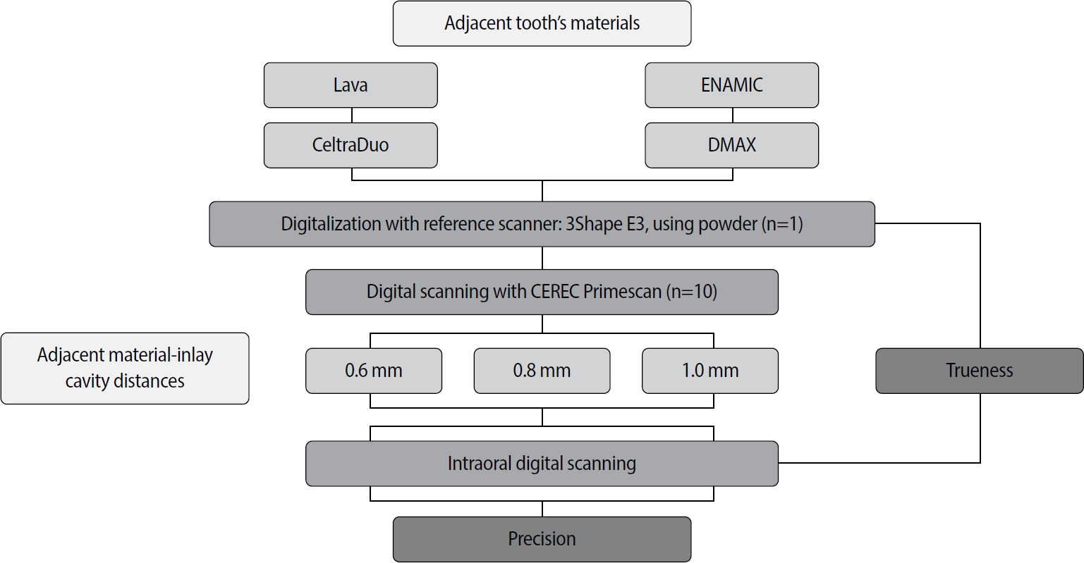
This study aimed to evaluate the influence of adjacent restorative material and interproximal distance on the accuracy of digital impressions of inlay cavities obtained using an intraoral scanner.
Methods
A disto-occlusal inlay cavity was prepared on a mandibular right first molar model, and digital scans were performed using a CEREC Primescan (Dentsply Sirona). The adjacent restorative materials used were Lava (3M ESPE), ENAMIC (VITA Zahnfabrik), Celtra Duo (Dentsply Sirona), and DMAX (DMAX), and the interproximal distances were set to 0.6 mm, 0.8 mm, and 1.0 mm. The obtained scan data were analyzed using GOM Inspect software (GOM GmbH).
Results
Trueness, maximum positive and negative deviations, and precision were significantly influenced by both the adjacent restorative material and the interproximal distance, while their interaction showed a significant effect only on precision. Celtra Duo demonstrated the highest trueness, with mean deviation values decreasing from 7.8 μm at a 0.6 mm interproximal distance to 7.3 μm at 1.0 mm. ENAMIC showed the best precision, presenting mean deviations of 2.6 μm at 0.6 mm, 2.9 μm at 0.8 mm, and 2.4 μm at 1.0 mm. A narrow interproximal distance of 0.6 mm resulted in lower trueness, measured at 8.3 μm, and the highest precision deviation of 3.4 μm. In contrast, an interproximal distance of 1.0 mm yielded improved scan accuracy, with increased trueness and reduced precision variation.
Conclusions
Digital impression accuracy of inlay cavities was influenced by adjacent restorative material and interproximal distance, suggesting clinical consideration is needed in CAD/CAM workflows to optimize restoration fit. -
Citations
Citations to this article as recorded by
- 3D-SCANNING IN PROSTHETIC DENTISTRY: ADVANTAGES, DISADVANTAGES, AND DEVELOPMENT PROSPECTS
V. S. Kuz, O. I. Teslenko, H. M. Kuz, H. M. Balia, Yu. S. Lunkova, O. V. Shemetov, I. M. Martynenko
Bulletin of Problems Biology and Medicine.2026; 1(1): 98. CrossRef
- 3D-SCANNING IN PROSTHETIC DENTISTRY: ADVANTAGES, DISADVANTAGES, AND DEVELOPMENT PROSPECTS
- 2,206 View
- 125 Download
- 1 Crossref

Original Article
- Influence of microhardness and fluoride content of tooth structure by fluoride-containing restorative materials
- Su-Jong Lee, Young-Gon Cho, Jong-Uk Kim, Byung-Cheul Park
- J Korean Acad Conserv Dent 2004;29(1):36-43. Published online January 31, 2004
- DOI: https://doi.org/10.5395/JKACD.2004.29.1.036
-
Abstract
PDFPubReaderePub
The purpose of this study was to compare the microhardness and the fluoride content of enamel and dentin around fluoride- or non fluoride-containing restorations. Forty extracted human teeth were used and prepared cervical cavities on proximal surface. Experimental teeth were divided into five groups. Group 1 : Prime & Bond NT and Z100, Group 2 : Prime & Bond NT and F2000, Group 3 : Scotchbond Multi-Purpose and Z100, Group 4 : Scothcbond Multi-purpose and F2000, Group 5 : Fuji II LC. The cavities were filled with dentin adhesives and restorative materials. After each tooth was bisected, one half was tested microhardness and the other half was analyzed the fluoride at the enamel and dentin by an EPMA-WDX device. The results were as follows:
1. There was no statistical difference among the microhardness of enamel surface in all group.
2. The microhardness at dentin of 100 µm point in Group 2 and 20 µm point in Group 4 was lower than that of normal dentin (p>0.05).
3. There was no statistical difference among the fluoride content of enamel surface in all group.
4. The fluoride content at the dentin of 30 µm point in Group 2 and 5 were higher than those at 100 µm and 200 µm point in Group 2 and normal dentin (p<0.05).
5. At the dentin of 30 µm point, Group 2 showed higher fluoride content than Group 1 and 3, and Group 5 showed higher fluoride content than other groups.
- 1,311 View
- 2 Download
 First
First Prev
Prev

