
Search
- Page Path
- HOME > Search
Research Articles
- Effect of quality of radiographs taken during root canal treatment on technical quality of root canal fillings and endodontic outcome
- Jia Min Ng, Yan Yee Lee, Prashanti Chippagiri, Elaheh Ahanin, Abhishek Parolia
- Restor Dent Endod 2025;50(1):e3. Published online January 7, 2025
- DOI: https://doi.org/10.5395/rde.2025.50.e3
-
 Abstract
 PDF
PDF PubReader
 ePub
ePub - Objectives
This study evaluated the number and quality of working length (WL) and master cone (MC) radiographs taken during root canal treatment by dental undergraduates, and their associations with the technical quality of root canal fillings (TQRCF) and endodontic outcomes (EO).
Methods
A retrospective evaluation of radiographs from 303 root canal-treated teeth in 231 patients was conducted, with 72 patients attending recall visits to assess EO. The chi-square and one-way analysis of variance tests were performed.
Results
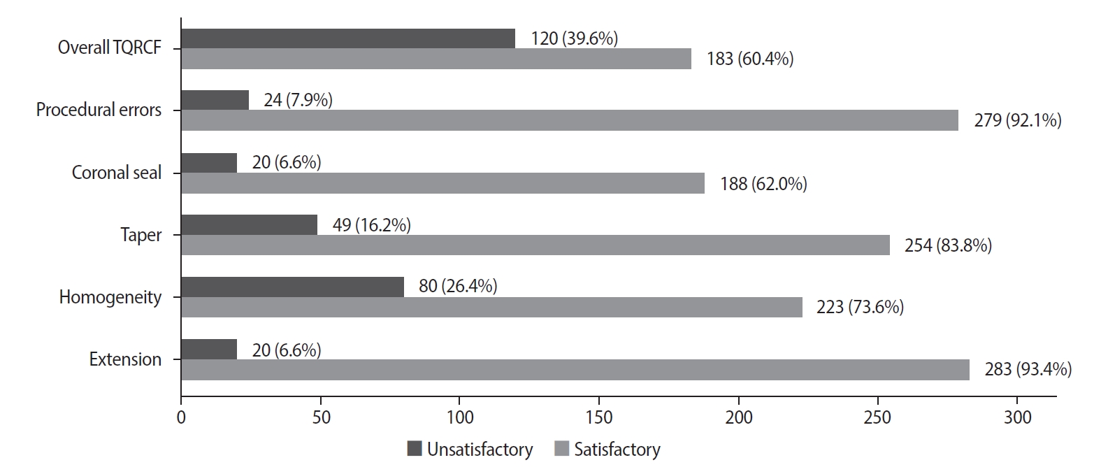
A total of 505 WL and 557 MC radiographs were reviewed, with 72.9% and 75% deemed satisfactory, respectively. Satisfactory TQRCF was achieved in 60.4% of cases. Significant associations were found between the extension of the file in WL and gutta-percha in MC radiographs and TQRCF (p = 0.000). Misinterpretation of these radiographs resulted in poor TQRCF. Furthermore, 64.2% of teeth had satisfactory EO. A significant relationship was noted between the quality of MC radiographs and both TQRCF (p = 0.043) and EO (p = 0.003).
Conclusions
Unsatisfactory MC radiographs were linked to poor TQRCF and unfavorable EO. Regular radiographic training is recommended to enhance EO. -
Citations
Citations to this article as recorded by
- Radiographic evaluation of root canal fillings: can undergraduate dental students perform it?
Emine Odabaşı Tezer, Fadi Nahas, Alhabab Shbitah, İrem Dilara Kılıç, Ahmet Bölük, Meltem Öztan
BMC Medical Education.2026;[Epub] CrossRef - Assessment of radiographic errors and repetition rates in undergraduate endodontic education: a retrospective clinical study
Marwa Ameen, Abdul Rahman Saleh, Dunia Alhadi, Manal Almaslamani
The Saudi Dental Journal.2025;[Epub] CrossRef - Application of Periapical Radiography in Root Canal Treatment: A Literature Review
Jennifer Lois Violita Malau, Keizha Allysia Nabila, Widiani Harrista, Regina Amara Ginting, Tassa Kusuma Arya Putri, Jatu Rachel Keshena
Acta Odontologica Indonesia.2025; 1(2): 49. CrossRef
- Radiographic evaluation of root canal fillings: can undergraduate dental students perform it?
- 12,934 View
- 291 Download
- 2 Web of Science
- 3 Crossref

-
In vivo assessment of accuracy of Propex II, Root ZX II, and radiographic measurements for location of the major foramen - Fernanda Garcia Tampelini, Marcelo Santos Coelho, Marcos de Azevêdo Rios, Carlos Eduardo Fontana, Daniel Guimarães Pedro Rocha, Sergio Luiz Pinheiro, Carlos Eduardo da Silveira Bueno
- Restor Dent Endod 2017;42(3):200-205. Published online May 16, 2017
- DOI: https://doi.org/10.5395/rde.2017.42.3.200
-
Abstract
PDFPubReaderePub
Objectives The aim of this
in vivo study was to assess the accuracy of 2 third-generation electronic apex locators (EALs), Propex II (Dentsply Maillefer) and Root ZX II (J. Morita), and radiographic technique for locating the major foramen (MF).Materials and Methods Thirty-two premolars with single canals that required extraction were included. Following anesthesia, access, and initial canal preparation with size 10 and 15 K-flex files and SX and S1 rotary ProTaper files, the canals were irrigated with 2.5% sodium hypochlorite. The length of the root canal was verified 3 times for each tooth using the 2 apex locators and once using the radiographic technique. Teeth were extracted and the actual WL was determined using size 15 K-files under a × 25 magnification. The Biostat 4.0 program (AnalystSoft Inc.) was used for comparing the direct measurements with those obtained using radiographic technique and the apex locators. Pearson's correlation analysis and analysis of variance (ANOVA) were used for statistical analyses.
Results The measurements obtained using the visual method exhibited the strongest correlation with Root ZX II (
r = 0.94), followed by Propex II (r = 0.90) and Ingle's technique (r = 0.81;p < 0.001). Descriptive statistics using ANOVA (Tukey'spost hoc test) revealed significant differences between the radiographic measurements and both EALs measurements (p < 0.05).Conclusions Both EALs presented similar accuracy that was higher than that of the radiographic measurements obtained with Ingle's technique. Our results suggest that the use of these EALs for MF location is more accurate than the use of radiographic measurements.
-
Citations
Citations to this article as recorded by- How Do Different Image Modules Impact the Accuracy of Working Length Measurements in Digital Periapical Radiography? An In Vitro Study
Vahide Hazal Abat, Rabia Figen Kaptan
Diagnostics.2025; 15(3): 305. CrossRef - Influence of maintaining apical patency in post-endodontic pain
Snigdha Shubham, Manisha Nepal, Ravish Mishra, Kishor Dutta
BMC Oral Health.2021;[Epub] CrossRef
- How Do Different Image Modules Impact the Accuracy of Working Length Measurements in Digital Periapical Radiography? An In Vitro Study
- 2,182 View
- 10 Download
- 2 Crossref
Original Article
- A study on the C-shaped root canal system of mandibular second molar
- Dong-Gyun Lee, Jun-Mo Park, Ho-Keel Hwang
- J Korean Acad Conserv Dent 2007;32(4):335-342. Published online July 31, 2007
- DOI: https://doi.org/10.5395/JKACD.2007.32.4.335
-
Abstract
PDFPubReaderePub
C-shaped canals are known to present a complex canal anatomy with numerous fins connecting individual canals, thus requiring supplementary effort to accomplish a successful root canal treatment. This study examined the frequency of the C-shaped mandibular second molars and interrelation between the clinical records and radiographs to recognize them treated in the Department of Conservative Dentistry of the Chosun University Dental Hospital during a six-year period (1998 - 2004). This study reviewed the clinical records of 227 patients who underwent root canal treatment of the mandibular second molars. After opening the chamber, those cases with C-shaped orifices in the pulpal floor were selected, and the C-shaped root canal types were classified according to Melton's criteria. Three experienced dentists evaluated the radiographs of the C-shaped mandibular second molar on a viewer using a magnifying glass in order to determine if the root apex was fused or separated, the distal root canal was either centered or mesial shifted in the distal root, and if there was bilateral symmetry in a panorama. In conclusion, there is a high frequency of C-shaped mandibular second molars in Koreans. Simultaneous interpretation of the root shape and distal root canal using the preoperative, working length and post-treatment radiographs is important for diagnosing a C-shaped mandibular second molar.
-
Citations
Citations to this article as recorded by- An evaluation of canal curvature at the apical one third in type II mesial canals of mandibular molars
Hye-Rim Yun, Dong-Kyun Lee, Ho-Keel Hwang
Restorative Dentistry & Endodontics.2012; 37(2): 104. CrossRef - A retrospective study on incidence of C-shaped canals in mandibular second molars
Hee-Sun Kim
Journal of Korean Academy of Conservative Dentistry.2009; 34(4): 346. CrossRef
- An evaluation of canal curvature at the apical one third in type II mesial canals of mandibular molars
- 2,549 View
- 13 Download
- 2 Crossref
 First
First Prev
Prev

