
Search
- Page Path
- HOME > Search
Research Article
- Effects of dentin surface preparations on bonding of self-etching adhesives under simulated pulpal pressure
- Chantima Siriporananon, Pisol Senawongse, Vanthana Sattabanasuk, Natchalee Srimaneekarn, Hidehiko Sano, Pipop Saikaew
- Restor Dent Endod 2022;47(1):e4. Published online December 28, 2021
- DOI: https://doi.org/10.5395/rde.2022.47.e4
-
 Abstract
 PDF
PDF PubReader
 ePub
ePub Objectives This study evaluated the effects of different smear layer preparations on the dentin permeability and microtensile bond strength (µTBS) of 2 self-etching adhesives (Clearfil SE Bond [CSE] and Clearfil Tri-S Bond Universal [CTS]) under dynamic pulpal pressure.
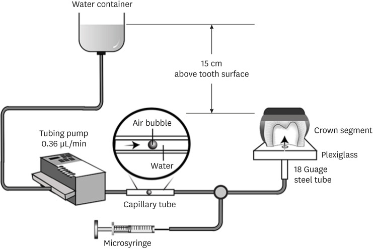
Materials and Methods Human third molars were cut into crown segments. The dentin surfaces were prepared using 4 armamentaria: 600-grit SiC paper, coarse diamond burs, superfine diamond burs, and carbide burs. The pulp chamber of each crown segment was connected to a dynamic intra-pulpal pressure simulation apparatus, and the permeability test was done under a pressure of 15 cmH2O. The relative permeability (%P) was evaluated on the smear layer-covered and bonded dentin surfaces. The teeth were bonded to either of the adhesives under pulpal pressure simulation, and cut into sticks after 24 hours water storage for the µTBS test. The resin-dentin interface and nanoleakage observations were performed using a scanning electron microscope. Statistical comparisons were done using analysis of variance and
post hoc tests.Results Only the method of surface preparation had a significant effect on permeability (
p < 0.05). The smear layers created by the carbide and superfine diamond burs yielded the lowest permeability. CSE demonstrated a higher µTBS, with these values in the superfine diamond and carbide bur groups being the highest. Microscopic evaluation of the resin-dentin interface revealed nanoleakage in the coarse diamond bur and SiC paper groups for both adhesives.Conclusions Superfine diamond and carbide burs can be recommended for dentin preparation with the use of 2-step CSE.
-
Citations
Citations to this article as recorded by
- Effect of smear layer pretreatment with EDTA and sodium hypochlorite on the dentin bond durability of universal adhesives
Thanawat Ruaydee, Chantida Pawaputanon Na Mahasarakham, Vanthana Sattabanasuk, Pipop Saikaew
Frontiers in Dental Medicine.2026;[Epub] CrossRef - Determination of marginal permeability of restorations in the cervical region using a universal adhesive system: a randomized controlled open-label laboratory study
Svetlana N. Razumova, Anzhela S. Brago, Oxana R. Ruda, Artur G. Talandis, Lamara M. Khaskhanova, Ruzanna M. Bragunova, Bohdan O. Pecherskyi
Russian Journal of Dentistry.2026; 30(2): 113. CrossRef - Catechol–Phosphonate–Augmented Universal Adhesive for Hydrolysis-Resistant Dentin Bonds: A µTBS and Spectroscopic Study
Rabeia J. Khalil, Suha K. Ibrahim, Athraa H. Madhat, Ali H. Tawfieq
European Journal of Dentistry.2026;[Epub] CrossRef - The effect of different adhesive strategies and diamond burs on dentin bond strength of universal resin cements
Chavakorn Atsavathavornset, Pipop Saikaew, Choltacha Harnirattisai, Hidehiko Sano
Clinical Oral Investigations.2025;[Epub] CrossRef - Universal adhesive systems in dentistry: A narrative review
Svetlana N. Razumova, Anzhela S. Brago, Oxana R. Ruda, Zoya A. Guryeva, Elvira V. Adzhieva
Russian Journal of Dentistry.2024; 28(5): 512. CrossRef - Delayed light activation of resin composite affects the bond strength of adhesives under dynamic simulated pulpal pressure
Nattaporn Sukprasert, Choltacha Harnirattisai, Pisol Senawongse, Hidehiko Sano, Pipop Saikaew
Clinical Oral Investigations.2022; 26(11): 6743. CrossRef
- Effect of smear layer pretreatment with EDTA and sodium hypochlorite on the dentin bond durability of universal adhesives
- 4,325 View
- 65 Download
- 4 Web of Science
- 6 Crossref

Original Article
- Micro-shear bond strength to dentin under simulated pulpal pressure
- Yun-Jung Song, Sung-Ho Park
- J Korean Acad Conserv Dent 2004;29(4):339-345. Published online July 31, 2004
- DOI: https://doi.org/10.5395/JKACD.2004.29.4.339
-
Abstract
PDFPubReaderePub
The aim of this study was to measure and compare the micro shear bond strengths of the following dentin bonding systems to the dentin surfaces under simulated pulpal pressure; All Bond 2®, Second®, AdheSE®, Adper Prompt L-Pop®. The occlusal surfaces of 180 extracted human molars were prepared so the dentin bonding surfaces could be exposed. The teeth were randomly assigned to 3 equal groups of 60 each and subdivided. The dentin surfaces were treated with the above mentioned bonding system and resin composite cylinders were built up under a simulated pulpal pressure when saline (Group II) or diluted bovine serum (Group III) was used as the pulpal fluid. As a control, the same procedures were performed in the dried dentin surfaces (Group I). After one day of storage in water, the micro shear bond strengths were measured using an EZ tester. Group II and III showed significantly lower shear bond strength than Group I statistically (p < 0.05). SEbond® and AdheSE® showed no difference among the different dentin condition. In the Adper Prompt L-Pop®, a simulated pulpal pressure were applied to the specimens using diluted bovine serum, which showed a higher strength than the specimens in which saline was used (p < 0.05).
- 1,154 View
- 2 Download
 First
First Prev
Prev

