
Search
- Page Path
- HOME > Search
Research Article
- Resolvin E1 incorporated carboxymethyl chitosan scaffold accelerates repair of dental pulp stem cells under inflammatory conditions: a laboratory investigation
- Hemalatha P Balasubramanian, Nandini Suresh, Vishnupriya Koteeswaran, Velmurugan Natanasabapathy
- Restor Dent Endod 2025;50(4):e40. Published online November 28, 2025
- DOI: https://doi.org/10.5395/rde.2025.50.e40
-
 Abstract
 PDF
PDF Supplementary Material
 PubReader
 ePub
ePub - Objectives
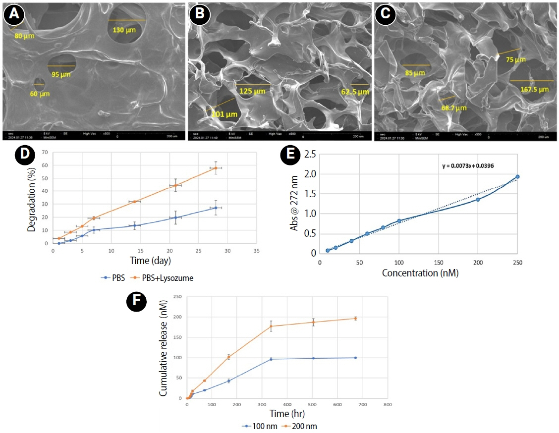
This study fabricated and characterized a resolvin E1 (RvE1)-loaded carboxymethyl chitosan (CMC) scaffold and determined its cytotoxicity and mineralization potential on inflamed human dental pulp stem cells (hDPSCs).
Methods
CMC scaffold incorporated with two concentrations of RvE1 (100 and 200 nM) was fabricated and characterized. The scaffolds’ porosity, drug release kinetics, and degradation were assessed. The impact of RvE1 on inflamed hDPSCs proliferation, proinflammatory gene expression (tumor necrosis factor alpha [TNF-α]), alkaline phosphatase activity, and alizarin red S staining was evaluated.
Results
Scanning electron microscopy analysis demonstrated a highly porous interconnected microstructure. Release kinetics showed gradual RvE1 release peaking at day 14. Cumulative degradation of the CMC scaffold at 28 days was 57.35%. Inflamed hDPSCs exposed to 200 nM RvE1-CMC scaffold exhibited significantly improved viability compared to 100 nM. Both RvE1-CMC scaffolds significantly suppressed the expression of TNF-α at 7 days. Alkaline phosphatase activity was enhanced by both RvE1 concentrations on days 7 and 14. Alizarin red staining revealed superior mineralization potential of 200 nM RvE1 on days 14 and 21.
Conclusions
This study concludes 200 nM RvE1-CMC scaffold is a promising therapy for inflamed pulp conditions, enhancing cell proliferation and biomineralization potential in inflamed hDPSCs.
- 1,477 View
- 63 Download

Basic Research
- Pulp response of beagle dog to direct pulp capping materials: Histological study
- Ji-Hyun Bae, Young-Gyun Kim, Pil-Young Yoon, Byeong-Hoon Cho, Yong-Hoon Choi
- J Korean Acad Conserv Dent 2010;35(1):5-12. Published online January 31, 2010
- DOI: https://doi.org/10.5395/JKACD.2010.35.1.005
-
Abstract
PDFPubReaderePub
The purpose of this study was to evaluate the pulp tissue reaction to direct pulp capping of mechanically exposed beagle dogs'pulp with several capping materials. A total of 36 teeth of 2 healthy beagle dongs were used. The mechanically exposed pulps were capped with one of the followings: (1) Mineral Trioxide Aggregate (MTA: ProRoot® MTA, Dentsply, Tulsa, USA), (2) Clearfil SE Bond (Dentin adhesive system: Kuraray, Osaka, Japan), (3) Ultra-Blend (Photo-polymerized Calcium hydroxide: Ultradent, South Jordan, USA), (4) Dycal (Quick setting Calcium hydroxide: LD Caulk Co., Milford, USA) at 7, 30, and 90 days before sacrificing. The cavities were restored with Z350 flowable composite resin (3M ESPE, St. Paul. MN, USA). After the beagle dogs were sacrificed, the extracted teeth were fixed, decalcified, prepared for histological examination and stained with HE stain. The pulpal tissue responses to direct pulp capping materials were assessed.
In MTA, calcium hydroxide, and photo-polymerized calcium hydroxide groups, initial mild inflammatory cell infiltration, newly formed odontoblast-like cell layer and hard tissue bridge formation were observed. Compared with dentin adhesive system, these materials were biocompatible and good for pulp tissue regeneration.
In dentin adhesive system group, severe inflammatory cell infiltration, pulp tissue degeneration and pulp tissue necrosis were observed. It seemed evident that application of dentin adhesive system in direct pulp capping of beagle dog teeth cannot lead to acceptable repair of the pulp tissue with dentine bridge formation.
-
Citations
Citations to this article as recorded by
- Experimental Study of Pulp Capping Using Xenogenic Demineralized Dentin Paste
Ji-Young Yun, Yong-Hoon Choi, Young-Kyun Kim, In-Woong Um, Joo-Cheol Park, Ji-Yoon Kim
Journal of Hard Tissue Biology.2016; 25(3): 321. CrossRef - Comparison of gene expression profiles of human dental pulp cells treated with mineral trioxide aggregate and calcium hydroxide
Yong-Beom Kim, Won-Jun Shon, Woocheol Lee, Kee-Yeon Kum, Seung-Ho Baek, Kwang-Shik Bae
Journal of Korean Academy of Conservative Dentistry.2011; 36(5): 397. CrossRef - Gene expression profiling in human dental pulp cells treated with mineral trioxide aggregate
Yong-Beom Kim, Won-Jun Shon, WooCheol Lee, Kee-Yeon Kum, Seung-Ho Baek, Kwang-Shik Bae
Journal of Korean Academy of Conservative Dentistry.2010; 35(3): 152. CrossRef - Histology of dental pulp healing after tooth replantation in rats
Eun-Jin Go, Han-Seong Jung, Eui-Seong Kim, Il-Young Jung, Seung-Jong Lee
Journal of Korean Academy of Conservative Dentistry.2010; 35(4): 273. CrossRef
- Experimental Study of Pulp Capping Using Xenogenic Demineralized Dentin Paste
- 1,949 View
- 13 Download
- 4 Crossref
 First
First Prev
Prev

