
Search
- Page Path
- HOME > Search
Research Articles
-
Evaluation of mineral induction ability and cytotoxicity of carbonated hydroxyapatite for pulp tissue regeneration: an
in vitro study - S. Swathi Priyadharshini, Chinnasamy Ragavendran, Anand Sherwood, J. Ramana Ramya, Jogikalmat Krithikadatta
- Restor Dent Endod 2024;49(4):e40. Published online October 29, 2024
- DOI: https://doi.org/10.5395/rde.2024.49.e40
-
 Abstract
 PDF
PDF PubReader
 ePub
ePub Objectives This study aimed to evaluate carbonated hydroxyapatite (CHA)’s ability for mineral induction and its
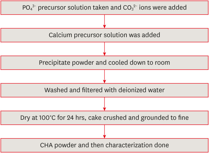
in vitro cytotoxicity with human dental pulp cells.Materials and Methods Precursors for the study include di-ammonium hydrogen phosphate and calcium nitrate tetrahydrate, with sodium hydrogen carbonate added to achieve different levels of carbonate substitution. The synthesized CHA samples are characterized using X-ray diffraction, Fourier transform infrared spectroscopy, and Raman spectroscopy. Scanning electron microscopy (SEM) was used to observe morphology. For 14 days at 37°C, samples were submerged in simulated body fluid to assess their mineral induction capabilities. SEM was used to confirm apatite formation on sample surfaces. The cytotoxicity assay was used to assess the vitality of the cells following their exposure to various concentrations of CHA.
Results The Joint Committee on Powder Diffraction Standards data for HA aligned well with the results from X-ray diffraction analysis of CHA across 3 different concentrations, indicating strong agreement. Fourier transform infrared spectra indicated the presence of phosphate, hydroxyl, and carbonate groups within the samples. SEM and Energy-dispersive X-ray analysis show agglomerated and flaky nanoparticles. All the samples are bioactive, but the formation of apatite differs from one another.
In vitro cytotoxicity assay showed that over 70% of cells maintain viability.Conclusions The results of this study may provide insight into the potential use of carbonated HA as a dental pulp-capping material for vital pulp therapy.
-
Citations
Citations to this article as recorded by
- Smart Nanomaterials: Current State and Future Prospects in Drug Delivery and Tissue Engineering
E. Elizabeth Rani, D. Sakthi Sanjana, E. Karthikeyan, J. Nandhini
Biomedical Materials & Devices.2026; 4(2): 1455. CrossRef - Thermoresponsive Nanomaterials: Revolutionizing Cancer Theranostics
Bellarmin Michael, Mohanakrishnan Srinivasan, Karthikeyan Elumalai, Lokeshwar Ravikumar, Sivaprakash Kathiresan, Nandhini Jayaprakash
Biomedical Materials & Devices.2026; 4(3): 2697. CrossRef - Physicochemical and antibacterial evaluation of novel nano α-TCP–AgNPs biocomposites for direct pulp-capping applications
Selviana Wulansari, Hendra Dian Adhita Dharsono, Nasrul Wathoni, Rosalina Tjandrawinata, Arief Cahyanto, Moehamad Orliando Roeslan
Frontiers in Oral Health.2026;[Epub] CrossRef - Physicochemical effects of nano type-B bone substitute on pulp protective cement formulations
Njwan Fadhel SHEHAB
Dental Materials Journal.2026; 45(1): 92. CrossRef - Recycling waste for sustainability: The green synthesis of silver nanoparticles from Bougainvillea glabra green waste, and the evaluation of their antioxidant, cytotoxic, catalytic, antibacterial and in-silico molecular docking properties
Hafsa Naleem, Mathivathani Kandiah, Beneli Gunaratne, Ominda Perera
Next Research.2026; 11: 101990. CrossRef - Comparative evaluation of compressive strength and morphological interface of carbonated hydroxyapatite with other pulp capping materials: An in vitro analysis
S. Swathi Priyadharshini, Chinnasamy Ragavendran, I. Anand Sherwood, Ramanaramya Jeyapalan
Endodontology.2025; 37(1): 90. CrossRef - Bioactive Dioxo-Phosphobetaines derived from the reaction of Dichlorodinitrobenzofuroxane with various phosphines
Irina V. Galkina, Haiyan Fan, Semen R. Romanov, Dmitriy I. Bakhtiyarov, Luisa M. Usupova, Svetlana N. Egorova, Yulia V. Bakhtiyarova, Enrico Benassi
Bioorganic Chemistry.2025; 163: 108695. CrossRef - Near-infrared laser-activated PLGA-PDA core-shell nanohybrids for synergistic photothermal antibacterial therapy and sustained ion release in orthodontic white spot lesions prevention
Zezhou Feng, Yujiang Liu, Silu Sun, Minmin Si, Di Huang, Zhiyuan Feng
Journal of Dentistry.2025; 162: 106078. CrossRef - Formation and utilization of soluble microbial products in denitrifying biofilters at different carbon-to-nitrogen ratios: Microbial community characteristics
Fangyuan Jiang, Xianyang Shi
Journal of Environmental Chemical Engineering.2025; 13(6): 119554. CrossRef - Bioactivity and biocompatibility of bioceramic-based pulp capping materials in laboratory and animal models
Rafiqul Islam, Md. Refat Readul Islam, Kenta Tsuchiya, Yu Toida, Hidehiko Sano, Monica Yamauti, Hany Mohamed Aly Ahmed, Atsushi Tomokiyo
Journal of Materials Science: Materials in Medicine.2025;[Epub] CrossRef - Physical, Chemical, and Biological Properties of Graphene Nanoparticle-added Tricalcium Silicate Formulations: A Systematic Review
Soundaria Srinivasan, Deepa Gurunathan, Lakshmi Thangavelu
Journal of International Oral Health.2025; 17(6): 453. CrossRef - Advanced structural and compositional profiling of mineral trioxide aggregate incorporated with nano-carbonated hydroxyapatite: a comprehensive X-ray diffraction and energy dispersive X-ray investigation
Njwan Fadhel Shehab, Nadia Hameed Hasan, Alaa Edrees Dawood, Nawal Atiya Khalaf
Biomaterial Investigations in Dentistry.2025; 12: 216. CrossRef
- Smart Nanomaterials: Current State and Future Prospects in Drug Delivery and Tissue Engineering
- 4,751 View
- 156 Download
- 9 Web of Science
- 12 Crossref

- Changes in SIRT gene expression during odontoblastic differentiation of human dental pulp cells
- Young-Eun Jang, Su-Hee Go, Bin-Na Lee, Hoon-Sang Chang, In-Nam Hwang, Won-Mann Oh, Yun-Chan Hwang
- Restor Dent Endod 2015;40(3):223-228. Published online July 15, 2015
- DOI: https://doi.org/10.5395/rde.2015.40.3.223
-
Abstract
PDFPubReaderePub
Objectives The aim of this study was to investigate the expression of 7 different sirtuin genes (SIRT1-SIRT7) in human dental pulp cells (HDPCs), and to determine the role of SIRTs in the odontoblastic differentiation potential of HDPCs.
Materials and Methods HDPCs were isolated from freshly extracted third molar teeth of healthy patients and cultulred in odontoblastic differentiation inducing media. Osteocalcin (OCN) and dentin sialophosphoprotein (DSPP) expression was analyzed to evaluate the odontoblastic differentiation of HDPCs by reverse transcription-polymerase chain reaction (RT-PCR), while alizarin red staining was used for the mineralization assay. To investigate the expression of SIRTs during odontoblastic differentiation of HDPCs, real time PCR was also performed with RT-PCR.
Results During the culture of HDPCs in the differentiation inducing media, OCN, and DSPP mRNA expressions were increased. Mineralized nodule formation was also increased in the 14 days culture. All seven SIRT genes were expressed during the odontogenic induction period. SIRT4 expression was increased in a time-dependent manner.
Conclusions Our study identified the expression of seven different SIRT genes in HDPCs, and revealed that SIRT4 could exert an influence on the odontoblast differentiation process. Further studies are needed to determine the effects of other SIRTs on the odontogenic potential of HDPCs.
-
Citations
Citations to this article as recorded by- Biodegradable Zn‐5Dy Alloy with Enhanced Osteo/Angio‐Genic Activity and Osteointegration Effect via Regulation of SIRT4‐Dependent Mitochondrial Function
Yue Han, Xian Tong, Runqi Zhou, Yilin Wang, Yuge Chen, Liang Chen, Xinhua Hong, Linmei Wu, Zhiqiang Lin, Yichi Zhang, Xuejia Zhang, Chaoming Hu, Bin Li, Yifan Ping, Zelin Cao, Zhou Ye, Zhongchen Song, Yuncang Li, Cuie Wen, Yongsheng Zhou, Jixing Lin, Shen
Advanced Science.2024;[Epub] CrossRef - The Role of Histone Acetylation Modification in Dental Tissue-Derived Mesenchymal Stem Cells and Odontogenesis
Haoling Chen, Zijing Huang, Chuxiao Chen
Cellular Reprogramming.2023; 25(1): 11. CrossRef - Metabolic Remodeling Impacts the Epigenetic Landscape of Dental Mesenchymal Stem Cells
Haiyun Luo, Yachuan Zhou, Wenjing Liu, Jun Wang
Stem Cells International.2022; 2022: 1. CrossRef - SIRT4 regulates rat dental papilla cell differentiation by promoting mitochondrial functions
Haoling Chen, Jun Kang, Fuping Zhang, Tong Yan, Wenguo Fan, Hongwen He, Fang Huang
The International Journal of Biochemistry & Cell Biology.2021; 134: 105962. CrossRef - Sirtuins as Interesting Players in the Course of HIV Infection and Comorbidities
Karolina Jurkowska, Beata Szymańska, Brygida Knysz, Amadeusz Kuźniarski, Agnieszka Piwowar
Cells.2021; 10(10): 2739. CrossRef - Robust expression of SIRT6 inhibits pulpitis via activation of the TRPV1 channel
Jia Hu, Weiran Chen, Zailing Qiu, Hongbing Lv
Cell Biochemistry and Function.2020; 38(5): 676. CrossRef - Downregulation of microRNA‐143‐5p is required for the promotion of odontoblasts differentiation of human dental pulp stem cells through the activation of the mitogen‐activated protein kinases 14‐dependent p38 mitogen‐activated protein kinases signaling pa
Bao‐Liang Wang, Zhi Wang, Xi Nan, Qing‐Cai Zhang, Wei Liu
Journal of Cellular Physiology.2019; 234(4): 4840. CrossRef - A potential role for the silent information regulator 2 homologue 1 (SIRT1) in periapical periodontitis
H. Kudo, O. Takeichi, K. Hatori, K. Makino, K. Himi, B. Ogiso
International Endodontic Journal.2018; 51(7): 747. CrossRef - Overexpressed Sirt1 in MSCs Promotes Dentin Formation in Bmi1-Deficient Mice
H. Wang, C. Lv, Y. Gu, Q. Li, L. Xie, H. Zhang, D. Miao, W. Sun
Journal of Dental Research.2018; 97(12): 1365. CrossRef - Expression of silent information regulator 2 homolog 1 (SIRT1) in periapical granulomas
Hiroshi Kudo, Osamu Takeichi, Kosuke Makino, Keisuke Hatori, Bunnai Ogiso
Journal of Oral Science.2018; 60(3): 411. CrossRef - TET1 knockdown inhibits the odontogenic differentiation potential of human dental pulp cells
Li-Jia Rao, Bai-Cheng Yi, Qi-Meng Li, Qiong Xu
International Journal of Oral Science.2016; 8(2): 110. CrossRef
- Biodegradable Zn‐5Dy Alloy with Enhanced Osteo/Angio‐Genic Activity and Osteointegration Effect via Regulation of SIRT4‐Dependent Mitochondrial Function
- 2,469 View
- 8 Download
- 11 Crossref
Review Article
- Epigenetics: general characteristics and implications for oral health
- Ji-Yun Seo, Yoon-Jung Park, Young-Ah Yi, Ji-Yun Hwang, In-Bog Lee, Byeong-Hoon Cho, Ho-Hyun Son, Deog-Gyu Seo
- Restor Dent Endod 2015;40(1):14-22. Published online November 13, 2014
- DOI: https://doi.org/10.5395/rde.2015.40.1.14
-
Abstract
PDFPubReaderePub
Genetic information such as DNA sequences has been limited to fully explain mechanisms of gene regulation and disease process. Epigenetic mechanisms, which include DNA methylation, histone modification and non-coding RNAs, can regulate gene expression and affect progression of disease. Although studies focused on epigenetics are being actively investigated in the field of medicine and biology, epigenetics in dental research is at the early stages. However, studies on epigenetics in dentistry deserve attention because epigenetic mechanisms play important roles in gene expression during tooth development and may affect oral diseases. In addition, understanding of epigenetic alteration is important for developing new therapeutic methods. This review article aims to outline the general features of epigenetic mechanisms and describe its future implications in the field of dentistry.
-
Citations
Citations to this article as recorded by- Embracing change: Chemical modifications of nucleic acid bases as epigenetic marks
Nishu Nain, Shoaib Khan, Priyanka Phogat, Aparna Bansal, Shrikant Kukreti
Next Research.2026; 5: 101292. CrossRef - Holobiontic Intercellular Relationships Between the Oral Cavity and the Rest of the Human Organism: A Narrative Review
Vasile Burlui, Daniela Luminița Ichim, Daniela Ivona Tomița, Malina Visternicu, Alin Ciobica, Mihaela Diana Gheban
Medicina.2026; 62(7): 1365. CrossRef - Conversation between skin microbiota and the host: from early life to adulthood
Jimin Cha, Tae-Gyun Kim, Ji-Hwan Ryu
Experimental & Molecular Medicine.2025; 57(4): 703. CrossRef - Identification of two novel variants in homeodomain of
MSX1 associated with oligodontia
Ting Zeng, Xiuyou Wang, Li Xu, Xin Dong, Xili Qiu, Zhiyuan Deng, Saimin Pei, Rong Lei, Yuehong Wang, Ling Peng
Oral Science and Homeostatic Medicine.2025; 1(2): 9610029. CrossRef - DNA Methylation of COX‐2, IFN‐γ, TNF‐α, and LINE‐1 in Clinically Stable Periodontal Tissues Following Periodontal Therapy
Giulio Rasperini, Koki Yoshida, Alessandro Martinotti, Valentina Bollati, Letizia Tarantini, Farah Asa'ad
Clinical and Experimental Dental Research.2025;[Epub] CrossRef - Effect of Long Non-coding RNA and DNA Methylation on Gene Expression in Dental Fluorosis
Xiaoyan Hu, Huiru Li, Minzhi Yang, Yujiong Chen, Ailin Zeng, Jiayuan Wu, Jian Zhang, Yuan Tian, Jing Tang, Shengyan Qian, Mingsong Wu
Biological Trace Element Research.2024; 202(1): 221. CrossRef - MicroRNAs: Mighty Mite RNAs in Oral Diseases
Devapriya Appukuttan, P. S. G. Prakash
Journal of Interdisciplinary Dentistry.2024; 14(3): 145. CrossRef - Role of epigenetics in OSCC: an understanding above genetics
Priyanka P. Vatsa, Yogita Jindal, Janhavi Bhadwalkar, Ambika Chamoli, Vinal Upadhyay, Amit Mandoli
Medical Oncology.2023;[Epub] CrossRef - Downregulation of miRNA‐26 in chronic periodontitis interferes with innate immune responses and cell migration by targeting phospholipase C beta 1
Juhi R. Uttamani, Afsar R. Naqvi, Araceli Maria Valverde Estepa, Varun Kulkarni, Maria F. Brambila, Gloria Martínez, Gabriela Chapa, Christine D. Wu, Wei Li, Sona Rivas‐Tumanyan, Salvador Nares
Journal of Clinical Periodontology.2023; 50(1): 102. CrossRef - The Potential Role of Epigenetic Modifications on Different Facets in the Periodontal Pathogenesis
Samuel Laberge, Daniel Akoum, Piotr Wlodarczyk, Jean-Daniel Massé, Dominique Fournier, Abdelhabib Semlali
Genes.2023; 14(6): 1202. CrossRef - The Role of Histone Acetylation Modification in Dental Tissue-Derived Mesenchymal Stem Cells and Odontogenesis
Haoling Chen, Zijing Huang, Chuxiao Chen
Cellular Reprogramming.2023; 25(1): 11. CrossRef - Your health is in your mouth: A comprehensive view to promote general wellness
Antonia Barranca-Enríquez, Tania Romo-González
Frontiers in Oral Health.2022;[Epub] CrossRef - A Brief Landscape of Epigenetic Mechanisms in Dental Pathologies
Wojciech Tynior, Joanna Katarzyna Strzelczyk
Cytology and Genetics.2022; 56(5): 475. CrossRef - Influence of epigenetics on periodontitis and peri‐implantitis pathogenesis
Lena Larsson, Nolan M. Kavanagh, Trang V. N. Nguyen, Rogerio M. Castilho, Tord Berglundh, William V. Giannobile
Periodontology 2000.2022; 90(1): 125. CrossRef - DNA methylation alterations and their potential influence on macrophage in periodontitis
Yiyang Jiang, Jingfei Fu, Juan Du, Zhenhua Luo, Lijia Guo, Junji Xu, Yi Liu
Oral Diseases.2022; 28(2): 249. CrossRef - Stabilizing and Anti-Repressor Elements Effectively Increases Transgene Expression in Transfected CHO Cells
Qin Li, Rui-Fang Yan, Yong-Xiao Yang, Chun-liu Mi, Yan-long Jia, Tian-Yun Wang
Frontiers in Bioengineering and Biotechnology.2022;[Epub] CrossRef - Synthesis and Anticancer Potential of New Hydroxamic Acid Derivatives as Chemotherapeutic Agents
Işıl Nihan Korkmaz, Hasan Özdemir
Applied Biochemistry and Biotechnology.2022; 194(12): 6349. CrossRef - Impact of Epigenetic Alterations in the Development of Oral Diseases
Rodopi Emfietzoglou, Evangelos Pachymanolis, Christina Piperi
Current Medicinal Chemistry.2021; 28(6): 1091. CrossRef - Basics of Epigenetics and Role of Epigenetics in Diabetic Complications
Andamuthu Yamunadevi, Ramani Pratibha, Muthusamy Rajmohan, Sengottaiyan Mahendraperumal, Nalliappan Ganapathy
Journal of Pharmacy and Bioallied Sciences.2021; 13(Suppl 1): S336. CrossRef - Effects of Epigenetic Regulation on Cancer
Muhammet Mesut Nezir ENGİN, Esra ÖZEN ENGİN, Recep ERÖZ, Gorkem DULGER, Hüseyin YÜCE
Journal of Biotechnology and Strategic Health Research.2021; 5(1): 1. CrossRef - Photobiomodulation therapy improves human dental pulp stem cell viability and migration in vitro associated to upregulation of histone acetylation
Ivana M. Zaccara, Letícia B. Mestieri, Emily F. S. Pilar, Maria S. Moreira, Fabiana S. Grecca, Manoela D. Martins, Patrícia Maria Poli Kopper
Lasers in Medical Science.2020; 35(3): 741. CrossRef - The Biology of Social Adversity Applied to Oral Health
N. Gomaa, H. Tenenbaum, M. Glogauer, C. Quiñonez
Journal of Dental Research.2019; 98(13): 1442. CrossRef - The effect of DNA methylation on the miRNA expression pattern in lipopolysaccharide-induced inflammatory responses in human dental pulp cells
Zehuan Mo, Qimeng Li, Luhui Cai, Minkang Zhan, Qiong Xu
Molecular Immunology.2019; 111: 11. CrossRef - One-Carbon Metabolism Links Nutrition Intake to Embryonic Development via Epigenetic Mechanisms
Si Wu, Jun Zhang, Feifei Li, Wei Du, Xin Zhou, Mian Wan, Yi Fan, Xin Xu, Xuedong Zhou, Liwei Zheng, Yachuan Zhou
Stem Cells International.2019; 2019: 1. CrossRef - Epigenetic regulation in dental pulp inflammation
T Hui, C Wang, D Chen, L Zheng, D Huang, L Ye
Oral Diseases.2017; 23(1): 22. CrossRef - Current Concepts of Epigenetics and Its Role in Periodontitis
Lena Larsson
Current Oral Health Reports.2017; 4(4): 286. CrossRef - The periodontal war: microbes and immunity
Jeffrey L. Ebersole, Dolph Dawson, Pinar Emecen‐Huja, Radhakrishnan Nagarajan, Katherine Howard, Martha E. Grady, Katherine Thompson, Rebecca Peyyala, Ahmad Al‐Attar, Kathryn Lethbridge, Sreenatha Kirakodu, Octavio A. Gonzalez
Periodontology 2000.2017; 75(1): 52. CrossRef - Epigenetic regulatory elements: Recent advances in understanding their mode of action and use for recombinant protein production in mammalian cells
Niamh Harraghy, David Calabrese, Igor Fisch, Pierre‐Alain Girod, Valérie LeFourn, Alexandre Regamey, Nicolas Mermod
Biotechnology Journal.2015; 10(7): 967. CrossRef - Protocol for assessing maternal, environmental and epigenetic risk factors for dental caries in children
Surani Fernando, David J. Speicher, Mahmoud M. Bakr, Miles C. Benton, Rodney A. Lea, Paul A. Scuffham, Gabor Mihala, Newell W. Johnson
BMC Oral Health.2015;[Epub] CrossRef
- Embracing change: Chemical modifications of nucleic acid bases as epigenetic marks
- 3,555 View
- 31 Download
- 29 Crossref
Research Article
- Analysis of gene expression during odontogenic differentiation of cultured human dental pulp cells
- Min-Seock Seo, Kyung-Gyun Hwang, Hyongbum Kim, Seung-Ho Baek
- Restor Dent Endod 2012;37(3):142-148. Published online August 29, 2012
- DOI: https://doi.org/10.5395/rde.2012.37.3.142
-
Abstract
PDFPubReaderePub
Objectives We analyzed gene-expression profiles after 14 day odontogenic induction of human dental pulp cells (DPCs) using a DNA microarray and sought candidate genes possibly associated with mineralization.
Materials and Methods Induced human dental pulp cells were obtained by culturing DPCs in odontogenic induction medium (OM) for 14 day. Cells exposed to normal culture medium were used as controls. Total RNA was extracted from cells and analyzed by microarray analysis and the key results were confirmed selectively by reverse-transcriptase polymerase chain reaction (RT-PCR). We also performed a gene set enrichment analysis (GSEA) of the microarray data.
Results Six hundred and five genes among the 47,320 probes on the BeadChip differed by a factor of more than two-fold in the induced cells. Of these, 217 genes were upregulated, and 388 were down-regulated. GSEA revealed that in the induced cells, genes implicated in Apoptosis and Signaling by wingless MMTV integration (Wnt) were significantly upregulated.
Conclusions Genes implicated in Apoptosis and Signaling by Wnt are highly connected to the differentiation of dental pulp cells into odontoblast.
-
Citations
Citations to this article as recorded by- iPSC-derived cranial neural crest-like cells can replicate dental pulp tissue with the aid of angiogenic hydrogel
Yoshifumi Kobayashi, Julie Nouet, Erdenechimeg Baljinnyam, Zain Siddiqui, Daniel H. Fine, Diego Fraidenraich, Vivek A. Kumar, Emi Shimizu
Bioactive Materials.2022; 14: 290. CrossRef - The role of sclerostin and dickkopf-1 in oral tissues – A review from the perspective of the dental disciplines
Mohammad Samiei, Klara Janjić, Barbara Cvikl, Andreas Moritz, Hermann Agis
F1000Research.2019; 8: 128. CrossRef - The Influence of Pro-Inflammatory Factors on Sclerostin and Dickkopf-1 Production in Human Dental Pulp Cells Under Hypoxic Conditions
Klara Janjić, Mohammad Samiei, Andreas Moritz, Hermann Agis
Frontiers in Bioengineering and Biotechnology.2019;[Epub] CrossRef - Do hypoxia and L-mimosine modulate sclerostin and dickkopf-1 production in human dental pulp-derived cells? Insights from monolayer, spheroid and tooth slice cultures
Klara Janjić, Barbara Cvikl, Christoph Kurzmann, Andreas Moritz, Hermann Agis
BMC Oral Health.2018;[Epub] CrossRef - The effects of dexamethasone on the apoptosis and osteogenic differentiation of human periodontal ligament cells
Sung-Mi Kim, Yong-Gun Kim, Jin-Woo Park, Jae-Mok Lee, Jo-Young Suh
Journal of Periodontal & Implant Science.2013; 43(4): 168. CrossRef
- iPSC-derived cranial neural crest-like cells can replicate dental pulp tissue with the aid of angiogenic hydrogel
- 2,406 View
- 3 Download
- 5 Crossref
Basic Researchs
- Comparison of gene expression profiles of human dental pulp cells treated with mineral trioxide aggregate and calcium hydroxide
- Yong-Beom Kim, Won-Jun Shon, Woocheol Lee, Kee-Yeon Kum, Seung-Ho Baek, Kwang-Shik Bae
- J Korean Acad Conserv Dent 2011;36(5):397-408. Published online September 14, 2011
- DOI: https://doi.org/10.5395/JKACD.2011.36.5.397
-
Abstract
PDFPubReaderePub
Abstract Objectives: This study investigated changes in gene expressions concerning of differentiation, proliferation, mineralization and inflammation using Human-8 expression bead arrays when white Mineral Trioxide Aggregate and calcium hydroxide-containing cement were applied
in vitro to human dental pulp cells (HDPCs).Materials and Methods: wMTA (white ProRoot MTA, Dentsply) and Dycal (Dentsply Caulk) in a Teflon tube (inner diameter 10 mm, height 1 mm) were applied to HDPCs. Empty tube-applied HDPCs were used as negative control. Total RNA was extracted at 3, 6, 9 and 24 hr after wMTA and Dycal application. The results of microarray were confirmed by reverse transcriptase polymerase chain reaction.
Results: Out of the 24,546 genes, 43 genes (e.g., BMP2, FOSB, THBS1, EDN1, IL11, COL10A1, TUFT1, HMOX1) were up-regulated greater than two-fold and 25 genes (e.g., SMAD6, TIMP2, DCN, SOCS2, CEBPD, KIAA1199) were down-regulated below 50% by wMTA. Two hundred thirty nine genes (e.g., BMP2, BMP6, SMAD6, IL11, FOS, VEGFA, PlGF, HMOX1, SOCS2, CEBPD, KIAA1199) were up-regulated greater than two-fold and 358 genes (e.g., EDN1, FGF) were down-regulated below 50% by Dycal.
Conclusions: Both wMTA and Dycal induced changes in gene expressions related with differentiation and proliferation of pulp cells. wMTA induced changes in gene expressions related with mineralization, and Dycal induced those related with angiogenesis. The genes related with inflammation were more expressed by Dycal than by wMTA. It was confirmed that both wMTA and Dycal were able to induce gene expression changes concerned with the pulp repair in different ways.
-
Citations
Citations to this article as recorded by- Analysis of gene expression during odontogenic differentiation of cultured human dental pulp cells
Min-Seock Seo, Kyung-Gyun Hwang, Hyongbum Kim, Seung-Ho Baek
Restorative Dentistry & Endodontics.2012; 37(3): 142. CrossRef
- Analysis of gene expression during odontogenic differentiation of cultured human dental pulp cells
- 2,014 View
- 1 Download
- 1 Crossref
- Biocompatibility of bioaggregate cement on human pulp and periodontal ligament (PDL) derived cells
- Choo-Ryung Chung, Euiseong Kim, Su-Jung Shin
- J Korean Acad Conserv Dent 2010;35(6):473-478. Published online November 30, 2010
- DOI: https://doi.org/10.5395/JKACD.2010.35.6.473
-
Abstract
PDFPubReaderePub
Objectives This study was performed to investigate the biocompatibility of newly introduced Bioaggregate on human pulp and PDL cells.
Materials and Methods Cells were collected from human pulp and PDL tissue of extracted premolars. Cell culture plate was coated either with Bioaggregate or white MTA, then the same number of cells were poured to cell culture dishes. Cell attachment and growth was examined under a phase microscope after 1,3 and 7 days of seeding. Cell viability was measured and the data was analyzed using Student
t -test and one way ANOVA.Results Both types of cells used in this study were well attached and grew healthy on Bioaggregate and MTA coated culture dishes. No cell inhibition zone was observed in Bioaggregate group. There was no statistical difference of viable cells between bioaggreagte and MTA groups.
Conclusions Bioaggregate appeared to be biocompatible compared with white MTA on human pulp and PDL cells.
-
Citations
Citations to this article as recorded by- Evaluation of bioactivity, biocompatibility, and antibacterial properties of tricalcium silicate bone cement modified with wollastonite/ fluorapatite glass and glass-ceramic
H.K. Abd El-Hamid, A.M. Fayad, R.L. Elwan
Ceramics International.2024; 50(14): 25322. CrossRef - Influence of insulin on the healing of exposed dental pulp after pulp capping: An experimental study in a dog model
Mokhtar A. Al‐Anesi, Ashraf M. Abu‐Seida, Salma H. El Ashry, Abeer H. Mahran, Ehab S. Abd‐Elhamid
Special Care in Dentistry.2021; 41(1): 49. CrossRef - ROOT END FILLING MATERIALS – A REVIEW
Bynagari Chandra Shekar, Veerendra Uppin, Madhu Pujar
GLOBAL JOURNAL FOR RESEARCH ANALYSIS.2021; : 5. CrossRef - Effects of two fast-setting calcium-silicate cements on cell viability and angiogenic factor release in human pulp-derived cells
Chooryung J. Chung, Euiseong Kim, Minju Song, Jeong-Won Park, Su-Jung Shin
Odontology.2016; 104(2): 143. CrossRef - Cytotoxicity and physical properties of tricalcium silicate-based endodontic materials
Young-Eun Jang, Bin-Na Lee, Jeong-Tae Koh, Yeong-Joon Park, Nam-Eok Joo, Hoon-Sang Chang, In-Nam Hwang, Won-Mann Oh, Yun-Chan Hwang
Restorative Dentistry & Endodontics.2014; 39(2): 89. CrossRef - Biocompatibility of root-end filling materials: recent update
Payal Saxena, Saurabh Kumar Gupta, Vilas Newaskar
Restorative Dentistry & Endodontics.2013; 38(3): 119. CrossRef
- Evaluation of bioactivity, biocompatibility, and antibacterial properties of tricalcium silicate bone cement modified with wollastonite/ fluorapatite glass and glass-ceramic
- 2,190 View
- 6 Download
- 6 Crossref
- Gene expression profiling in human dental pulp cells treated with mineral trioxide aggregate
- Yong-Beom Kim, Won-Jun Shon, WooCheol Lee, Kee-Yeon Kum, Seung-Ho Baek, Kwang-Shik Bae
- J Korean Acad Conserv Dent 2010;35(3):152-163. Published online May 31, 2010
- DOI: https://doi.org/10.5395/JKACD.2010.35.3.152
-
Abstract
PDFPubReaderePub
This study investigated the changes in gene expression when mineral trioxide aggregate (MTA) was applied
in vitro to human dental pulp cells (HDPCs). MTA in a teflon tube (diameter 10 mm, height 2 mm) was applied to HDPCs. Empty tube-applied HDPCs were used as negative control. For microarray analysis, total RNA was extracted at 6, 24, and 72 hrs after MTA application. The results were confirmed selectively by performing reverse transcriptase polymerase chain reaction for genes that showed changes of more than two-fold or less than half. Of the 24,546 genes, 109 genes were up-regulated greater than two-fold (e.g., FOSB, THBS1, BHLHB2, EDN1, IL11, FN1, COL10A1, and TUFT1) and 69 genes were down-regulated below 50% (e.g., SMAD6 and DCN). These results suggest that MTA, rather than being a bio-inert material, may have potential to affect the proliferation and differentiation of pulp cells in various ways.-
Citations
Citations to this article as recorded by- Analysis of gene expression during odontogenic differentiation of cultured human dental pulp cells
Min-Seock Seo, Kyung-Gyun Hwang, Hyongbum Kim, Seung-Ho Baek
Restorative Dentistry & Endodontics.2012; 37(3): 142. CrossRef - Comparison of gene expression profiles of human dental pulp cells treated with mineral trioxide aggregate and calcium hydroxide
Yong-Beom Kim, Won-Jun Shon, Woocheol Lee, Kee-Yeon Kum, Seung-Ho Baek, Kwang-Shik Bae
Journal of Korean Academy of Conservative Dentistry.2011; 36(5): 397. CrossRef
- Analysis of gene expression during odontogenic differentiation of cultured human dental pulp cells
- 1,847 View
- 5 Download
- 2 Crossref
Original Articles
- The comparison of gene expression from human dental pulp cells and periodontal ligament cells
- Hyoun So, Sang-Hyuk Park, Gi-Woon Choi
- J Korean Acad Conserv Dent 2009;34(5):430-441. Published online September 30, 2009
- DOI: https://doi.org/10.5395/JKACD.2009.34.5.430
-
Abstract
PDFPubReaderePub
The purpose of this study was to characterize functional distinction between human dental pulp cells(PC) and periodontal ligament cells(PDLC) using cDNA microarray assay and to confirm the results of the microarray assay using RT-PCR. 3 genes out of 51 genes which were found to be more expressed(>2 fold) in PC were selected, and 3 genes out of 19 genes which were found to be more expressed(>2 fold) in PDLC were selected for RT-PCR as well.
According to this study, the results were as follows:
1. From the microarray assay, 51 genes were more expressed (2 fold) from PC than PDLC.
2. RT-PCR confirmed that ITGA4 and TGF β2 were more expressed in PC than in PDLC.
3. From the microarray assay, 19 genes were more expressed (2 fold) from PDLC than PC.
4. RT-PCR confirmed that LUM, WISP1, and MMP1 were more expressed in PDLC than in PC.
From the present study, different expression of the genes between the PC and PDLC were characterized to show the genes which play an important role in dentinogenesis were more expressed from PC than PDLC, while the genes which were related with collagen synthesis were more expressed from PDLC than PC.
-
Citations
Citations to this article as recorded by- Gene expression profiling in human dental pulp cells treated with mineral trioxide aggregate
Yong-Beom Kim, Won-Jun Shon, WooCheol Lee, Kee-Yeon Kum, Seung-Ho Baek, Kwang-Shik Bae
Journal of Korean Academy of Conservative Dentistry.2010; 35(3): 152. CrossRef
- Gene expression profiling in human dental pulp cells treated with mineral trioxide aggregate
- 1,758 View
- 1 Download
- 1 Crossref
- A bioactivity study of Portland cement mixed with β-glycerophosphosphate on human pulp cell
- Young-Hwan Oh, Young-Joo Jang, Yong-Bum Cho
- J Korean Acad Conserv Dent 2009;34(5):415-423. Published online September 30, 2009
- DOI: https://doi.org/10.5395/JKACD.2009.34.5.415
-
Abstract
PDFPubReaderePub
The purpose of this study is to investigate the response of human pulp cell on Portland cement mixed with β-glycerophosphate. To investigate the effect of β-glycerophosphate and/or dexamethasone on human pulp cell, ALP activity on various concentration of β-glycerophosphate and dexamethasone was measured and mineral nodule of human pulp cell was stained with Alizarin red S. MTS assay and ALP activity of human pulp cell on Portland cement mixed with various concentration of β-glycerophosphate (10 mM, 100mM, 1M) was measured and the specimens were examined under SEM.
Addition of β-glycerophosphate or dexamethasone alone had no effect however, the addition of 5 mM β-glycerophosphate and 100 nM dexamethasone had the largest increasement in ALP activity. There was no toxicity in all samples and the data showed that Portland cement mixed with 10 mM β-glycerophosphate had more increase in ALP activity compared with control.
In conclusion, Portland cement mixed with β-glycerophosphate has no toxicity and promotes differentiation and mineralization of pulp cell compared with additive-free Portland cement. This implicated that application of Portland cement mixed with β-glycerophosphate might form more reparative dentin and in turn it would bring direct pulp capping to success.
-
Citations
Citations to this article as recorded by- Dentinogenic potential of human adult dental pulp cells during the extended primary culture
Jin-Hee Min, Seon-Yle Ko, Yong-Bum Cho, Chun-Jeih Ryu, Young-Joo Jang
Human Cell.2011; 24(1): 43. CrossRef
- Dentinogenic potential of human adult dental pulp cells during the extended primary culture
- 1,610 View
- 1 Download
- 1 Crossref
- Anti-inflammatory effects of PPARγ on human dental pulp cells
- Jeong-Hee Kim
- J Korean Acad Conserv Dent 2006;31(3):203-214. Published online May 31, 2006
- DOI: https://doi.org/10.5395/JKACD.2006.31.3.203
-
Abstract
PDFPubReaderePub
Dental pulp is a loose, mesenchymal tissue almost entirely enclosed in the dentin. It consists of cells, ground substance, and neural and vascular supplies. Damage to the dental pulp by mechanical, chemical, thermal, and microbial irritants can provoke various types of inflammatory response. Pulpal inflammation leads to the tissue degradation, which is mediated in part by Matrix metalloproteinase leads to accelerate extracellular matrix degradation with pathological pathway. We have now investigated the induction of MMPs and inflammatory cytokines by Lipopolysaccharide (LPS) control of inflammatory mediators by peroxisome proliferator-activated receptors (PPARs).
Human dental pulp cells exposed to various concentrations of LPS (1-10 µg/ml) revealed elevated levels of MMP-2 and MMP-9 at 24 hrs of culture. LPS also stimulated the production of ICAM-1, VCAM-1, IL-1β, and TNF-α. Adenovirus PPARγ (Ad/PPARγ) and PPARγ agonist rosiglitazone reduced the synthesis of MMPs, adhesion molecules and pro-inflammatory cytokines. The inhibitory effect of Ad/PPARγ was higher than that of PPARγ agonist.
These result offer new insights in regard to the anti-inflammatory potential of PPARγ in human dental pulp cell.
- 1,777 View
- 3 Download
- Tissue engineering of dental pulp on type I collagen
- Gwang-Hee Lee, Sung-Yoon Huh, Sang-Hyuk Park
- J Korean Acad Conserv Dent 2004;29(4):370-377. Published online July 31, 2004
- DOI: https://doi.org/10.5395/JKACD.2004.29.4.370
-
Abstract
PDFPubReaderePub
The purpose of this study was to regenerate human dental pulp tissues similar to native pulp tissues. Using the mixture of type I collagen solution, primary cells collected from the different tissues (pulp, gingiva, and skin) and NIH 3T3 (1 × 10⁵ cells/ml/well) were cultured at 12-well plate at 37℃ for 14 days. Standardized photographs were taken with digital camera during 14 days and the diameter of the contracted collagen gel matrix was measured and statistically analyzed with student t-test. As one of the pulp tissue engineering, normal human dental pulp tissue and collagen gel matrix cultured with dental pulp cells for 14 days were fixed and stained with Hematoxyline & Eosin.
According to this study, the results were as follows:
1. The contraction of collagen gel matrix cultured with pulp cells for 14 days was significantly higher than other fibroblasts (gingiva, skin) (p < 0.05).
2. The diameter of collagen gel matrix cultured with pulp cells was reduced to 70.4% after 7 days, and 57.1% after 14 days.
3. The collagen gel without any cells did not contract, whereas the collagen gel cultured with gingiva and skin showed mild contraction after 14 days (88.1% and 87.6% respectively).
4. The contraction of the collagen gel cultured with NIH 3T3 cells after 14 days was higher than those cultured with gingival and skin fibroblasts, but it was not statistically significant (72.1%, p > 0.05).
5. The collagen gel matrix cultured with pulp cells for 14 days showed similar shape with native pulp tissue without blood vessels.
This approach may provide a means of engineering a variety of other oral tissue as well and these cell behaviors may provide information needed to establish pulp tissue engineering protocols.
-
Citations
Citations to this article as recorded by- Human amniotic membrane extracellular matrix scaffold for dental pulp regeneration in vitro and in vivo
Hengameh Bakhtiar, Azin Ashoori, Sarah Rajabi, Mohamad Pezeshki‐Modaress, Alireza Ayati, Mohammad Reza Mousavi, Mohammad Reza Ellini, Amir Kamali, Amir Azarpazhooh, Anil Kishen
International Endodontic Journal.2022; 55(4): 374. CrossRef
- Human amniotic membrane extracellular matrix scaffold for dental pulp regeneration in vitro and in vivo
- 2,475 View
- 4 Download
- 1 Crossref
 First
First Prev
Prev

