
Search
- Page Path
- HOME > Search
Research Articles
- Does the use of different root canal sealers and adhesive resin cements impact the bond strength of glass fiber posts?
- Ália Regina Neves de Paula Porto, Rudá França Moreira, Felipe Gonçalves Belladonna, Victor Talarico Leal Vieira, Emmanuel João Nogueira Leal da Silva
- Restor Dent Endod 2025;50(3):e29. Published online August 29, 2025
- DOI: https://doi.org/10.5395/rde.2025.50.e29
-
 Abstract
 PDF
PDF PubReader
 ePub
ePub - Objectives
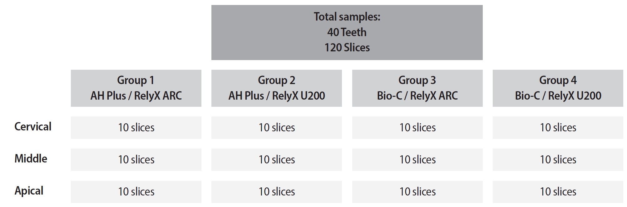
This study aimed to assess the influence of two endodontic sealers on the bond strength of glass fiber posts using conventional and self-adhesive resin cement through a push-out test. Methods: Forty central human incisors were randomly divided into four groups (n = 10) based on sealer (epoxy resin- based or calcium silicate-based) and cement (conventional and self-adhesive resin) types: AH Plus (Dentsply De- Trey)/RelyX ARC (3M ESPE), AH Plus/RelyX U200 (3M ESPE), Bio-C Sealer (Angelus)/RelyX ARC, and Bio-C Sealer/RelyX U200. After canal filling and post cementation, roots were sectioned to obtain one specimen per root third. A pushout test and failure pattern assessment were conducted, with bond strength analyzed using the one-way analysis of variance and Tukey test. Results: AH Plus/RelyX ARC showed the highest bond strength values, with a significant difference in the middle third. The most common failure was mixed (55%), while adhesive failures made up 45%, with 23.5% at the cement/post interface and 21.5% at the cement/dentin interface. Conclusions: AH Plus/RelyX ARC provided the highest bond strength values for glass fiber posts to dentin. -
Citations
Citations to this article as recorded by
- Effect of Endodontic Sealers on the Bond Strength of Glass Fibre Posts: A Systematic Review
Thiago Bessa Marconato Antunes, Juliana D. Bronzato, Vanessa Gallego Arias Pecorari, Jennifer Santos Pereira, Talita Tartari, Adriana de Jesus Soares, Brenda P. F. A. Gomes, Marina Angélica Marciano
Australian Endodontic Journal.2026;[Epub] CrossRef
- Effect of Endodontic Sealers on the Bond Strength of Glass Fibre Posts: A Systematic Review
- 2,928 View
- 176 Download
- 1 Web of Science
- 1 Crossref

- Impact of post adhesion on stress distribution: an in silico study
- Kkot-Byeol Bae, Jae-Yoon Choi, Young-Tae Cho, Bin-Na Lee, Hoon-Sang Chang, Yun-Chan Hwang, Won-Mann Oh, In-Nam Hwang
- Restor Dent Endod 2025;50(2):e19. Published online May 21, 2025
- DOI: https://doi.org/10.5395/rde.2025.50.e19
-
Abstract
PDFPubReaderePub
- Objectives
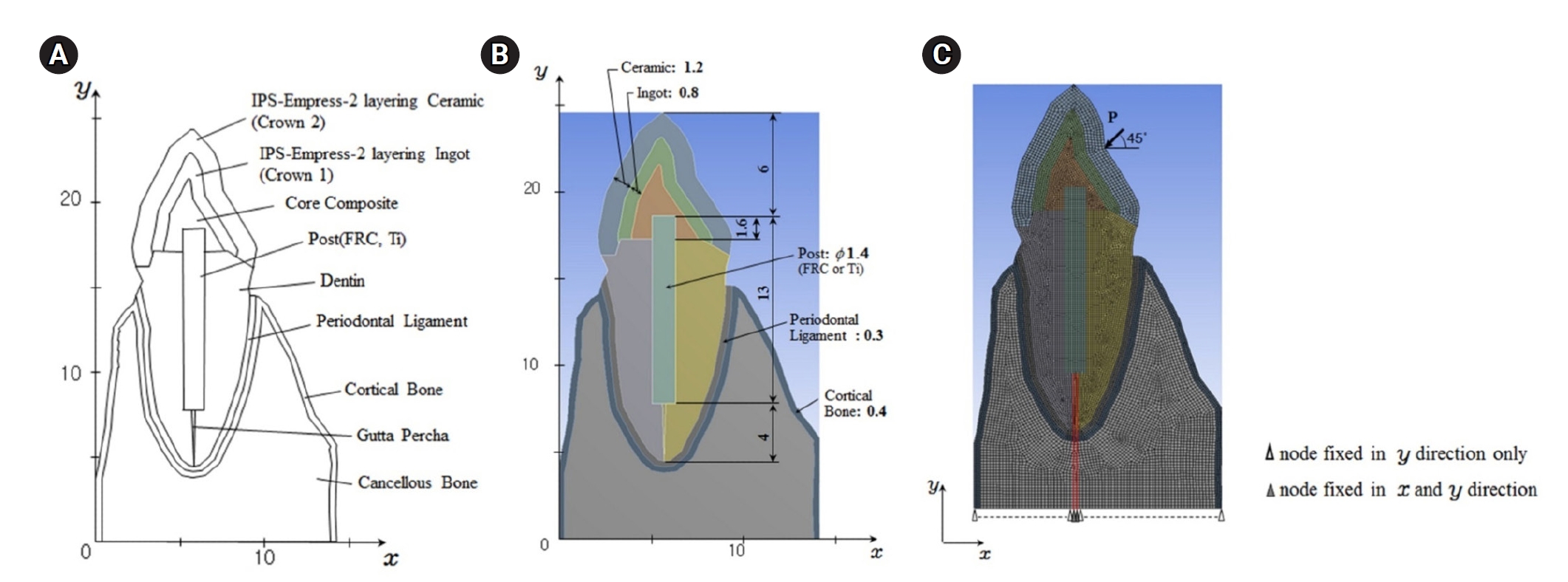
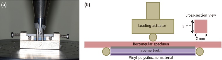
This study aimed to evaluate the stress distribution in teeth restored with different post materials and bonding conditions using finite element analysis (FEA).
Methods
A two-dimensional FEA model of a maxillary central incisor restored with IPS-Empress-2 crown (Ivoclar Vivadent), composite resin core, and posts were created. The model simulated bonded and non-bonded conditions for both fiber-reinforced composite (FRC) and titanium (Ti) posts. Stress distribution was analyzed using ANSYS 14.0 software under a 100-N load applied at a 45° angle to the long axis of the tooth.
Results
The results revealed that stress concentration was significantly higher in non-bonded posts compared to bonded ones. FRC posts exhibited stress values closer to those of dentin, whereas Ti posts demonstrated higher stress concentration, particularly in non-bonded states, increasing the potential risk of damage to surrounding tissues.
Conclusions
FRC posts, with elastic properties similar to dentin and proper adhesion, minimize stress concentration and potential damage to surrounding tissues. Conversely, materials with higher elastic modulus like Ti, can cause unfavorable stress concentrations if not properly bonded, emphasizing the importance of post adhesion in tooth restoration. -
Citations
Citations to this article as recorded by- Advances in Ecofriendly and High-Strength Dental Composites: Structural and Functional Perspectives
Sayem A. Mulla, Amit Patil, Himmat Jaiswal, Bhavani Sangala Nagendra, Ashima Jakhar, Waseem Z. Khan
European Journal of General Dentistry.2026;[Epub] CrossRef
- Advances in Ecofriendly and High-Strength Dental Composites: Structural and Functional Perspectives
- 3,322 View
- 114 Download
- 1 Crossref
- Fracture resistance and failure modes of endodontically-treated permanent teeth restored with Ribbond posts vs other post systems: a systematic review and meta-analysis of in vitro studies
- Meghana Aditya Vartak, Vibha Rahul Hegde, Sanitra Rahul Hegde, Ushaina Fanibunda
- Restor Dent Endod 2025;50(1):e5. Published online February 17, 2025
- DOI: https://doi.org/10.5395/rde.2025.50.e5

-
Abstract
PDFPubReaderePub
- Objectives
This systematic review aimed to investigate the fracture resistance and mode of failure of endodontically-treated permanent teeth restored with Ribbond posts (Ribbond, Inc.) compared with endodontically-treated permanent teeth restored with other post systems.
Methods
A comprehensive, systematic literature search was carried out using several electronic databases: MEDLINE/PubMed, Google Scholar, and Cochrane Library. Two separate researchers were appointed to identify the studies meeting the eligibility criteria, and to perform the data extraction, risk of bias, and quality assessment.
Results
Twelve studies were included in the quantitative analysis. Meta-analysis was performed with 11 of the 12 included articles. The meta-analysis showed that Ribbond posts have a fracture strength less than prefabricated metal posts, cast metal posts, and prefabricated fiber posts and greater than custom e-glass fiber posts. Mode of failure analysis revealed that Ribbond posts have the most favorable non-catastrophic fractures.
Conclusions
Although Ribbond posts have lower fracture resistance, their favorable mode of failure makes them potentially the most biomimetic post system. -
Citations
Citations to this article as recorded by- Clinical Outcomes of Nonmetallic Customized Post-and-Core Systems: A Systematic Review
Jonathan Jun Xian Yuen, Yew Hin Beh, Zhi Kuan Saw, Hock Siang Chua
Journal of Endodontics.2026; 52(4): 525. CrossRef - Fracture Resistance of Extensively Compromised Anterior Teeth Restored With Fiberglass Posts and Biomimetic Protocols: An In Vitro Study
Chiu Tzyy Haur, Emanuel Ewerton Mendonça Vasconcelos, Natália Gomes de Oliveira, Gabriela Queiroz de Melo Monteiro, Luís Felipe Espíndola‐Castro
Journal of Esthetic and Restorative Dentistry.2026; 38(4): 874. CrossRef - Effect of Short and Long Fiber-Reinforced Composite Resins Used as Post and Core on Fracture Resistance of Premolars: An in vitro Study
Manal Hussian Abd-alla, Tuqa Jameel Ebrahim, Ahmed Sleibi Mustafa
Al-Rafidain Journal of Medical Sciences ( ISSN 2789-3219 ).2026; 10(1): 66. CrossRef - Study of the Effect of the Resin Matrix Type of Glass Fiber Posts on their Retention Within the Root Canal “An In Vitro Study”
Tarek Sheikh Salem, Maysam Khaddam, Nadim Sleman
The Open Dentistry Journal.2026;[Epub] CrossRef - Effect of three post systems on the fracture resistance of teeth with extensive coronal structure loss and internal resorption
Melis Oya Ateş, Esma Dinger, Kubra Degirmenci, Furkan Yılmaz, Dilaycan Uğureli
The Journal of Advanced Prosthodontics.2026; 18(2): 70. CrossRef - Polyethylene fiber reinforcement in resin composite restorations: A systematic review of clinical trials
Rim Bourgi, Ahmed A. Holiel, António HS Delgado, Abigailt Flores-Ledesma, Souheir Khafaja, Carlos Enrique Cuevas-Suárez
Dentistry Review.2026; 6(2): 100422. CrossRef - Fracture resistance of endodontically treated teeth restored with polyetheretherketone (PEEK) versus glass fiber posts: A comparative in vitro analysis
Manduwada Vishal, Neha Mehra, Mamta Kaushik, Prabakaran Saravanan
Endodontology.2026; 38(2): 172. CrossRef - Influence of Different Post-core Restorative Modalities on Fracture Characteristics of Immature Endodontically Treated Premolars
Wafa H Alaajam, Khalid M Abdelaziz, Malaz M Mustafa, Mohammed S Al-Ak'hali, Ashraf A Khalil, Mohammed M Al Moaleem, Hoda L Abouzeid
The Journal of Contemporary Dental Practice.2026; 27(4): 399. CrossRef - Biomimetic rehabilitation of a structurally compromised endodontically treated tooth using deep margin elevation and polyethylene fiber–reinforced post and core: a case report
Derek Shaji Pious, Chitharanjan M. Shetty, Maria Anna Geevarghis, Sunheri Bajpe, Rashi Shroff
Frontiers in Dental Medicine.2026;[Epub] CrossRef - Análise comparativa dos aspectos biomecânicos dos pinos de fibra de vidro e fibra de polietileno (RIBBOND) - revisão de literatura
Ana Kamily da Cunha Silva, Tânia Regina Carvalho de Sá, Livia Duarte Santos Lopes de Carvalho, Lilian Gomes Soares Pires, Marconi Raphael de Siqueira Rego, Matheus Araújo Brito Santos Lopes
RCMOS - Revista Científica Multidisciplinar O Saber.2025;[Epub] CrossRef - Biomimetic Strategies for the Rehabilitation of Compromised Anterior Teeth
Aakansha Puri, M.S. Prathap
Contemporary Clinical Dentistry.2025; 16(3): 218. CrossRef - Evaluation of fracture resistance and crack propensity of bulk-fill composite restorations reinforced by polyethylene fiber
Ayşe Aslı Şenol, Aybike Manav, Bengü Doğu Kaya, Pınar Yılmaz Atalı, Erkut Kahramanoğlu, Bilge Tarçın, Cafer Türkmen
BMC Oral Health.2025;[Epub] CrossRef - A Comparative Study on the Fracture Resistance of CAD/CAM–Fabricated Single‐Piece Post‐Crowns
Ali Erdem, Mehmet Selim Bilgin, Ibrahim Ersoy, Erhan Dilber, Ebru Nur Işık, Tan Fırat Eyüboğlu, Mutlu Özcan
Clinical and Experimental Dental Research.2025;[Epub] CrossRef - CAD/CAM Technologies in Post and Core Restoration of Endodontically Treated Teeth: Current Evidence, Clinical Applications, and Interdisciplinary Perspectives
Rawabi Abdulrahman Ahmed, Faris Ali Aseri, Ahmed Saleh Alammari, Zaher Saleh Asiri, Fahad Oudah Al Matir, Sami Safar Al Shahrani, Abdullah Ali Alharthi, Abdulaziz Ahmed Alfaifi, Hassan Yahya Hassan Asiri, Hassan Manea Ali Al Fotais, Amal Mali Almutairi, A
Journal of Clinical Practice and Medical Research.2025; 1(3): 178. CrossRef
- Clinical Outcomes of Nonmetallic Customized Post-and-Core Systems: A Systematic Review
- 15,339 View
- 671 Download
- 9 Web of Science
- 14 Crossref
- The status of clinical trials regarding root canal sealers
- Ahmad AL Malak, Yasmina EL Masri, Mira Al Ziab, Nancy Zrara, Tarek Baroud, Pascale Salameh
- Restor Dent Endod 2024;49(1):e5. Published online January 15, 2024
- DOI: https://doi.org/10.5395/rde.2024.49.e5
-
Abstract
PDFPubReaderePub
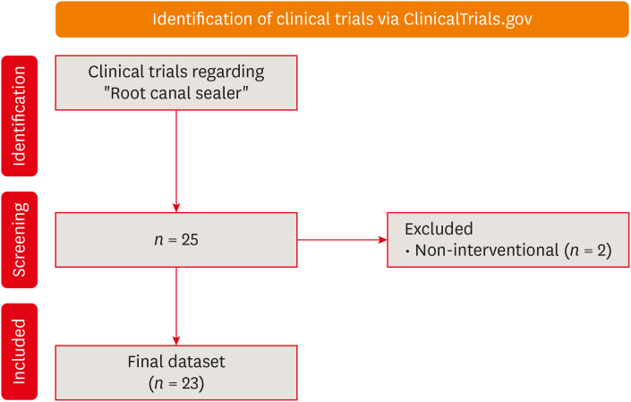
Objectives This study aimed to present the results and analyses of clinical trials, including updates on the different functions of root canal sealers.
Materials and Methods In June 2023, we performed a comprehensive search of ClinicalTrials.gov to identify interventional clinical trials pertaining to root canal sealers. In total, 23 clinical trials conducted up to June 2023 were included in this study.
Results Approximately half of the trials (11 out of 23) were completed, while none were terminated or withdrawn. Each included trial had a minimum of 10 participants, with 11 trials having more than 100 participants. None of the assessed trials provided outcomes, and the majority (17 out of 23) lacked associated publications. In terms of geographic distribution, the USA and Canada did not contribute to any root canal sealer trials.
Conclusions This study highlights the lack of diversity in trial locations, the absence of reported results, and a scarcity of clinical trials examining the physicochemical properties of different sealers. Most published trials primarily focused on assessing the post-operative pain effect of these sealers, but no significant difference was found regarding post-operative pain control.
-
Citations
Citations to this article as recorded by- Antimicrobial additives in resin-based endodontic sealers: a scoping review of antimicrobial efficacy, physicochemical properties, and cytotoxicity
Faisal Alharamlah, Maha I. AlGhannam, Wejdan Almutairi, Faisal Alonaizan, Theeb A. Alquria, Mary Anne S. Melo, Abdulrahman A. Balhaddad
Frontiers in Dental Medicine.2026;[Epub] CrossRef
- Antimicrobial additives in resin-based endodontic sealers: a scoping review of antimicrobial efficacy, physicochemical properties, and cytotoxicity
- 5,596 View
- 76 Download
- 1 Crossref
-
Effect of cryotherapy duration on experimentally induced connective tissue inflammation
in vivo - Jorge Vera, Mayra Alejandra Castro-Nuñez, María Fernanda Troncoso-Cibrian, Ana Gabriela Carrillo-Varguez, Edgar Ramiro Méndez Sánchez, Viviana Sarmiento, Lourdes Lanzagorta-Rebollo, Prasanna Neelakantan, Monica Romero, Ana Arias
- Restor Dent Endod 2023;48(3):e29. Published online August 2, 2023
- DOI: https://doi.org/10.5395/rde.2023.48.e29
-
Abstract
PDFPubReaderePub
Objectives This study tested the hypothesis that cryotherapy duration influences lipopolysaccharide (LPS)-induced inflammation in a rat model.
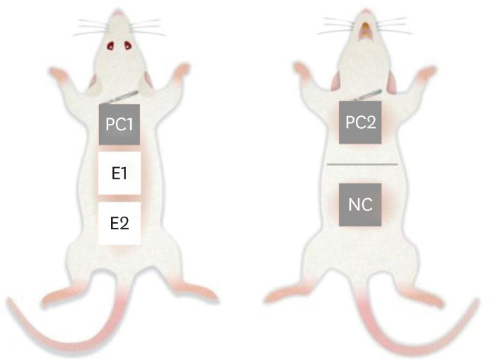
Materials and Methods Six Wistar rats (
Rattus norvegicus albinus ) were used. Five sites were selected per animal and divided into 5 groups: a negative control group (NC), 2 positive control groups (PC1 and PC2), and 2 experimental groups (E1 and E2). Cryotherapy was applied for 1 minute (E1) or 5 minutes (E2). An acute inflammatory response was induced in the PC and E groups via subcutaneous administration of 0.5 mL/kg. In the PC2 group, a catheter was inserted without additional treatment. For the E1 and E2 groups, 2.5°C saline solution was administered through the implanted catheters for 1 and 5 minutes, respectively. The rats were sacrificed, and samples were obtained and processed for histological analysis, specifically examining the presence of polymorphonuclear neutrophils and hemorrhage. The χ2 test was used to compare the presence of acute inflammation across groups. Dependent variables were compared using the linear-by-linear association test.Results Inflammation and hemorrhage varied significantly among the groups (
p = 0.001). A significantly higher degree of acute inflammation was detected (p = 0.0002) in the PC and E1 samples than in the E2 group, in which cryotherapy was administered for 5 minutes. The PC and E1 groups also exhibited significantly greater numbers of neutrophils (p = 0.007), which were essentially absent in both the NC and E2 groups.Conclusions Cryotherapy administration for 5 minutes reduced the acute inflammation associated with LPS and catheter implantation.
-
Citations
Citations to this article as recorded by- The impact of using cold irrigation on postoperative endodontic pain and substance P level: a randomized clinical trial
Reem Mohammed Amr Sharaf, Tariq Yehia Abdelrahman, Maram Farouk Obeid
Odontology.2026; 114(2): 527. CrossRef - Cryotherapy as a supplementary aid to inferior alveolar nerve block in patients with symptomatic irreversible pulpitis: A randomized controlled trial
Setu Katyal, Poonam Bogra, Rajinder Bansal, Vishakha Grover, Saurabh Gupta, Saru Gupta
Medicine International.2025; 5(5): 1. CrossRef - Determining Efficacy of Intracanal Cryotherapy on Post Endodontic Pain in Irreversible Pulpitis
Anam Fayyaz Bashir, Ussamah Waheed Jatala, Moeen ud din Ahmad, Muhammad Talha Khan, Saima Razzaq Khan, Aisha Arshad Butt
Pakistan Journal of Health Sciences.2024; : 68. CrossRef
- The impact of using cold irrigation on postoperative endodontic pain and substance P level: a randomized clinical trial
- 5,401 View
- 67 Download
- 2 Web of Science
- 3 Crossref
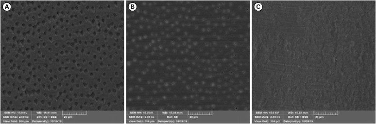
- Effect of irrigation protocols on smear layer removal, bond strength and nanoleakage of fiber posts using a self-adhesive resin cement
- Rodrigo Stadler Alessi, Renata Terumi Jitumori, Bruna Fortes Bittencourt, Giovana Mongruel Gomes, João Carlos Gomes
- Restor Dent Endod 2023;48(3):e28. Published online July 27, 2023
- DOI: https://doi.org/10.5395/rde.2023.48.e28
-
Abstract
PDFPubReaderePub
Objectives This study aimed to investigate the effect of the application method of 2% chlorhexidine (CHX) and its influence on the adhesion of fiberglass posts cemented with a self-adhesive resin cement.
Materials and Methods Sixty human mandibular premolars were endodontically treated and divided into 5 groups (
n = 12), according to the canal irrigant and its application method: 2 groups with conventional syringe irrigation (CSI)—2.5% sodium hypochlorite (NaOCl) (control) and 2% CHX— and 3 groups with 2% CHX irrigation/activation—by passive ultrasonic irrigation (PUI), Easy Clean file, and XP-Endo Finisher file. Two roots per group were evaluated for smear layer (SL) removal by scanning electron microscopy. For other roots, fiber posts were luted using a self-adhesive resin cement. The roots were sectioned into 6 slices for push-out bond strength (BS) (7/group) and nanoleakage (NL) (3/group). Data from SL removal were submitted to Kruskal-Wallis and Student-Newman-Keuls tests (α = 0.05). Data from BS and NL were evaluated by 2-way analysis of variance and Tukey’s test (α = 0.05).Results For SL removal and BS, the CHX irrigation/activation promoted better values than CSI with CHX (
p < 0.05), but it was not significantly different from CSI with NaOCl (p > 0.05). For NL, the lowest values were obtained by the chlorhexidine irrigation/activation groups (p < 0.05).Conclusions Active 2% CHX irrigation can be used to improve the post space cleaning and adhesion before fiber post cementation with self-adhesive resin cements.
-
Citations
Citations to this article as recorded by- Effects of radiotherapy dose and endodontic irrigants on universal resin cement bonding to root dentin: mechanical and interfacial analyses
Lívia Ribeiro, Luíz Carlos de Lima Dias-Júnior, Paulo Henrique dos Santos, Mariana Comparotto Minamisako, Paulo Marcelo Rodrigues, Vicente Ribeiro Netto, Bruno Alexandre Pacheco de Castro Henriques, Renata Gondo Machado, Cleonice da Silveira Teixeira, Luc
International Journal of Adhesion and Adhesives.2026; 146: 104252. CrossRef - Effect of Erbium: Yttrium–Aluminum Garnet Laser, XP-Endo Finisher, and Passive Ultrasonic Irrigation in the Cleaning of Post Spaces on Push-Out Bond Strength
Dilek Hançerlioğulları, Gökhan Karadağ
Current Research in Dental Sciences.2026; 36(2): 121. CrossRef - Current trends in irrigant activation techniques among Brazilian endodontists
Mônica Pagliarini Buligon , Natália Franco Brum , Carlos Alexandre Souza Bier, Renata Domelles Morgental
Brazilian Journal of Oral Sciences.2026; 25: e269686. CrossRef - Laser‐Activated Irrigation via Photon‐Induced Photoacoustic Streaming and Shock Wave Enhanced Emission on Smear Layer Removal Efficacy, Pushout Bond Strength, and Sealer Adaptation: A SEM Assessment
Basil Almutairi, Fahad Alkhudhairy
Microscopy Research and Technique.2025; 88(6): 1806. CrossRef - The impact of passive ultrasonic irrigation on the bond strength of two different self-etch adhesives to human pulp chamber dentine: a laboratory investigation
Mohammed Turky, Jukka Matinlinna, Monika Lukomska-Szymanska, Venkateshbabu Nagendrababu, Paul M. H. Dummer, Ahmad Abdel Hamid Elheeny, Nermin Alsayed Mahmoud
BMC Oral Health.2025;[Epub] CrossRef - The effect of nanoparticles incorporation titanium dioxide and zirconium oxide within self-adhesive resin cement on the push-out bond strength of the fiber post to the radicular dentin: An in vitro study
Sawsan Hameed Al-Jubori, Maha Anwer AL-Murad
Saudi Endodontic Journal.2025; 15(2): 162. CrossRef - The Effects of Different Post Space Conditioning Procedures and Different Endodontic Sealers on the Push-Out Bond Strengths of Fiber Posts
Leyla Ayranci, Ahmet Serkan Küçükekenci, Fatih Sarı, Ahmet Çetinkaya
Clinical and Experimental Health Sciences.2025; 15(3): 620. CrossRef - Evaluation of Microleakage Using Different Luting Cements in Kedo Zirconia Crowns: An In Vitro Assessment
Guru Vishnu, Ganesh Jeevanandan
Cureus.2024;[Epub] CrossRef
- Effects of radiotherapy dose and endodontic irrigants on universal resin cement bonding to root dentin: mechanical and interfacial analyses
- 4,684 View
- 91 Download
- 7 Web of Science
- 8 Crossref
Review Article
- Effect of endodontic sealer on postoperative pain: a network meta-analysis
- Cynthia Maria Chaves Monteiro, Ana Cristina Rodrigues Martins, Alessandra Reis, Juliana Larocca de Geus
- Restor Dent Endod 2023;48(1):e5. Published online December 29, 2022
- DOI: https://doi.org/10.5395/rde.2023.48.e5
-
Abstract
PDFPubReaderePub
This systematic review and network meta-analysis aimed to answer the following focused research question: “Does the type of endodontic sealer affect the postoperative pain in patients who received endodontic treatment?” Different databases and grey literature were surveyed. Only one randomized controlled trial were included. The risk of bias in the studies was evaluated by using the Cochrane Collaboration’s tool. A random-effects meta-analysis was conducted to compare the risk and intensity of postoperative pain. The quality of the body of evidence was assessed using the Grading of Recommendations Assessment, Development, and Evaluation approach. Out of 11,601 studies, 15 remained for qualitative analyses and 12 for meta-analysis. Seven studies were classified at high risk of bias, and 8 studies raised some concerns. No significant differences between the endodontic materials were observed in the direct comparisons, both in risk and in intensity of postoperative pain (pairwise comparisons with 2 studies: I2 = 0%;
p > 0.05 and 8 studies: I2 = 23%;p > 0.05, respectively). The certainty of the evidence was graded as low or moderate. There was no difference in the risk and intensity of postoperative pain after filling with different endodontic sealers. Further systematic reviews should be conducted.Trial Registration PROSPERO Identifier:
CRD42020215314 -
Citations
Citations to this article as recorded by- Does the Use of a Bioceramic Sealer Reduce Postoperative Pain Compared With an Epoxy Resin‐Based Sealer After Primary Root Canal Treatment and Retreatment?—An Umbrella Review
Lokhasudhan Govindaraju, Rajeswari Kalaiselvam, Mathan Rajan Rajendran, Aleksandar Jakovljevic, Jelena Jacimovic, Henry F. Duncan, Venkateshbabu Nagendrababu
International Endodontic Journal.2026; 59(3): 341. CrossRef - Evidence synthesis of postoperative pain with bioceramic vs. epoxy resin sealers: umbrella review of randomized trials within existing systematic reviews
Mrunali Dahikar, Ashish Mandwe, Kulvinder Singh Banga, Alexander Maniangat Luke, Suraj Arora, Unmesh Khanvilkar, Ajinkya M. Pawar
Frontiers in Dental Medicine.2026;[Epub] CrossRef - Factors Influencing Apical Extrusion of 2 Types of Endodontic Sealers with Different Delivery Systems
Shumaila Iqbal, Nicholas S. Adams, Josette Camilleri
Journal of Endodontics.2026; 52(5): 806. CrossRef - Evaluation of Postoperative Pain Frequency in Single‐Session Endodontic Treatments With Patency and Foraminal Enlargement
Viviane Barbosa Godoy, Ana Grasiela Limoeiro, Vanessa Sandini, Vini Mehta, Wayne Martins Nascimento, Marilia Fagury Videira Marceliano‐Alves, Marcos Frozoni
Clinical and Experimental Dental Research.2026;[Epub] CrossRef - Silicone vs. Silicon/Silica in Intraoral Healing: A Systematic Review
David Parker, Aditi Bopardikar, Georgios E. Romanos
Materials.2026; 19(7): 1425. CrossRef - Post-Operative Pain After Endodontic Instrumentation, Irrigation and Obturation: An Umbrella Review of Systematic Reviews Published from 2016 to 2025
Fausto Zamparini, Andrea Spinelli, Gioia Quadrini, Maria Giovanna Gandolfi, Carlo Prati
Journal of Clinical Medicine.2026; 15(12): 4775. CrossRef - Comparative Evaluation of Postoperative Pain Following Nonsurgical Endodontic Therapy with Calcium Silicate-Based Sealer and Traditional Sealers: A Systematic Review and Meta-Analysis
Guha Poulomi, Solete Pradeep, Antony Delphine, Arun Nishitha, Surendar Ramamoorthi, Choudhari Sahil, Hima Sandeep Adimulapu
Pesquisa Brasileira em Odontopediatria e Clínica Integrada.2026;[Epub] CrossRef - Effect of occlusal reduction on post-operative pain of symptomatic and asymptomatic molar teeth
Aysenur Kamacı Esen, Fatma Furuncuoğlu, Fatima Betul Basturk, Muhammet Nuri Taşcıoğlu, Masoud Parirokh
Acta Odontologica Scandinavica.2025; 84: 371. CrossRef - An Observational Study on Pain Occurrence After Root Canal Treatment: Role of Operator Experience When Using a Bioceramic Sealer
Mihai Merfea, Ioana Sofia Pop-Ciutrila, Mindra Eugenia Badea, Ada Gabriela Delean, Oana Cimponeriu, Razvan Corneliu Pop, Maria Peter, Iulia Clara Badea, Sanda Ileana Cimpean
Journal of Clinical Medicine.2025; 14(13): 4558. CrossRef - Assessment of Postoperative Pain After Single‐ or Multiple‐Visit Endodontic Therapy and Its Molecular Aspects: A Randomised Controlled Study
Larissa Nunes Rosa Bedene, Denise Piotto Leonardi, Joana Santana Couto, Bruno Marques‐da‐Silva, Marilisa Carneiro Leão Gabardo, João Arnando Brancher, Flávia Sens Fagundes Tomazinho
Australian Endodontic Journal.2025; 51(3): 668. CrossRef - Clinical and Radiographic Outcomes of Root Canal Obturation with Hydraulic Condensation and Tricalcium Silicate Bioceramic Sealer: A 12-Month Observational Study on Periapical Healing
Kostadin Zhekov, Vesela Stefanova
Journal of Functional Biomaterials.2025; 16(11): 412. CrossRef - Comparative evaluation of postoperative pain and periapical healing after root canal treatment using three different endodontic sealers: A randomized controlled clinical trial
Ruchika Pandey, Nitin Kararia, Deepak Kumar Sharma, Vishal Rathod, Anand Vilas Bansod, Dhaval Desai
Journal of Conservative Dentistry and Endodontics.2024; 27(9): 962. CrossRef - Effect of bioceramic-based and resin-based sealers on postoperative discomfort following root canal therapy: a systematic review and meta-analysis
Mansi Supare, Ajinkya M. Pawar, Kashmira Sawant, Dian Agustin Wahjuningrum, Suraj Arora, Firas Elmsmari, Mohmed Isaqali Karobari, Bhagyashree Thakur
PeerJ.2024; 12: e18198. CrossRef - Comparative Evaluation of Incidences of Post Operative Pain in Patient Treated in Single Visit Root Canal Treatment by Using Different Sealers: - An in-Vivo Study
Sadashiv Daokar, Aishwarya Ranjalkar, Kalpana Pawar, Komal Potfode, Dhanashri Padwal, Sana Khan
International Journal of Innovative Science and Research Technology (IJISRT).2024; : 2743. CrossRef
- Does the Use of a Bioceramic Sealer Reduce Postoperative Pain Compared With an Epoxy Resin‐Based Sealer After Primary Root Canal Treatment and Retreatment?—An Umbrella Review
- 8,404 View
- 163 Download
- 12 Web of Science
- 14 Crossref
Research Articles
- Effect of intracanal cryotherapy on postoperative pain after endodontic treatment: systematic review with meta-analysis
- Fernanda Garcias Hespanhol, Ludmila Silva Guimarães, Lívia Azeredo Alves Antunes, Leonardo Santos Antunes
- Restor Dent Endod 2022;47(3):e30. Published online July 4, 2022
- DOI: https://doi.org/10.5395/rde.2022.47.e30
-
Abstract
PDFPubReaderePub
Objectives This study aimed to evaluate the effectiveness of final irrigation with cold saline solution after endodontic treatment compared with saline solution at room temperature against postoperative pain following endodontic treatment.
Materials and Methods A broad search was performed in the PubMed, Web of Science, Scopus, Cochrane Library, Virtual Health Library (LILACS), and Grey Literature databases. Two independent reviewers performed data extraction, risk of bias using the Cochrane methodology, and certainty of evidence using the Grading of Recommendations, Assessment, Development and Evaluations (GRADE) approach.
Results Eight studies were included in qualitative synthesis. Intracanal cryotherapy favored the reduction of postoperative pain in the systematic review. Four studies were included in meta-analyses. The meta-analysis showed that intracanal cryotherapy reduced postoperative pain in teeth with symptomatic apical periodontitis (SAP) at 24 hours. There was no association between intracanal cryotherapy and control (room temperature) groups in teeth with normal periapical tissue with respect to postoperative pain at 24 hours and 48 hours.
Conclusions Intracanal cryotherapy was effective in reducing postoperative pain after endodontic treatment in teeth with SAP.
-
Citations
Citations to this article as recorded by- Postoperative Pain After Endodontic Treatment in HIV‐Positive Patients Under HAART: A Prospective Observational Cohort Study
Marcos Felipe Iparraguirre Nuñovero, Marco Antonio Hungaro Duarte, Luciana Reis Azevedo Alanis, Bruno Cavalini Cavenago, Ulisses Xavier da Silva Neto, Everdan Carneiro
International Endodontic Journal.2026; 59(5): 788. CrossRef - Effect of low-temperature intracanal sodium hypochlorite on root surface temperature reduction and organic tissue dissolution: an in vitro study
Marcos Felipe Iparraguirre Nuñovero, Marco Antonio Hungaro Duarte, Ulisses Xavier da Silva Neto, Vânia Portela Ditzel Westphalen, Pedro Cesar Gomes Titato, Bruno Cavalini Cavenago, Everdan Carneiro
Scientific Reports.2026;[Epub] CrossRef - Effectiveness of intracanal cryotherapy in reducing post-endodontic pain in irreversible pulpitis: a systematic review and meta-analysis
Maged Mohamed, Asmaa Abdelmajeed, Muhammad Salah-Uddin Anwar Laithy, Dina Abozaid
Scientific Reports.2026;[Epub] CrossRef - Post-Operative Pain After Endodontic Instrumentation, Irrigation and Obturation: An Umbrella Review of Systematic Reviews Published from 2016 to 2025
Fausto Zamparini, Andrea Spinelli, Gioia Quadrini, Maria Giovanna Gandolfi, Carlo Prati
Journal of Clinical Medicine.2026; 15(12): 4775. CrossRef - Impact of intracanal cryotherapy on postoperative pain in symptomatic apical periodontitis: A systematic review and meta-analysis of randomized clinical trials
Nishtha K. Patel, Prerak Doshi, Shaily R. Dalal, Pooja R. Kesharani, Shilpa S. Shah, Mohil H. Kale
Endodontology.2025; 37(2): 101. CrossRef - Evaluation of Post‐Endodontic Pain Reduction Using Intracanal Cryotherapy in Symptomatic Apical Periodontitis
Anam Fayyaz Bashir, Ussamah Waheed Jatala, Muhammad Amber Fareed, Sheryar Sheryar, Saadia Ahmad Chattha, Saima Razaq Khan, Shahzad Ahmad, Shazia Iqbal, Muhammad Sohail Zafar, Shahzad Ali
Australian Endodontic Journal.2025; 51(3): 677. CrossRef - Comparing cryotherapy and ketorolac tromethamine against room-temperature saline irrigation using interleukin-8 levels and post-operative pain within single-visit endodontic treatment of symptomatic irreversible pulpitis superimposed by apical periodontit
Yousra Khaled Ezzat, Alaa Diab, Olfat Shaker, Sarah Abouelenien
BMC Oral Health.2025;[Epub] CrossRef - Determining Efficacy of Intracanal Cryotherapy on Post Endodontic Pain in Irreversible Pulpitis
Anam Fayyaz Bashir, Ussamah Waheed Jatala, Moeen ud din Ahmad, Muhammad Talha Khan, Saima Razzaq Khan, Aisha Arshad Butt
Pakistan Journal of Health Sciences.2024; : 68. CrossRef - The effect of intracanal cryotherapy with and without foraminal enlargement on pain prevention after endodontic treatment: a randomized clinical trial
Marcos Felipe Iparraguirre Nuñovero, Marco Antonio Hungaro Duarte, André Vinícius Kaled Segato, Ulisses Xavier da Silva Neto, Vania Portela Ditzel Westphalen, Everdan Carneiro
Scientific Reports.2024;[Epub] CrossRef - Effect of cryotherapy duration on experimentally induced connective tissue inflammationin vivo
Jorge Vera, Mayra Alejandra Castro-Nuñez, María Fernanda Troncoso-Cibrian, Ana Gabriela Carrillo-Varguez, Edgar Ramiro Méndez Sánchez, Viviana Sarmiento, Lourdes Lanzagorta-Rebollo, Prasanna Neelakantan, Monica Romero, Ana Arias
Restorative Dentistry & Endodontics.2023;[Epub] CrossRef - Evaluation of knowledge and awareness of pediatric oral health among school teachers of Hazaribag before and after oral health education.
Vipin Ahuja, Annapurna Ahuja, Nilima Thosar
F1000Research.2023; 12: 1292. CrossRef
- Postoperative Pain After Endodontic Treatment in HIV‐Positive Patients Under HAART: A Prospective Observational Cohort Study
- 4,780 View
- 103 Download
- 10 Web of Science
- 11 Crossref
- A 3-year retrospective study of clinical durability of bulk-filled resin composite restorations
- Muhittin Ugurlu, Fatmanur Sari
- Restor Dent Endod 2022;47(1):e5. Published online December 30, 2021
- DOI: https://doi.org/10.5395/rde.2022.47.e5
-
Abstract
PDFPubReaderePub
Objectives This study aimed to assess the clinical longevity of a bulk-fill resin composite in Class II restorations for 3-year.
Materials and Methods Patient record files acquired from the 40 patients who were treated due to needed 2 similar sizes Class II composite restorations were used for this retrospective study. In the experimental cavity, the flowable resin composite SDR was inserted in the dentinal part as a 4 mm intermediate layer. A 2 mm coverage layer with a nano-hybrid resin composite (CeramX) was placed on SDR. The control restoration was performed by an incremental technique of 2 mm using the nano-hybrid resin composite. The restorations were blindly assessed by 2 calibrated examiners using modified United States Public Health Service criteria at baseline and 1, 2, and 3 years. The data were analyzed using non-parametric tests (
p = 0.05).Results Eighty Class II restorations were evaluated. After 3-years, 4 restorations (5%) failed, 1 SDR + CeramX, and 3 CeramX restorations. The annual failure rate (AFR) of the restorations was 1.7%. The SDR + CeramX group revealed an AFR of 0.8%, and the CeramX group an AFR of 2.5% (
p > 0.05). Regarding anatomical form and marginal adaptation, significant alterations were observed in the CeramX group after 3-years (p < 0.05). The changes in the color match were observed in each group over time (p < 0.05).Conclusions The use of SDR demonstrated good clinical durability in deep Class II resin composite restorations.
-
Citations
Citations to this article as recorded by- Bond Strength of Bulk-Fill Resin Repairs: Impact of Surface and Adhesive Protocols
Samuel Eleutério Paiva Sousa, Fiorella Elizabeth Arévalo Tarrillo, Maria Paula Novaes Camargo Manna, Sandra Ribeiro de Barros da Cunha, Maria Ângela Pita Sobral
Odovtos - International Journal of Dental Sciences.2026; 28(2): 204. CrossRef - Evaluation of Surface Roughness and Microhardness of New Generation Bulk-Fill Composites
Zehra SÜSGÜN YILDIRIM, Ezgi SONKAYA, Zeliha Gonca BEK KÜRKLÜ
Cumhuriyet Dental Journal.2023; 26(2): 180. CrossRef - Damping Behaviour and Mechanical Properties of Restorative Materials for Primary Teeth
Thomas Niem, Roland Frankenberger, Stefanie Amend, Bernd Wöstmann, Norbert Krämer
Materials.2022; 15(21): 7698. CrossRef
- Bond Strength of Bulk-Fill Resin Repairs: Impact of Surface and Adhesive Protocols
- 5,033 View
- 45 Download
- 2 Web of Science
- 3 Crossref
Case Report
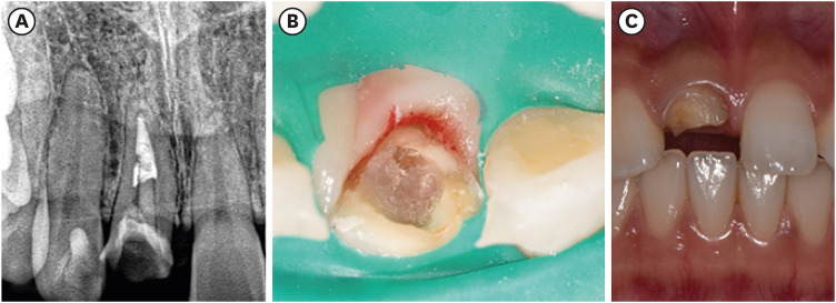
- Fiber-reinforced composite post removal using guided endodontics: a case report
- Changgi Cho, Hyo Jin Jo, Jung-Hong Ha
- Restor Dent Endod 2021;46(4):e50. Published online September 23, 2021
- DOI: https://doi.org/10.5395/rde.2021.46.e50
-
Abstract
PDFPubReaderePub
Although several techniques have been proposed to remove fiber-reinforced composite (FRC) post, no safe and efficient technique has been established. Recently, a guided endodontics technique has been introduced in cases of pulp canal obliteration. This study describes 2 cases of FRC post removal from maxillary anterior teeth using this guided endodontics technique with a dental operating microscope. Optically scanned data set from plaster cast model was superimposed with the data set of cone-beam computed tomography. By implant planning software, the path of a guide drill was selected. Based on them, a customized stent was fabricated and utilized to remove the FRC post. Employing guided endodontics, the FRC post was removed quickly and safely with minimizing the loss of the remaining tooth structure. The guided endodontics was a useful option for FRC post removal.
-
Citations
Citations to this article as recorded by- Comparing the Effectiveness of a Robotic and Dynamic Navigation System in Fiber Post removal: An In Vitro Study
Duo Zhou, Fulu Xu, Jiayun Dai, Xingyang Wang, Yifan Ping, Juan Wang
Journal of Endodontics.2026; 52(2): 261. CrossRef - Robot-assisted haptic guidance in endodontics: A pilot study evaluating efficiency and tooth structure preservation
Roshanak Momen, Joshua Dale, Mithun Suresh, Theodore D. Ravenel, Julie Marshall, Chin-Lo Hahn
Journal of Dentistry.2026; 173: 106854. CrossRef - Deviation analysis of guided fiber post removal using an assembled sleeveless guide system: A case series
Jingqi Zhu, Siyi Mo, Yuan Li, Yaojun Zhang, Xutong Song, Jingwen Liu, Ye Cao, Xiaoxiang Xu
The Journal of Prosthetic Dentistry.2026;[Epub] CrossRef - Static‐Guided Endodontics for Complex Intracanal Obstructions and Zirconia Post Removal: Report on Three Cases
Xinxuan Wang, Xuguang Li, Baicheng Yi
Clinical Case Reports.2026;[Epub] CrossRef - Application of 3D-printed resin guides for the removal of molar fiber posts
Yumin Wu, Lumei Huang, Bing Ge, Yuhang Zhang, Juan Zhang, Haifeng Xie, Ye Zhu, Chen Chen
Journal of Dentistry.2025; 153: 105462. CrossRef - Guided Removal of Long and Short Fiber Posts Using Endodontic Static Guides: A Case Report
Sahar Shafagh, Mamak Adel, Atiyeh Sabzpai
Clinical Case Reports.2025;[Epub] CrossRef - Guided versus non-guided fiber post removal: A systematic review and meta-analysis of the accuracy, efficiency, and dentin preservation of static navigation techniques in the removal of fiber posts
Mohamad Elabdalla, Farshad Khosraviani, Shahryar Irannejadrankouhi, Niloofar Ghadimi, Turgut Yağmur Yalçın, Shaheen Wathiq Tawfeeq Al Hajaj, Mahmood Dashti
The Journal of Prosthetic Dentistry.2025; 134(3): 630.e1. CrossRef - Top 100 Most-cited Scientific Articles in Guided Endodontic 2018–2024: A Bibliometric Analysis
Gustavo Adrián Morales Valladares, Raquel Esmeralda Guillén Guillén, Martha Elena Gallegos Intriago, Mary Yussely Burgos Barreiro, Claudia Jhelissa Campos Vélez, Andrés Alexander Castillo Chacón, Silvana Beatriz Terán Ayala
The Open Dentistry Journal.2025;[Epub] CrossRef - Nonsurgical Management of a Tooth With Intracanal Fiber Post and Periapical Lesion Using Guided Endodontic Technique
Mamak Adel, Zohreh Asgari
Clinical Case Reports.2025;[Epub] CrossRef - Impact of Guided Endodontics on the Success of Endodontic Treatment: An Umbrella Review of Systematic Reviews and Meta-Analyses
Aakansha Puri, Dax Abraham, Alpa Gupta
Cureus.2024;[Epub] CrossRef - Endodontia guiada por tomografia computadorizada de feixe cônico
Maysa Gaudereto Laurindo, Celso Neiva Campos, Anamaria Pessoa Pereira Leite, Paola Cantamissa Rodrigues Ferreira
Cadernos UniFOA.2024; 19(54): 1. CrossRef - Removal of fiber posts using conventional versus guided endodontics: a comparative study of dentin loss and complications
R. Krug, F. Schwarz, C. Dullin, W. Leontiev, T. Connert, G. Krastl, F. Haupt
Clinical Oral Investigations.2024;[Epub] CrossRef - Accuracy and Efficiency of the Surgical-Guide-Assisted Fiber Post Removal Technique for Anterior Teeth: An Ex Vivo Study
Ryota Ito, Satoshi Watanabe, Kazuhisa Satake, Ryuma Saito, Takashi Okiji
Dentistry Journal.2024; 12(10): 333. CrossRef - Endodontic management of severely calcified mandibular anterior teeth using guided endodontics: A report of a case and a review of the literature
Mina Davaji, Sahar Karimpour
Saudi Endodontic Journal.2024; 14(2): 245. CrossRef - A laboratory study comparing the static navigation technique using a bur with a conventional freehand technique using ultrasonic tips for the removal of fibre posts
Francesc Abella Sans, Zeena Tariq Alatiya, Gonzalo Gómez Val, Venkateshbabu Nagendrababu, Paul Michael Howell Dummer, Fernando Durán‐Sindreu Terol, Juan Gonzalo Olivieri
International Endodontic Journal.2024; 57(3): 355. CrossRef - A three‐dimensional printed assembled sleeveless guide system for fiber‐post removal
Yang Xue, Lei Zhang, Ye Cao, Yongsheng Zhou, Qiufei Xie, Xiaoxiang Xu
Journal of Prosthodontics.2023; 32(2): 178. CrossRef - Accuracy of a 3D printed sleeveless guide system used for fiber post removal: An in vitro study
Siyi Mo, Yongwei Xu, Lei Zhang, Ye Cao, Yongsheng Zhou, Xiaoxiang Xu
Journal of Dentistry.2023; 128: 104367. CrossRef - Expert consensus on digital guided therapy for endodontic diseases
Xi Wei, Yu Du, Xuedong Zhou, Lin Yue, Qing Yu, Benxiang Hou, Zhi Chen, Jingping Liang, Wenxia Chen, Lihong Qiu, Xiangya Huang, Liuyan Meng, Dingming Huang, Xiaoyan Wang, Yu Tian, Zisheng Tang, Qi Zhang, Leiying Miao, Jin Zhao, Deqin Yang, Jian Yang, Junqi
International Journal of Oral Science.2023;[Epub] CrossRef - Knowledge, attitude, practice and perception survey on post and core restorations
Aruna Kumari Veronica, Shamini Sai, Anand V Susila
Endodontology.2023; 35(3): 228. CrossRef
- Comparing the Effectiveness of a Robotic and Dynamic Navigation System in Fiber Post removal: An In Vitro Study
- 6,142 View
- 123 Download
- 14 Web of Science
- 19 Crossref
Research Articles
- Postoperative pain after endodontic treatment of necrotic teeth with large intentional foraminal enlargement
- Ricardo Machado, Daniel Comparin, Sérgio Aparecido Ignácio, Ulisses Xavier da Silva Neto
- Restor Dent Endod 2021;46(3):e31. Published online May 31, 2021
- DOI: https://doi.org/10.5395/rde.2021.46.e31
-
Abstract
PDFPubReaderePub
Objectives To evaluate postoperative pain after endodontic treatment of necrotic teeth using large intentional foraminal enlargement (LIFE).
Materials and Methods The sample included 60 asymptomatic necrotic teeth (with or without chronic apical periodontitis), and a periodontal probing depth of 3 mm, previously accessed and referred to perform endodontic treatment. After previous procedures, the position and approximate size of the apical foramen (AF) were determined by using an apex locator and K flexo-files, respectively. The chemomechanical preparation was performed with Profile 04 files 2 mm beyond the AF to achieve the LIFE, using 2.5 mL of 2.5% NaOCl at each file change. The filling was performed by Tagger's hybrid technique and EndoFill sealer. Phone calls were made to all the patients at 24, 48 and 72 hours after treatment, to classify postoperative pain. Statistical analysis was performed by different tests with a significance level of 5%.
Results Age, gender, periradicular status and tooth type did not influence postoperative pain (
p > 0.05). Only 1 patient (1.66%) reported severe pain after 72 hours. Moderate pain was reported by 7, 4 and 3 patients after 24, 48 and 72 hours, respectively (p = 0.0001). However, paired analyses showed a statistically significant difference only between 24 and 72 hours (p = 0.04). Sealer extrusion did not influence the postoperative pain (p > 0.05).Conclusions Acute or moderate postoperative pain was uncommon after endodontic treatment of necrotic teeth with LIFE.
Trial Registration The Brazilian Clinical Trials Registry Identifier:
RBR-3r967t -
Citations
Citations to this article as recorded by- Postoperative Pain After Endodontic Treatment in HIV‐Positive Patients Under HAART: A Prospective Observational Cohort Study
Marcos Felipe Iparraguirre Nuñovero, Marco Antonio Hungaro Duarte, Luciana Reis Azevedo Alanis, Bruno Cavalini Cavenago, Ulisses Xavier da Silva Neto, Everdan Carneiro
International Endodontic Journal.2026; 59(5): 788. CrossRef - Does maintaining apical patency reduce early postoperative pain after root canal treatment? A randomized controlled trial in asymptomatic vital single-rooted teeth
Ozan Arda Deger, Sehnaz Yilmaz, Kübra Gürler
Clinical Oral Investigations.2026;[Epub] CrossRef - Evaluation of Postoperative Pain Frequency in Single‐Session Endodontic Treatments With Patency and Foraminal Enlargement
Viviane Barbosa Godoy, Ana Grasiela Limoeiro, Vanessa Sandini, Vini Mehta, Wayne Martins Nascimento, Marilia Fagury Videira Marceliano‐Alves, Marcos Frozoni
Clinical and Experimental Dental Research.2026;[Epub] CrossRef - Assessment of apical extrusion in regenerative endodontics: a comparative study of different irrigation methods using three-dimensional immature tooth models
Vahide Hazal Abat, Gökçen Deniz Bayrak, Mustafa Gündoğar
Odontology.2025; 113(1): 213. CrossRef - Clinical Advances in Calcium Phosphate for Maxillomandibular Bone Regeneration: From Bench to Bedside
Seyed Ali Mostafavi Moghaddam, Hamid Mojtahedi, Amirhossein Bahador, Lotfollah Kamali Hakim, Hamid Tebyaniyan
Ceramics.2025; 8(4): 129. CrossRef - Postoperative pain after single-visit root canal treatments in necrotic teeth comparing instruments’ kinematics and apical instrumentation limits – a prospective randomized multicenter clinical trial
Ricardo Machado, Guilherme Moreira, Daniel Comparin, Arthur Pimentel Barroso, Jaqueline Nascimento, Caio Cézar Randi Ferraz, Sérgio Aparecido Ignácio, Lucas da Fonseca Roberti Garcia, Rodrigo Rodrigues Amaral, David Shadid, Ulisses Xavier da Silva Neto
BMC Oral Health.2024;[Epub] CrossRef - Assessment of mechanical allodynia in healthy teeth adjacent and contralateral to endodontically diseased teeth: a clinical study
Vaishnavi Ratnakar Patankar, Ashish K Jain, Rahul D Rao, Prajakta R Rao
Restorative Dentistry & Endodontics.2024;[Epub] CrossRef - A systematic review and meta-analysis on the effects of phototherapy on postoperative pain in conventional endodontic reintervention
Larissa Pereira Nunes, Gabriel Pereira Nunes, Túlio Morandin Ferrisse, Henrico Badaoui Strazzi-Sahyon, Eloi Dezan-Júnior, Luciano Tavares Angelo Cintra, Gustavo Sivieri-Araujo
Clinical Oral Investigations.2024;[Epub] CrossRef - The effect of intracanal cryotherapy with and without foraminal enlargement on pain prevention after endodontic treatment: a randomized clinical trial
Marcos Felipe Iparraguirre Nuñovero, Marco Antonio Hungaro Duarte, André Vinícius Kaled Segato, Ulisses Xavier da Silva Neto, Vania Portela Ditzel Westphalen, Everdan Carneiro
Scientific Reports.2024;[Epub] CrossRef - Clinical determination of anatomical diameter in different dental groups correlating them with gender, age, tooth/canal and pulpoperiradicular diagnosis: an observational clinical study
Ricardo Machado, Gabriel Filipe Pamplona, Claudemir de Souza Júnior, Jaqueline Nascimento, Eduardo Donato Eing Elgelke Back, Daniel Comparin, Sérgio Aparecido Ignácio, Stella Maria Glaci Reinke, Ana Cristina Kovalik, Ulisses Xavier da Silva Neto
Scientific Reports.2023;[Epub] CrossRef - How much to enlarge? A letter to the editor
Krishnamachari Janani, Kavalipurapu Venkata Teja, Kumar Chandan Srivatsava
Saudi Endodontic Journal.2023; 13(3): 288. CrossRef - Efficiency of diode laser in control of post-endodontic pain: a randomized controlled trial
Hend H. Ismail, Maram Obeid, Ehab Hassanien
Clinical Oral Investigations.2023; 27(6): 2797. CrossRef - Periapical Healing following Root Canal Treatment Using Different Endodontic Sealers: A Systematic Review
Akshay Khandelwal, Krishnamachari Janani, KavalipurapuVenkata Teja, Jerry Jose, Gopi Battineni, Francesco Riccitiello, Alessandra Valletta, Ajitha Palanivelu, Gianrico Spagnuolo, Vincenzo Grassia
BioMed Research International.2022;[Epub] CrossRef
- Postoperative Pain After Endodontic Treatment in HIV‐Positive Patients Under HAART: A Prospective Observational Cohort Study
- 6,469 View
- 72 Download
- 11 Web of Science
- 13 Crossref
- Effect of post space preparation drills on the incidence of root dentin defects
- Thaíse Ayres Bezerra Zuli, Orlando Aguirre Guedes, Gislaine Figueiredo Zarza Arguello Gonçalves, Aurélio Rosa da Silva Júnior, Álvaro Henrique Borges, Andreza Maria Fábio Aranha
- Restor Dent Endod 2020;45(4):e53. Published online October 16, 2020
- DOI: https://doi.org/10.5395/rde.2020.45.e53
-
Abstract
PDFPubReaderePub
Objectives This study investigated the incidence of root dentin defects after the use of different post space preparation (PSP) drills.
Materials and Methods Seventy-two bovine incisors were selected and obtained 14-mm-long root sections. Twelve roots served as controls with no intervention (G1). The 60 root canals remaining were instrumented using the crown-down technique with the ProTaper Next system and obturated using the lateral condensation technique. Specimens were randomly distributed into 5 groups (
n = 12) according to the operative steps performed: G2, root canal instrumentation and filling (I+F); G3, I+F and PSP with Gates-Glidden drills; G4, I+F and PSP with Largo-Peeso reamers; G5, I+F and PSP with Exacto drill; and G6, I+F and PSP with WhitePost drill. Roots were sectioned at 3, 6, 9, and 12 mm from the apex, and digital images were captured. The presence of root dentin defects was recorded. Data were analyzed by the χ2 test, withp < 0.05 considered to indicate statistical significance.Results Root dentin defects were observed in 39.6% of the root sections. No defects were observed in G1. G5 had significantly more cracks and craze lines than G1, G2, and G3 (
p < 0.05), and more fractures than G1, G2, G3, and G4 (p < 0.05). When all root sections were analyzed together, significantly more defects were observed at the 12-mm level than at the 3-mm level (p < 0.05).Conclusions PSP drills caused defects in the root dentin. Gates-Glidden drills caused fewer root defects than Largo-Peeso reamers and Exacto drills.
-
Citations
Citations to this article as recorded by- Fracture Strength of CAD/CAM Endocrown and Post-Core Restorations with Fiber Strip Reinforcement in Mandibular Premolars
Kerem Yılmaz, Hakan Aydın, Zeynep Soylu, Özge Çiloğlu, Esma Fatıma Delican, Mehmet Mustafa Özarslan, Fehmi Gönüldaş
Journal of Functional Biomaterials.2026; 17(5): 248. CrossRef - Evaluation of dentinal crack formation during post space preparation using different fiber post systems with micro-computed tomography
Ayşe Nur Kuşuçar, Damla Kırıcı
BMC Oral Health.2025;[Epub] CrossRef - Fracture and Crack Behavior of Weakened Incisors Restored With Fiber Posts, Polyethylene Reinforcement, or 3D-Printed Endocrowns
Diana Codas-Duarte, Laís L Pelozo, Jardel F Mazzi-Chaves, Fabiane C Lopes-Olhê, Manoel D Sousa-Neto, Aline E Souza-Gabriel
Cureus.2025;[Epub] CrossRef - Selecting drill size for post space preparation based on final endodontic radiographs: An in vitro study
Farzaneh Farid, Julfikar Haider, Marjan Sadeghpour Shahab, Nika Rezaeikalantari
Technology and Health Care.2024; 32(4): 2575. CrossRef - Cone Beam Computed Tomography Analysis of Post Space in Bifurcated Premolars Using ParaPost and Peeso Reamer Drills
Abdulaziz Saleh Alqahtani, Omar Nasser Almonabhi, Abdulmajeed Moh. Almutairi, Reem R. Alnatsha
The Open Dentistry Journal.2024;[Epub] CrossRef - A Comparative Evaluation of Real-Time Guided Dynamic Navigation and Conventional Techniques for Post Space Preparation During Post Endodontic Management: An In Vitro Study
Sherifa Shervani, Sihivahanan Dhanasekaran, Vijay Venkatesh
Cureus.2024;[Epub] CrossRef - The effect of ultrasonic vibration protocols for cast post removal on the incidence of root dentin defects
Giulliano C. Serpa, Orlando A. Guedes, Neurinelma S. S. Freitas, Julio A. Silva, Carlos Estrela, Daniel A. Decurcio
Journal of Oral Science.2023; 65(3): 190. CrossRef
- Fracture Strength of CAD/CAM Endocrown and Post-Core Restorations with Fiber Strip Reinforcement in Mandibular Premolars
- 3,737 View
- 42 Download
- 7 Crossref
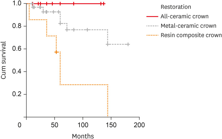
- Retrospective clinical and radiographic evaluation of restored endodontically treated teeth
- Paula Pontes Garcia, Aline Cappoani, Ricardo Susin Schelbauer, Gisele Maria Correr, Carla Castiglia Gonzaga
- Restor Dent Endod 2020;45(4):e49. Published online October 7, 2020
- DOI: https://doi.org/10.5395/rde.2020.45.e49
-
Abstract
PDFPubReaderePub
Objectives The aim of this study was to perform a clinical and radiographic analysis of endodontically treated teeth (ETT) restored with cast metal posts (CMPs) or prefabricated glass fiber posts (GFPs) and crowns.
Materials and Methods Fifty ETT were restored with 25 CMPs and 25 GFPs at a private dental clinic between 2001 and 2016. The restorations consisted of 12 all-ceramic crowns, 31 metal-ceramic crowns, and 7 composite resin crowns. Demographic data, type of teeth, type of post-and-core system, time of placement, crown restorations, the number of proximal contacts, the type of antagonist, and reports of any complications after post-and-core placement were recorded for each patient. Assessments were performed at baseline (radiographic) and follow-up (radiographic and clinical). Data were analyzed by the McNemar test, the Pearson χ2 test, and Kaplan-Meier survival curves (α = 0.05). The mean follow-up was 67.6 months.
Results No significant difference was observed for any of the radiographic parameters when the baseline and final radiographs were compared. In the clinical evaluation, anatomical form (
p = 0.009) and occlusion (p = 0.001) showed significant differences according to the type of crown restoration; specifically, metal-ceramic and all-ceramic crowns outperformed composite resin crowns.Conclusions CMPs and GFPs showed favorable results for restoring ETT after 6 years of follow-up. All-ceramic and metal-ceramic crowns showed higher survival rates and better clinical outcomes.
-
Citations
Citations to this article as recorded by- The Outcomes of Endodontically Treated Teeth Restored with Custom-Made Cast Post-and-Core Restorations: A Retrospective Cohort Study
Ahmed Ben Suleiman, Shivani Desai, Adam Tepperman, David Chvartszaid, Gevik Malkhassian, Effrat Habsha, Izchak Barzilay, Amir Azarpazhooh
Journal of Endodontics.2024; 50(3): 316. CrossRef - Effect of a circumferential ferrule on the survival and success of endodontically treated teeth restored with fiber posts: A systematic review and meta-analysis
Raghad A. Al-Dabbagh, Mohammed A. Sindi, Mohammed A. Sanari, Alaa I. Manna, Mona A. Al-Dabbagh
The Journal of Prosthetic Dentistry.2024; 132(6): 1251. CrossRef - Effect of Luting Cement Film Thickness on the Pull-Out Bond Strength of Endodontic Post Systems
Khalil Aleisa, Syed Rashid Habib, Abdul Sadekh Ansari, Ragad Altayyar, Shahad Alharbi, Sultan Ali S. Alanazi, Khalid Tawfik Alduaiji
Polymers.2021; 13(18): 3082. CrossRef
- The Outcomes of Endodontically Treated Teeth Restored with Custom-Made Cast Post-and-Core Restorations: A Retrospective Cohort Study
- 3,845 View
- 48 Download
- 3 Crossref
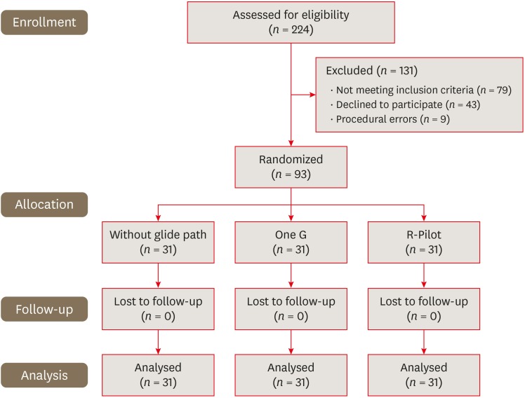
- Comparison of postoperative pain intensity after using reciprocating and continuous rotary glide path systems: a randomized clinical trial
- Mehmet Adıgüzel, Koray Yılmaz, Pelin Tüfenkçi
- Restor Dent Endod 2019;44(1):e9. Published online February 12, 2019
- DOI: https://doi.org/10.5395/rde.2019.44.e9
-
Abstract
PDFPubReaderePub
Objectives The aim of this study was to compare postoperative pain intensity after root canal treatment with One G (OG) vs. R-Pilot (RP) files used for glide path preparation.
Materials and Methods Ninety-three single-canaled mandibular premolar teeth with asymptomatic non-vital pulp were randomly assigned into 3 groups (
n = 31): OG, RP, or without glide path (WGP). After creating the glide path, the root canals were prepared using sequential Mtwo rotary files to size 30/0.05. One endodontic specialist carried out single-visit endodontic treatment. The patients were asked to rate the severity of postoperative pain on a visual analogue scale at 24, 48, and 72 hours after the visit. They were also asked to record their intake of prescribed analgesics taken. The data were analyzed using the χ2, Friedman, Kruskal-Wallis, and Mann-WhitneyU tests.Results In all 3 groups, postoperative pain decreased significantly at each time interval (
p < 0.05). At 24 hours, the OG group had less postoperative pain than the WGP group (p < 0.05). However, no significant difference was found between the RP group and the others. No statistically significant difference was found among the WGP, OG, and RP groups in postoperative pain intensity at 48 or 72 hours or in analgesic tablet intake at the 3 assessed time intervals.Conclusions The OG group had less postoperative pain than the WGP group in the first 24 hours. The OG and RP systems were similar regarding postoperative pain intensity and analgesic intake.
-
Citations
Citations to this article as recorded by- Effect of glide path preparation on postoperative pain using TruNatomy and Reciproc Blue in single-visit root canal therapy: A randomized clinical trial
Fatima Siddiqui, Sajid Ali, Huma Iftekhar, Rajendra Kumar Tewari, Ashok Kumar, Sharique Alam
Journal of Conservative Dentistry and Endodontics.2025; 28(12): 1222. CrossRef - Postoperative pain in patients following endodontic treatment by XP-endo Shaper files: A systematic review and meta-analysis
Henal Nilesh Dedhia, Vibha R. Hegde, Maitri B. Bhayani, Sanitra R. Hegde
Journal of Conservative Dentistry and Endodontics.2024; 27(11): 1168. CrossRef - Postoperative Pain Following Single Visit Root Canal Treatment With Reciproc Blue And Hyflex EDM Instrumentation; A Prospective Randomized Clinical Trial
Nimet Gençoğlu, Anıl Özgün Karatekin, Mustafa Gündoğar
Meandros Medical And Dental Journal.2024; 25(1): 78. CrossRef - Glide Path in Endodontics: A Literature Review of Current Knowledge
Vlad Mircea Lup, Giulia Malvicini, Carlo Gaeta, Simone Grandini, Gabriela Ciavoi
Dentistry Journal.2024; 12(8): 257. CrossRef - Post-Operative Pain in Reciprocating Versus Rotary Kinematics Post-Endodontic Treatment: A Systematic Review
Youssef Algarni
Archives of Pharmacy Practice.2024; 15(2): 53. CrossRef - Evaluation of Pain Following the Use of Different Single-file Glide Path Systems: A Randomized Clinical Trial
Zeliha Danaci, Kübra Yeşildal Yeter
Journal of Endodontics.2024; 50(2): 120. CrossRef - Incidence of postoperative pain after using single continuous, single reciprocating, and full sequence continuous rotary file system: a prospective randomized clinical trial
Umesh Kumar, Pragnesh Parmar, Ruchi Vashisht, Namita Tandon, Charan Kamal Kaur
Journal of Dental Anesthesia and Pain Medicine.2023; 23(2): 91. CrossRef - Impact of kinematics on the efficiency and safety of an engine-driven file for glide path preparation in MB2 canals of maxillary molars
Larissa B. B. Araújo, Pedro H. S. Calefi, Murilo P. Alcalde, Giulio Gavini, Rodrigo R. Vivan, Marco Antonio Hungaro Duarte
Clinical Oral Investigations.2022; 27(3): 1153. CrossRef - Evaluation of Postoperative Pain after Pulpectomy using Different File Systems in Primary Teeth
Lakshimi Lakshmanan, Sujatha Somasundaram, Ganesh Jeevanandan, EMG Subramanian
Contemporary Clinical Dentistry.2021; 12(1): 3. CrossRef - Evaluation of type of kinematics on glide path procedures and torsional fatigue resistance after preparation of moderately curved canals
Murilo Priori Alcalde, Marco Antonio Hungaro Duarte, Pedro Henrique Souza Calefi, Victor de Moraes Cruz, Bruno Carvalho de Vasconcelos, Marcus Vinícius Reis Só, Rodrigo Ricci Vivan
Brazilian Oral Research.2021;[Epub] CrossRef - Influence of glide path kinematics during endodontic treatment on the occurrence and intensity of intraoperative and postoperative pain: a systematic review of randomized clinical trials
Thaís Christina Cunha, Felipe de Souza Matos, Luiz Renato Paranhos, Ítalo de Macedo Bernardino, Camilla Christian Gomes Moura
BMC Oral Health.2020;[Epub] CrossRef
- Effect of glide path preparation on postoperative pain using TruNatomy and Reciproc Blue in single-visit root canal therapy: A randomized clinical trial
- 4,169 View
- 29 Download
- 11 Crossref
- Smear layer removal by different chemical solutions used with or without ultrasonic activation after post preparation
- Daniel Poletto, Ana Claudia Poletto, Andressa Cavalaro, Ricardo Machado, Leopoldo Cosme-Silva, Cássia Cilene Dezan Garbelini, Márcio Grama Hoeppner
- Restor Dent Endod 2017;42(4):324-331. Published online November 1, 2017
- DOI: https://doi.org/10.5395/rde.2017.42.4.324
-
Abstract
PDFPubReaderePub
Objectives This study evaluated smear layer removal by different chemical solutions used with or without ultrasonic activation after post preparation.
Materials and Methods Forty-five extracted uniradicular human mandibular premolars with single canals were treated endodontically. The cervical and middle thirds of the fillings were then removed, and the specimens were divided into 9 groups: G1, saline solution (NaCl); G2, 2.5% sodium hypochlorite (NaOCl); G3, 2% chlorhexidine (CHX); G4, 11.5% polyacrylic acid (PAA); G5, 17% ethylenediaminetetraacetic acid (EDTA). For the groups 6, 7, 8, and 9, the same solutions used in the groups 2, 3, 4, and 5 were used, respectively, but activated with ultrasonic activation. Afterwards, the roots were analyzed by a score considering the images obtained from a scanning electron microscope.
Results EDTA achieved the best performance compared with the other solutions evaluated regardless of the irrigation method (
p < 0.05).Conclusions Ultrasonic activation did not significantly influence smear layer removal.
-
Citations
Citations to this article as recorded by- Cerium Oxide Nanoparticle Loaded with Toluidine Blue as Cavity Disinfectant Activated via Light-Emitting Diode on the Shear Bond Strength and Resin Tag Length of Universal Adhesive: A Scanning Electron Microscope-EDX Study
Amer M. Alanazi, Syed Hussain Askary, Ibrahim Warsi, Aamir Afzal, Muhammad Omar Niaz, Ambrina Qureshi
Photobiomodulation, Photomedicine, and Laser Surgery.2026;[Epub] CrossRef - O papel do ultrassom no tratamento e retratamento de canais radiculares: Revisão de literatura
Carlos Roberto Souza Hipp, Joaquim Carlos Fest da Silveira, Luiz Felipe Gilson de Oliveira Rangel, Tatiana Federici de Souza Fest da Silveira, Carla Minozzo Mello, Rodrigo Simões de Oliveira
Research, Society and Development.2025; 14(8): e1314849323. CrossRef - Effect of sodium hypochlorite, ethylenediaminetetraacetic acid, and dual-rinse irrigation on dentin adhesion using an etch-and-rinse or self-etch approach
Matej Par, Tobias Steffen, Selinay Dogan, Noah Walser, Tobias T. Tauböck
Scientific Reports.2024;[Epub] CrossRef - Evaluation of Effect of Poloxamer on Smear Layer Removal Using Apical Negative Pressure: An In Vitro Scanning Electron Microscopy Study
Chandra Prabha, Chitharanjan Shetty, Aditya Shetty
Journal of International Oral Health.2024; 16(6): 498. CrossRef - Laboratory Assessment of Antibacterial Efficacy of Five Different Herbal-based Potential Endodontic Irrigants
Anjali A Oak, Kailash Attur, Kamal Bagda, Nitish Mathur, Lubna Mohammad, Nikhat M Attar
Advances in Human Biology.2023; 13(4): 350. CrossRef - Dental Surface Conditioning Techniques to Increase the Micromechanical Retention to Fiberglass Posts: A Literature Review
Paulina Leticia Moreno-Sánchez, Maricela Ramírez-Álvarez, Alfredo del Rosario Ayala-Ham, Erika de Lourdes Silva-Benítez, Miguel Ángel Casillas-Santana, Diana Leyva del Rio, León Francisco Espinosa-Cristóbal, Erik Lizárraga-Verdugo, Mariana Melisa Avendaño
Applied Sciences.2023; 13(14): 8083. CrossRef - Effect of irrigation protocols on smear layer removal, bond strength and nanoleakage of fiber posts using a self-adhesive resin cement
Rodrigo Stadler Alessi, Renata Terumi Jitumori, Bruna Fortes Bittencourt, Giovana Mongruel Gomes, João Carlos Gomes
Restorative Dentistry & Endodontics.2023;[Epub] CrossRef - Effects of using different root canal sealers and protocols for cleaning intraradicular dentin on the bond strength of a composite resin used to reinforce weakened roots
Luiz Pascoal Vansan, Ricardo Machado, Celso Bernardes de Souza, Ricardo Gariba, Antônio Miranda da Cruz, Cinara Muniz, Jardel FranciscoX Jardel Francisco Mazzi-Chaves, Lucas da Fonseca Roberti Garcia
Journal of Oral Research.2022; 11(6): 1. CrossRef - Influence of the use of chelating agents as final irrigant on the push‐out bond strength of epoxy resin‐based root canal sealers: A systematic review
Carla M. Augusto, Miguel A. Cunha Neto, Karem P. Pinto, Ana Flavia A. Barbosa, Emmanuel J. N. L. Silva, Ana Paula P. dos Santos, Luciana M. Sassone
Australian Endodontic Journal.2022; 48(2): 347. CrossRef - Adhesion and whitening efficacy of P11-4 self-assembling peptide and HAP suspension after using NaOCl as a pre-treatment agent
Niloofar Hojabri, Karl-Heinz Kunzelmann
BMC Oral Health.2022;[Epub] CrossRef - Influence of resin cements and root canal disinfection techniques on the adhesive bond strength of fibre reinforced composite post to radicular dentin
Zaid A. Al Jeaidi
Photodiagnosis and Photodynamic Therapy.2021; 33: 102108. CrossRef - The Antibacterial Efficacy and In Vivo Toxicity of Sodium Hypochlorite and Electrolyzed Oxidizing (EO) Water-Based Endodontic Irrigating Solutions
Sung-Chih Hsieh, Nai-Chia Teng, Chia Chun Chu, You-Tai Chu, Chung-He Chen, Liang-Yu Chang, Chieh-Yun Hsu, Ching-Shuan Huang, Grace Ying-Wen Hsiao, Jen-Chang Yang
Materials.2020; 13(2): 260. CrossRef
- Cerium Oxide Nanoparticle Loaded with Toluidine Blue as Cavity Disinfectant Activated via Light-Emitting Diode on the Shear Bond Strength and Resin Tag Length of Universal Adhesive: A Scanning Electron Microscope-EDX Study
- 3,483 View
- 20 Download
- 12 Crossref
- The effects of non-thermal plasma and conventional treatments on the bond strength of fiber posts to resin cement
- Maíra do Prado, Eduardo Moreira da Silva, Juliana das Neves Marques, Caroline Brum Gonzalez, Renata Antoun Simão
- Restor Dent Endod 2017;42(2):125-133. Published online April 11, 2017
- DOI: https://doi.org/10.5395/rde.2017.42.2.125
-
Abstract
PDFPubReaderePub
Objectives This study compared the effect of hexamethyldisiloxane (HMDSO) and ammonia (NH3) plasmas on the bond strength of resin cement to fiber posts with conventional treatments.
Materials and Methods Sixty-five fiber posts were divided into 5 groups: Control (no surface treatment); H2O2 (24% hydrogen peroxide for 1 min); Blasting (blasting with aluminum oxide for 30 sec); NH3 (NH3 plasma treatment for 3 min); HMDSO (HMDSO plasma treatment for 15 min). After the treatments, the Ambar adhesive (FGM Dental Products) was applied to the post surface (
n = 10). The fiber post was inserted into a silicon matrix that was filled with the conventional resin cement Allcem Core (FGM). Afterwards, the post/cement specimens were cut into discs and subjected to a push-out bond strength (POBS) test. Additionally, 3 posts in each group were evaluated using scanning electron microscopy. The POBS data were analyzed by one-way analysis of variance and the Tukey's honest significant differencepost hoc test (α = 0.05).Results The Blasting and NH3 groups showed the highest POBS values. The HMDSO group showed intermediate POBS values, whereas the Control and H2O2 groups showed the lowest POBS values.
Conclusion Blasting and NH3 plasma treatments were associated with stronger bonding of the conventional resin cement Allcem to fiber posts, in a procedure in which the Ambar adhesive was used.
-
Citations
Citations to this article as recorded by- Optimization of Bond Strength Between Heat-Polymerized PMMA and Contemporary CAD/CAM Framework Materials: A Comparative In Vitro Study
Başak Topdağı
Polymers.2025; 17(11): 1488. CrossRef - Enhancement of Composite Resin Bonding on Dental Tissues Using Cold Atmospheric Plasma Discharge: A Systematic Literature Review and Proposal for a Redaction Grid
Thibault Canceill, Cristina Canal, Alison Prosper, Djakaou Iya‐Sou, Antoine Dubuc, Nofel Merbahi, Sarah Cousty
Plasma Processes and Polymers.2025;[Epub] CrossRef - In Vitro Enhanced Bonding of Silane‐Modified Adhesive Systems in Fiber Post Cementation
Thais Pantoja de França, João Victor Frazão Câmara, Leonardo Queiroz Athias, Renata Antoun Simão, Maíra Prado, Murilo Baena Lopes
International Journal of Dentistry.2025;[Epub] CrossRef - Comparative analysis of the push-out bond strength of fiber posts: Immediate vs. delayed post-space preparation with two obturation techniques
Weilin Long, Xiongjun Xu, Li Tang, Hongwei Jiang, Yihua Huang, Miriam Fatima Zaccaro Scelza
PLOS One.2025; 20(10): e0333880. CrossRef -
Effect of Cold Atmospheric Plasma Treatment on the Bond Strength of Glass Fiber Posts
Elif Şeyma Kaban, Gizem Dilara Özdemir, Ilgın İlgenli, Utku Kürşat Ercan
Plasma Medicine.2024; 14(1): 17. CrossRef - Effect of non-thermal argon plasma on the shear strength of adhesive systems
Isabella de Almeida Guimarães Passos, Juliana das Neves Marques, João Victor Frazão Câmara, Renata Antoun Simão, Maíra do Prado, Gisele Damiana da Silveira Pereira
Polímeros.2022;[Epub] CrossRef - The Oleofobization of Paper via Plasma Treatment
Matic Resnik, Eva Levičnik, Žiga Gosar, Rok Zaplotnik, Janez Kovač, Jernej Ekar, Miran Mozetič, Ita Junkar
Polymers.2021; 13(13): 2148. CrossRef - Analysis of physical properties of facial silicones with different pigmentations submitted to nonthermal plasma treatment and accelerated aging
Marcela Borghi Paulini, Daniela Micheline dos Santos, Clóvis Lamartine de Moraes Melo Neto, Sandro Basso Bitencourt, Emily Vivianne Freitas da Silva, Fernanda Pereira de Caxias, Rafael Parra Ribeiro, Elidiane Cipriano Rangel, Mariana Vilela Sônego, Marcel
The Journal of Prosthetic Dentistry.2020; 124(6): 815.e1. CrossRef - Effect of different surface treatments on the shear bond strength of luting cements used with implant-supported prosthesis: Anin vitrostudy
Kubra Degirmenci, Serkan Saridag
The Journal of Advanced Prosthodontics.2020; 12(2): 75. CrossRef - Non-thermal plasma treatment to enhance the adhesion between enamel surface and orthodontic bracket
Salem Almoammar, Ibrahim AlShahrani, Moshabab A. Asiry, Simone Duarte, Malvin Janal, Edmund Khoo
Bio-Medical Materials and Engineering.2019; 30(4): 439. CrossRef
- Optimization of Bond Strength Between Heat-Polymerized PMMA and Contemporary CAD/CAM Framework Materials: A Comparative In Vitro Study
- 2,334 View
- 7 Download
- 10 Crossref
- Post space preparation timing of root canals sealed with AH Plus sealer
- Hae-Ri Kim, Young Kyung Kim, Tae-Yub Kwon
- Restor Dent Endod 2017;42(1):27-33. Published online December 19, 2016
- DOI: https://doi.org/10.5395/rde.2017.42.1.27
-
Abstract
PDFPubReaderePub
Objectives To determine the optimal timing for post space preparation of root canals sealed with epoxy resin-based AH Plus sealer in terms of its polymerization and influence on apical leakage.
Materials and Methods The epoxy polymerization of AH Plus (Dentsply DeTrey) as a function of time after mixing (8, 24, and 72 hours, and 1 week) was evaluated using Fourier transform infrared (FTIR) spectroscopy and microhardness measurements. The change in the glass transition temperature (
Tg ) of the material with time was also investigated using differential scanning calorimetry (DSC). Fifty extracted human single-rooted premolars were filled with gutta-percha and AH Plus, and randomly separated into five groups (n = 10) based on post space preparation timing (immediately after root canal obturation and 8, 24, and 72 hours, and 1 week after root canal obturation). The extent of apical leakage (mm) of the five groups was compared using a dye leakage test. Each dataset was statistically analyzed by one-way analysis of variance and Tukey'spost hoc test (α = 0.05).Results Continuous epoxy polymerization of the material with time was observed. Although the
Tg values of the material gradually increased with time, the specimens presented no clearTg value at 1 week after mixing. When the post space was prepared 1 week after root canal obturation, the leakage was significantly higher than in the other groups (p < 0.05), among which there was no significant difference in leakage.Conclusions Poor apical seal was detected when post space preparation was delayed until 1 week after root canal obturation.
-
Citations
Citations to this article as recorded by- Bacterial microleakage in endodontically treated teeth following two methods of postspace preparation at two-time intervals: An in vitro study
AzamS Mostafavi, Mahsa Rasoulzadehsheikh, Naghmeh Meraji, Maryam Pourhajibagher
The Journal of Indian Prosthodontic Society.2022; 22(3): 233. CrossRef - Comparison of the effect of post space preparation time on the apical seal of two different sealers
Neda Hajihassani, Navid Mohammadi, Ahmad Karimi Kelayeh, Shima Aalaei
BMC Oral Health.2022;[Epub] CrossRef - Immediate and Delayed Post Space Preparations in Endodontically Treated Teeth: A Scoping Review
Sadaf Mahmoudi, Pedram Iranmanesh, Saber Khazaei, Maryam Zare Jahromi
BMC Oral Health.2022;[Epub] CrossRef - Physicochemical properties of a novel bioceramic silicone-based root canal sealer
Wei-Jia Lyu, Wei Bai, Xiao-Yan Wang, Yu-Hong Liang
Journal of Dental Sciences.2022; 17(2): 831. CrossRef - Impact of Immersion Media on Physical Properties and Bioactivity of Epoxy Resin-Based and Bioceramic Endodontic Sealers
Thais Gomes de Moraes, Alan Silva de Menezes, Renata Grazziotin-Soares, Rafael Ubaldo Moreira e Moraes, Paulo Vitor Campos Ferreira, Ceci Nunes Carvalho, Jose Bauer, Edilausson Moreno Carvalho
Polymers.2022; 14(4): 729. CrossRef - The effect of two endodontic sealers and interval before post-preparation and cementation on the bond strength of fiber posts
He Yuanli, Wu Juan, Ji Mengzhen, Chen Xuan, Xiong Kaixin, Yang Xueqin, Qiao Xin, Hu Hantao, Gao Yuan, Zou Ling
Clinical Oral Investigations.2021; 25(11): 6211. CrossRef - Sealing Ability of Various Types of Root Canal Sealers at Different Levels of Remaining Gutta Percha After Post Space Preparation at Two Time Intervals
Wiaam M O Al-Ashou, Rasha M Al-Shamaa, Shaymaa S Hassan
Journal of International Society of Preventive and Community Dentistry.2021; 11(6): 721. CrossRef - Comparison between immediate and delayed post space preparations: a systematic review and meta-analysis
Alexandre Henrique dos Reis-Prado, Lucas Guimarães Abreu, Warley Luciano Fonseca Tavares, Isabella Faria da Cunha Peixoto, Ana Cecília Diniz Viana, Elen Marise Castro de Oliveira, Juliana Vilela Bastos, Antônio Paulino Ribeiro-Sobrinho, Francine Benetti
Clinical Oral Investigations.2021; 25(2): 417. CrossRef - Apical Displacement and Residual Root Canal Filling with Single-Cone After Post Space Preparation: A Micro-CT Analysis
Camila Maria Peres de Rosatto, Lilian Vieira Oliveira, Danilo Cassiano Ferraz, Priscilla Barbosa Ferreira Soares, Carlos José Soares, Camilla Christian Gomes Moura
Brazilian Dental Journal.2020; 31(1): 25. CrossRef - Do Contaminating Substances Influence the Rheological Properties of Root Canal Sealers?
Jéssica Vavassori de Freitas, Johannes Ebert, Jardel Francisco Mazzi-Chaves, Manoel Damião de Sousa-Neto, Ulrich Lohbauer, Flares Baratto-Filho
Journal of Endodontics.2020; 46(2): 258. CrossRef
- Bacterial microleakage in endodontically treated teeth following two methods of postspace preparation at two-time intervals: An in vitro study
- 2,665 View
- 13 Download
- 10 Crossref
- Fracture resistance of upper central incisors restored with different posts and cores
- Maryam Rezaei Dastjerdi, Kamran Amirian Chaijan, Saeid Tavanafar
- Restor Dent Endod 2015;40(3):229-235. Published online July 24, 2015
- DOI: https://doi.org/10.5395/rde.2015.40.3.229
-
Abstract
PDFPubReaderePub
Objectives To determine and compare the fracture resistance of endodontically treated maxillary central incisors restored with different posts and cores.
Materials and Methods Forty-eight upper central incisors were randomly divided into four groups: cast post and core (group 1), fiber-reinforced composite (FRC) post and composite core (group 2), composite post and core (group 3), and controls (group 4). Mesio-distal and bucco-lingual dimensions at 7 and 14 mm from the apex were compared to ensure standardization among the groups. Twelve teeth were prepared for crown restoration (group 4). Teeth in other groups were endodontically treated, decoronated at 14 mm from the apex, and prepared for posts and cores. Resin-based materials were used for cementation in groups 1 and 2. In group 3, composite was used directly to fill the post space and for core build-up. All samples were restored by standard metal crowns using glass ionomer cement, mounted at 135° vertical angle, subjected to thermomechanical aging, and then fractured using a universal testing machine. Kruskal-Wallis and Mann-Whitney
U tests were used to analyze the data.Results Fracture resistance of the groups was as follows: Control (group 4) > cast post and core (group 1) > fiber post and composite core (group 2) > composite post and core (group 3). All samples in groups 2 and 3 fractured in restorable patterns, whereas most (58%) in group 1 were non-restorable.
Conclusions Within the limitations of this study, FRC posts showed acceptable fracture resistance with favorable fracture patterns for reconstruction of upper central incisors.
-
Citations
Citations to this article as recorded by- Can Coronal Restorative Choices Influence Root Strength After RET? A Fracture Resistance Evaluation in Simulated Immature Teeth In Vitro
Emel Uzunoglu Ozyurek, Betül Eren Kaya, Ceren Bayraktutan, Uzay Koç Vural
Dental Traumatology.2026; 42(3): 377. CrossRef - The impact of restoration protocols on the fracture resistance of root canal treated anterior teeth: an in vitro study
Samar Yasir Ahmed Mohamed, Mohamed Adel Eldemellawy, Fatma Adel Mohamed Ahmed
The Saudi Dental Journal.2026;[Epub] CrossRef - Fracture resistance of endodontically treated teeth restored with polyetheretherketone (PEEK) versus glass fiber posts: A comparative in vitro analysis
Manduwada Vishal, Neha Mehra, Mamta Kaushik, Prabakaran Saravanan
Endodontology.2026; 38(2): 172. CrossRef - Influence of Different Post-core Restorative Modalities on Fracture Characteristics of Immature Endodontically Treated Premolars
Wafa H Alaajam, Khalid M Abdelaziz, Malaz M Mustafa, Mohammed S Al-Ak'hali, Ashraf A Khalil, Mohammed M Al Moaleem, Hoda L Abouzeid
The Journal of Contemporary Dental Practice.2026; 27(4): 399. CrossRef - Effect of Ferrule Height on the Fracture Resistance of Endodontically Treated Teeth Restored With Glass Fiber Posts: An In Vitro Study
Sneha Rathaur, Pankaj K Gupta, Sonal Dhote, Kumari S Pravin, Komal Kishlay, Seema Gupta
Cureus.2025;[Epub] CrossRef - Influence of Contracted Endodontic Cavity Design on the Debridement Efficacy of Three Different Irrigant Activation Systems in Human Permanent Mandibular Molars: A Scanning Electron Microscopy Analysis
Srilekha Jayakumar, Vignesh Srinivasan, Janani Karunakaran, Jwaalaa Rajkumar, Vashni Solomon, Aarthi Thiagarajan
World Journal of Dentistry.2025; 16(1): 62. CrossRef - The Effect of Additional Silane Pre-Treatment on the Microtensile Bond Strength of Resin-Based Composite Post-and-Core Build-Up Material
Chia-Ying Wu, Keigo Nakamura, Aya Miyashita-Kobayashi, Akiko Haruyama, Yukiko Yokoi, Akihiro Kuroiwa, Nobuo Yoshinari, Atsushi Kameyama
Applied Sciences.2024; 14(15): 6637. CrossRef - The Influence on Fracture Resistance of Different Composite Resins and Prefabricated Posts to Restore Endodontically Treated Teeth
Saulo Pamato, Weber Adad Ricci, Milton Carlos Kuga, Eliane Cristina Gulin de Oliveira, João Carlos Silos Moraes, Marcus Vinicius Reis Só, Tamara Carolina Trevisan, Newton Fahl Júnior, Jefferson Ricardo Pereira
Polymers.2023; 15(1): 236. CrossRef - Comparison of the pull-out bond strength of endodontically treated anterior teeth with monolithic zirconia endocrown and post-and-core crown restorations
Durmus A. Bozkurt, Emine B. Buyukerkmen, Arslan Terlemez
Journal of Oral Science.2023; 65(1): 1. CrossRef - Minimally invasive access cavities in endodontics
Lubna A Abdulrazaq, Ahmed H Ali, Federico Foschi
Journal of Baghdad College of Dentistry.2023; 35(2): 65. CrossRef - Comparison of Fracture Resistance of Endodontically Treated Teeth With Traditional Endodontic Access Cavity, Conservative Endodontic Access Cavity, Truss Endodontic Access Cavity, and Ninja Endodontic Access Cavity Designs: An In Vitro Study
Prasad Patil , Pooja Newase, Swapnil Pawar, Hasmukh Gosai , Dharmendra Shah, Sameer M Parhad
Cureus.2022;[Epub] CrossRef - Effect of different access cavity designs on fracture toughness of endodontically treated teeth: a systematic review and network meta-analysis
Momina A. Motiwala, Meisha Gul, Robia Ghafoor
Evidence-Based Dentistry.2022;[Epub] CrossRef - Fracture resistance of polyetheretherketone, Ni-Cr, and fiberglass postcore systems: An in vitro study
Hossein Pourkhalili, Donya Maleki
Dental Research Journal.2022; 19(1): 20. CrossRef - The effect of bulk-fill composites: Activa and Smart Dentin Replacement on cuspal deflection in endodontically treated teeth with different access cavity designs
Rupali Karale, BJ Prathima, BR Prashanth, NS Shivaranjan, Neha Jain
Journal of Conservative Dentistry.2022; 25(4): 375. CrossRef - Efficacy of Root Canal Instrumentation and Fracture Strength Assessment in Primary Molars after Preparing Two Different Shapes of Access Cavity: An Ex Vivo Histological Study
Yashika Singhal, Nikhil Srivastava, Vivek Rana, Noopur Kaushik, Vandana Reddy
International Journal of Clinical Pediatric Dentistry.2021; 14(4): 518. CrossRef - MANAGEMENT OF ELISS CLASS IV FRACTURE USING FIBER POST : A CASE REPORT
Shivangi Shreya, Neha Verma
INTERNATIONAL JOURNAL OF SCIENTIFIC RESEARCH.2021; : 13. CrossRef - Effect of intracanal diode laser irradiation on fracture resistance of roots restored with CAD/CAM posts
Flavia Florentino Teixeira da Silva, André Hayato Saguchi, Sidnea Aparecida Freitas Paiva, Guilherme Espósito Pires, Mariana Isidoro, Aldo Brugnera Junior, Paulo Francisco Cesar, Ângela Toshie Araki
Brazilian Journal of Oral Sciences.2021;[Epub] CrossRef - Does ultraconservative access affect the efficacy of root canal treatment and the fracture resistance of two‐rooted maxillary premolars?
A. A. Silva, F. G. Belladonna, G. Rover, R. T. Lopes, E. J. L. Moreira, G. De‐Deus, E. J. N. L. Silva
International Endodontic Journal.2020; 53(2): 265. CrossRef - Effect of Glass Fiber Post Diameter on Fracture Resistance of Endodontically Treated Teeth
Saied Nokar, Mahsa Sadat Mortazavi, Somayeh Niakan
Pesquisa Brasileira em Odontopediatria e Clínica Integrada.2020;[Epub] CrossRef - One‐step fiber post cementation and core build‐up in endodontically treated tooth: A clinical case report
José Mauricio dos Santos Nunes Reis, Carlos R. de Moura Oliveira, Erica G. J. Reis, Bruno A. Mascaro, Filipe de Oliveira Abi‐Rached
Journal of Esthetic and Restorative Dentistry.2020; 32(1): 5. CrossRef - Impact of contracted endodontic cavities on fracture resistance of endodontically treated teeth: a systematic review of in vitro studies
Emmanuel João Nogueira Leal Silva, Gabriela Rover, Felipe Gonçalves Belladonna, Gustavo De-Deus, Cleonice da Silveira Teixeira, Tatiana Kelly da Silva Fidalgo
Clinical Oral Investigations.2018; 22(1): 109. CrossRef - Fracture Strength of Endodontically Treated Teeth with Different Access Cavity Designs
Gianluca Plotino, Nicola Maria Grande, Almira Isufi, Pietro Ioppolo, Eugenio Pedullà, Rossella Bedini, Gianluca Gambarini, Luca Testarelli
Journal of Endodontics.2017; 43(6): 995. CrossRef - Comparison of push-out bond strength of fiber-reinforced composite resin posts according to cement thickness
Jun-Seong Park, Jeong-Sub Lee, Jeong-Won Park, Won-Gyun Chung, Eun-Hee Choi, Yoon Lee
The Journal of Prosthetic Dentistry.2017; 118(3): 372. CrossRef - Tratamiento restaurador de lesiones dentales traumáticas. Reporte de tres casos clínicos
Johann Vladimir Uzcátegui Quintero, Alinne Hernández Ayala, Ricardo González Plata, Enrique Ríos Szalay
Revista Odontológica Mexicana.2017; 21(3): 185. CrossRef - The effects of post and core material combination on the surface strain of the 4-unit zirconia fixed partial denture margins
Yoko ISHIKAWA, Wataru KOMADA, Tasuku INAGAKI, Reina NEMOTO, Satoshi OMORI, Hiroyuki MIURA
Dental Materials Journal.2017; 36(6): 798. CrossRef - Restorative treatment of traumatic dental injuries. Report of three clinical cases
Johann Vladimir Uzcátegui Quintero, Alinne Hernández Ayala, Ricardo González Plata, Enrique Ríos Szalay
Revista Odontológica Mexicana.2017; 21(3): e179. CrossRef
- Can Coronal Restorative Choices Influence Root Strength After RET? A Fracture Resistance Evaluation in Simulated Immature Teeth In Vitro
- 3,710 View
- 27 Download
- 26 Crossref
Case Report
- Reattachment of a fractured fragment with relined fiber post using indirect technique: a case report
- Eun-Soo Kim, Kyung-San Min, Mi-Kyung Yu, Kwang-Won Lee
- Restor Dent Endod 2014;39(4):324-328. Published online September 5, 2014
- DOI: https://doi.org/10.5395/rde.2014.39.4.324
-
Abstract
PDFPubReaderePub
Although fiber-reinforced posts have been widely used, they sometimes fail to obtain sufficient retention because of an extremely large canal space. To address this, several techniques have been introduced including relining of the fiber-reinforced posts. Here, we used a relined glass-fiber post to increase retention and fitness to the root canal in a crown reattachment case. The relining procedure was performed by using an indirect method on the working cast. This case also highlights the esthetic concerns regarding dehydration of the attached crown fragment.
-
Citations
Citations to this article as recorded by- Clinical Outcomes of Nonmetallic Customized Post-and-Core Systems: A Systematic Review
Jonathan Jun Xian Yuen, Yew Hin Beh, Zhi Kuan Saw, Hock Siang Chua
Journal of Endodontics.2026; 52(4): 525. CrossRef
- Clinical Outcomes of Nonmetallic Customized Post-and-Core Systems: A Systematic Review
- 2,128 View
- 15 Download
- 1 Crossref
Research Articles
- Effect of antioxidants on push-out bond strength of hydrogen peroxide treated glass fiber posts bonded with two types of resin cement
- Maryam Khoroushi, Hamid Mazaheri, Pardis Tarighi, Pouran Samimi, Navid Khalighinejad
- Restor Dent Endod 2014;39(4):303-309. Published online September 2, 2014
- DOI: https://doi.org/10.5395/rde.2014.39.4.303
-
Abstract
PDFPubReaderePub
Objectives Hydrogen peroxide (H2O2) surface treatment of fiber posts has been reported to increase bond strength of fiber posts to resin cements. However, residual oxygen radicals might jeopardize the bonding procedure. This study examined the effect of three antioxidant agents on the bond strength of fiber posts to conventional and self-adhesive resin cements.
Materials and Methods Post spaces were prepared in forty human maxillary second premolars. Posts were divided into five groups of 8 each: G1 (control), no pre-treatment; G2, 10% H2O2 pre-treatment; G3, G4 and G5. After H2O2 application, Hesperidin (HES), Sodium Ascorbate (SA) or Rosmarinic acid (RA) was applied on each group respectively. In each group four posts were cemented with Duo-Link conventional resin cement and the others with self-adhesive BisCem cement. Push-out test was performed and data were analyzed using 2-way ANOVA and tukey's
post-hoc test (α = 0.05).Results There was a statistically significant interaction between the cement type and post surface treatment on push-out bond strength of fiber posts (
p < 0.001, F = 16). Also it was shown that different posts' surface treatments significantly affect the push-out bond strength of fiber posts (p = 0.001). H2O2 treated posts (G2) and control posts (G1) cemented with Duo-link showed the highest (15.96 ± 5.07MPa) and lowest bond strengths (6.79 ± 3.94) respectively.Conclusions It was concluded that H2O2 surface treatment might enhance the bond strength of fiber posts cemented with conventional resin cements. The effect of antioxidants as post's surface treatment agents depends on the characteristics of resin cements used for bonding procedure.
-
Citations
Citations to this article as recorded by- Effect of surface treatments on the adhesion of fiber post to resin composite core material
Jiyoon KWON, Hyo Jin JO, Jeong Hun LEE, Young Kyung KIM
Dental Materials Journal.2025; 44(6): 697. CrossRef - Comparison of the push-out bond strength of AH Plus sealer to dentin after using different herbal irrigation solutions as the final rinse
Mohammadreza Nabavizadeh, Fereshte Sobhnamayan, Mahdi Sedigh-Shams, Sepideh Liaghat, Ajinkya M. Pawar
PLOS ONE.2022; 17(11): e0276666. CrossRef - The influence of different treatments on fiber post and root canal to bond strength of fiber post
Deli Niu, Jinfang Xie, Chang Liu, Shanling Ni, Hong Liu
Journal of Adhesion Science and Technology.2021; 35(9): 928. CrossRef - Effect of surface treatment on the dislocation resistance of prefabricated esthetic fiber posts bonded with self-adhesive resin cement: A systematic review and meta-analysis
ShwetaElizabeth Jacob, SabahMohd Zubair, ManuelSebastian Thomas, Vinod Jathanna, Ramya Shenoy
Journal of Conservative Dentistry.2021; 24(2): 113. CrossRef - Dentin pretreatment with Er:YAG laser and sodium ascorbate to improve the bond strength of glass fiber post
Laís Lima Pelozo, Reinaldo Dias Silva-Neto, Silmara Aparecida Milori Corona, Regina Guenka Palma-Dibb, Aline Evangelista Souza-Gabriel
Lasers in Medical Science.2019; 34(1): 47. CrossRef - Test methods for bond strength of glass fiber posts to dentin: A review
F. C. Dos Santos, M. D. Banea, H. L. Carlo, S. De Barros
The Journal of Adhesion.2017; 93(1-2): 159. CrossRef - Effects of hydrogen peroxide pretreatment and heat activation of silane on the shear bond strength of fiber-reinforced composite posts to resin cement
Jung-Hoon Pyun, Tae-Bong Shin, Joo-Hee Lee, Kang-Min Ahn, Tae-Hyung Kim, Hyun-Suk Cha
The Journal of Advanced Prosthodontics.2016; 8(2): 94. CrossRef
- Effect of surface treatments on the adhesion of fiber post to resin composite core material
- 2,451 View
- 8 Download
- 7 Crossref
- Effect of ultrasonic tip designs on intraradicular post removal
- Anny Carine Barros Aguiar, Daniely Amorim de Meireles, André Augusto Franco Marques, Emílio Carlos Sponchiado Júnior, Angela Delfina Bitencourt Garrido, Lucas da Fonseca Roberti Garcia
- Restor Dent Endod 2014;39(4):265-269. Published online July 17, 2014
- DOI: https://doi.org/10.5395/rde.2014.39.4.265
-
Abstract
PDFPubReaderePub
Objectives To evaluate the effect of different ultrasonic tip designs on intraradicular post removal.
Materials and Methods The crowns of forty human canine teeth were removed, and after biomechanical preparation and filling, the roots were embedded in acrylic resin blocks. The post spaces were made, and root canal molding was performed with self-cured acrylic resin. After casting (Cu-Al), the posts were cemented with zinc phosphate cement. The specimens were randomly separated into 4 groups (
n = 10), as follows: G1 - no ultrasonic vibration (control); G2 - ultrasonic vibration using an elongated cylindrical-shaped and active rounded tip; G3 - ultrasonic vibration with a flattened convex and linear active tip; G4 - ultrasonic vibration with active semicircular tapered tip. Ultrasonic vibration was applied for 15 seconds on each post surface and tensile test was performed in a Universal Testing Machine (Instron 4444 - 1 mm/min).Results G4 presented the highest mean values, however, with no statistically significant difference in comparison to G3 (
P > 0.05). G2 presented the lowest mean values with statistically significant difference to G3 and G4 (P < 0.05).Conclusions Ultrasonic vibration with elongated cylindrical-shaped and active rounded tip was most effective in reducing force required for intraradicular post removal.
-
Citations
Citations to this article as recorded by- The effect of ultrasonic vibration protocols for cast post removal on the incidence of root dentin defects
Giulliano C. Serpa, Orlando A. Guedes, Neurinelma S. S. Freitas, Julio A. Silva, Carlos Estrela, Daniel A. Decurcio
Journal of Oral Science.2023; 65(3): 190. CrossRef - Activación ultrasónica durante la preparación bio químico mecánica del tratamiento endodóntico no quirúrgico. Revisión de la literatura
Gisselle Cantanzaro, Nelsin Villaroel, Diana Dorta
ODOUS Científica .2022; 22(2): 135. CrossRef - Endodontic Retreatment Using Dynamic Navigation: A Case Report
Jonathan Bardales-Alcocer, Marco Ramírez-Salomón, Elma Vega-Lizama, María López-Villanueva, Gabriel Alvarado-Cárdenas, Kenneth S. Serota, Jorgeraul Ramírez-Wong
Journal of Endodontics.2021; 47(6): 1007. CrossRef - Assessment of a Cavity to Optimize Ultrasonic Efficiency to Remove Intraradicular Posts
Izabela Araujo Aguiar Graça, Emílio Carlos Sponchiado Júnior, André Augusto Franco Marques, Leandro de Moura Martins, Ângela Delfina Bittencourt Garrido
Journal of Endodontics.2017; 43(8): 1350. CrossRef - REMOVAL ALLOY CAST ROOT INLAY BY LOWPOWER ULTRASONIC AND STANDARD TIP
L. D. Vejsgejm, T. N. Gomenjuk
Journal of Volgograd State Medical University.2017; 14(4): 37. CrossRef - Questioning the spot light on Hi-tech endodontics
Jojo Kottoor, Denzil Albuquerque
Restorative Dentistry & Endodontics.2016; 41(1): 80. CrossRef
- The effect of ultrasonic vibration protocols for cast post removal on the incidence of root dentin defects
- 2,353 View
- 19 Download
- 6 Crossref
-
Retentive strength of different intracanal posts in restorations of anterior primary teeth: an
in vitro study - Mahtab Memarpour, Fereshteh Shafiei, Maryam Abbaszadeh
- Restor Dent Endod 2013;38(4):215-221. Published online November 12, 2013
- DOI: https://doi.org/10.5395/rde.2013.38.4.215
-
Abstract
PDFPubReaderePub
Objectives To determine the retentive strength and failure mode of undercut composite post, glass fiber post and polyethylene fiber post luted with flowable composite resin and resin-cement.
Materials and Methods Coronal parts of 120 primary canine teeth were sectioned and specimens were treated endodontically. The teeth were randomly divided into 6 groups (
n = 20). Prepared root canals received intracanal retainers with a short composite post, undercut composite post, glass fiber post luted with flowable resin or resin-cement, and polyethylene fiber post luted with flowable resin or resin-cement. After crown reconstruction, samples were tested for retentive strength and failure mode. Statistical analysis was done with one-way ANOVA and Tukey tests (p < 0.05).Results There were statistically significant differences between groups (
p = 0.001). Mean bond strength in the undercut group was significantly greater than in the short composite post (p = 0.030), and the glass fiber post (p = 0.001) and the polyethylene fiber post group luted with resin-cement (p = 0.008). However, the differences between the undercut group and the groups with flowable composite as the luting agent were not significant (p = 0.068,p = 0.557). Adhesive failure was more frequent in the fiber post groups.Conclusions Although the composite post with undercutting showed the greatest resistance to dislodgement, fiber posts cemented with flowable composite resin provided acceptable results in terms of retentive strength and fracture mode.
-
Citations
Citations to this article as recorded by- Effect of Different Irrigating Solutions on Pushout Bond Strength of Composite Posts in Primary Teeth: An In Vitro Study
Faezeh Roshani, Kimia Jabarifard, Navid Babaei, Somayeh Hekmatfar
Journal of Research in Dental and Maxillofacial Sciences.2026; 11(1): 22. CrossRef - Evaluation of fracture resistance of three types of post utilized in restoration of root canal treated primary anterior teeth (an in-vitro study)
Doaa K. Hassan, Nagwa A. Ghoname, Arafa M. Khatab, Samy M. El Safty, Nahed S. Shoker
Tanta Dental Journal.2025; 22(1): 172. CrossRef - Comparative Evaluation of Compressive Strength and Modes of Failure of Unpolymerized Glass Fiber Post, Polyethylene Fiber Post, and Short Composite Post Used in Endodontically Treated Primary Anterior Teeth: An In Vitro Experimental Study
Gouri R Reddy, Bharath KP, Tejashree Rajanna, Praveen KS Bali, Nagaveni NB, Nivedita Bhovi
Dental Journal of Advance Studies.2025; 13(3): 120. CrossRef - Mechanical Properties and Clinical Success of Intracanal Posts in Primary Maxillary Anterior Teeth: A Systematic Review and Meta-analysis
Yamuna Shanmugam, Aksshaya Raghu, MS Muthu, Selvakumar Haridoss, Kavitha Swaminathan, Krishnapillai Chandrababu Vignesh, Sunil Babu Kotha
International Journal of Clinical Pediatric Dentistry.2024; 16(S3): S293. CrossRef - Comparison of shear bond strength of different types of intracanal posts in restoring extensively damaged primary anterior teeth
Shabnam Asghari Mollabashi, Shima Nourmohamadi, Afrooz Nakhostin
Journal of Dental Research, Dental Clinics, Dental Prospects.2024; 18(2): 95. CrossRef - Effects of glass fibers reinforced and non-reinforced composite resin on fracture behavior of severely destructed primary incisors and restored with post and core system
Rizk El Agamy
The Saudi Dental Journal.2024; 36(3): 451. CrossRef - Evaluation of the Effects of Different Types of Resin Cement Systems on the Push-Out Bond Strength of the Fiber Post to Intracanal Dentin in Anterior Primary Teeth
Ali Nozari, Boshra Rasoolzade, Zahra Jowkar, Seyed Ahmadreza Hamidi, Mohammad Jowkar, Murilo Baena Lopes
International Journal of Dentistry.2023; 2023: 1. CrossRef - Comparison of the Pull-Out Resistance of Grossly Decayed Primary Anterior Teeth Restored With Two Different Intracanal Posts: An In Vitro Study
Ayham Hijaz, Mohamed K Altinawi, Imad Katbeh, Eyad Gergos, Gharawi Alhamzah
Cureus.2023;[Epub] CrossRef - Comparison of fracture resistance of primary incisor teeth restored with glass fiber post and reversed-oriented metal post – an in vitro study
Hamideh Barghi, Samira Sharifi
Dental Research Journal.2023;[Epub] CrossRef - Clinical and radiographic comparative study to evaluate the efficacy of restoring destroyed primary incisors using two different techniques—A pilot study
Seba Ibrahim, Abdul Wahab Nourallah
Clinical and Experimental Dental Research.2020; 6(5): 537. CrossRef - Coronal Microleakage of Teeth Restored with Cast Posts and Cores Cemented with Four Different Luting Agents after Thermocycling
Maryam Mohajerfar, Kaveh Nadizadeh, Tabasom Hooshmand, Elaheh Beyabanaki, Hamid Neshandar Asli, Siamak Sabour
Journal of Prosthodontics.2019;[Epub] CrossRef - Evaluation of the effect of different post materials and adhesive systems on the bonding strength of short‐post technique for primary teeth
Nihal Beldüz Kara, Tunahan Kanyilmaz, Soner Çankaya, Cankat Kara
International Journal of Paediatric Dentistry.2018; 28(2): 239. CrossRef - Effect of ultrasonic tip designs on intraradicular post removal
Anny Carine Barros Aguiar, Daniely Amorim de Meireles, André Augusto Franco Marques, Emílio Carlos Sponchiado Júnior, Angela Delfina Bitencourt Garrido, Lucas da Fonseca Roberti Garcia
Restorative Dentistry & Endodontics.2014; 39(4): 265. CrossRef
- Effect of Different Irrigating Solutions on Pushout Bond Strength of Composite Posts in Primary Teeth: An In Vitro Study
- 2,852 View
- 18 Download
- 13 Crossref
Review Article
- Is stopping of anticoagulant therapy really required in a minor dental surgery? - How about in an endodontic microsurgery?
- Yong-Wook Cho, Euiseong Kim
- Restor Dent Endod 2013;38(3):113-118. Published online August 23, 2013
- DOI: https://doi.org/10.5395/rde.2013.38.3.113
-
Abstract
PDFPubReaderePub
Nowadays, oral anticoagulants are commonly prescribed to numerous patients for preventing cardiovascular accident such as thromboembolism. An important side effect of anticoagulant is anti-hemostasis. In a major surgery, the oral anticoagulant therapy (OAT) regimen must be changed before the surgery for proper post-operative bleeding control. However, in a minor dental surgery and endodontic surgery, the necessity for changing or discontinuing the OAT is open to debate. In this study, risks of the consequences were weighed and analyzed. In patients who stop the OAT, the occurrence of thromboembolic complication is rare but the result is fatal. In patients who continuing the OAT, post-operative bleeding can be controlled well with the local hemostatic measures. In the endodontic surgery, there are almost no studies about this issue. The intra-operative bleeding control is particularly important in the endodontic surgery because of its delicate and sensitive procedures such as inspection of resected root surface using dental microscope and retrograde filling. Further studies are necessary about this issue in the viewpoint of endodontic surgery.
-
Citations
Citations to this article as recorded by- Expert consensus on apical microsurgery
Hanguo Wang, Xin Xu, Zhuan Bian, Jingping Liang, Zhi Chen, Benxiang Hou, Lihong Qiu, Wenxia Chen, Xi Wei, Kaijin Hu, Qintao Wang, Zuhua Wang, Jiyao Li, Dingming Huang, Xiaoyan Wang, Zhengwei Huang, Liuyan Meng, Chen Zhang, Fangfang Xie, Di Yang, Jinhua Yu
International Journal of Oral Science.2025;[Epub] CrossRef - Management of Patients Receiving Anticoagulation Therapy in Dental Practice: A Systematic Review
Francesco Inchingolo, Angelo Michele Inchingolo, Fabio Piras, Laura Ferrante, Antonio Mancini, Andrea Palermo, Alessio Danilo Inchingolo, Gianna Dipalma
Healthcare.2024; 12(15): 1537. CrossRef - Hemostatic Alginate/Nano-Hydroxyapatite Composite Aerogel Loaded with Tranexamic Acid for the Potential Protection against Alveolar Osteitis
Mai El Halawany, Randa Latif, Mohamed H. H. AbouGhaly
Pharmaceutics.2022; 14(10): 2255. CrossRef - Perioperative Antiplatelet and Anticoagulant Management with Endodontic Microsurgical Techniques
Anita Aminoshariae, Mark Donaldson, Michael Horan, James C. Kulild, Dale Baur
Journal of Endodontics.2021; 47(10): 1557. CrossRef - Administration of Coagulation-Altering Therapy in the Patient Presenting for Oral Health and Maxillofacial Surgery
Thomas M. Halaszynski
Oral and Maxillofacial Surgery Clinics of North America.2016; 28(4): 443. CrossRef - Biologic response of local hemostatic agents used in endodontic microsurgery
Youngjune Jang, Hyeon Kim, Byoung-Duck Roh, Euiseong Kim
Restorative Dentistry & Endodontics.2014; 39(2): 79. CrossRef - Cardiovascular effect of epinephrine in endodontic microsurgery: a review
Youngjune Jang, Euiseong Kim
Restorative Dentistry & Endodontics.2013; 38(4): 187. CrossRef
- Expert consensus on apical microsurgery
- 2,977 View
- 6 Download
- 7 Crossref
Basic Researchs
- Effect of surface treatment of FRC-Post on bonding strength to resin cements
- Chan-Hyun Park, Se-Hee Park, Jin-Woo Kim, Kyung-Mo Cho
- J Korean Acad Conserv Dent 2011;36(2):125-131. Published online March 31, 2011
- DOI: https://doi.org/10.5395/JKACD.2011.36.2.125
-
Abstract
PDFPubReaderePub
Objectives The purpose of this study was to evaluate the effect of surface treatment of FRC-Post on bonding strength to resin cements.
Materials and Methods Pre-surface treated LuxaPost (DMG), Rely-X Fiber Post (3M ESPE) and self adhesive resin cement Rely-X Unicem (3M ESPE), conventional resin cement Rely-X ARC (3M ESPE), and Rely-X Ceramic Primer (3M ESPE) were used. After completing the surface treatments of the posts, posts and resin cement were placed in clear molds and photo-activation was performed. The specimens were sectioned perpendicular to the FRC-Post into 2 mm-thick segments, and push-out strength were measured. The results of bond strength value were statistically analyzed using independent samples
t -test and one-way ANOVA with multiple comparisons using Scheffe's test.Results Silanization of posts affect to the bond strength in LuxaPost, and did not affect in Rely-X Fiber Post. Rely-X ARC showed higher value than Rely-X Unicem.
Conclusions Silanization is needed to enhance the bond strength between LuxaPost and resin cements.
-
Citations
Citations to this article as recorded by- Comparative evaluation of fracture resistance and push-out bond strength of glass fiber post with composite core and single-unit post and core system luted with two different resin cements: An in vitro study
Ishika Garg, Pragya Kumar, Sonali Taneja
Endodontology.2024; 36(3): 240. CrossRef - Comparison of push-out bond strength of fiber-reinforced composite resin posts according to cement thickness
Jun-Seong Park, Jeong-Sub Lee, Jeong-Won Park, Won-Gyun Chung, Eun-Hee Choi, Yoon Lee
The Journal of Prosthetic Dentistry.2017; 118(3): 372. CrossRef - Currently there are so many fiber reinforced composite posts in the market. Some products are factory silanated but some products are not. Should I use silane for surface treatment of fiber reinforced composite posts?
Kyung-Mo Cho
Restorative Dentistry & Endodontics.2012; 37(2): 127. CrossRef
- Comparative evaluation of fracture resistance and push-out bond strength of glass fiber post with composite core and single-unit post and core system luted with two different resin cements: An in vitro study
- 1,722 View
- 5 Download
- 3 Crossref
- Comparison of push-out bond strength of post according to cement application methods
- Seo-Ryeong Kim, Jiwan Yum, Jeong-Kil Park, Bock Hur, Hyeon-Cheol Kim
- J Korean Acad Conserv Dent 2010;35(6):479-485. Published online November 30, 2010
- DOI: https://doi.org/10.5395/JKACD.2010.35.6.479
-
Abstract
PDFPubReaderePub
Objectives The aim of this study was to compare the push-out bond strengths of resin cement/fiber post systems to post space dentin using different application methods of resin cement.
Materials and Methods Thirty extracted human premolars were selected and randomly divided into 3 groups according to the technique used to place the cement into root canal: using lentulo-spiral instrument (group Lentulo), applying the cement onto the post surface (group Direct), and injecting the material using a specific elongation tip (group Elongation tip). After shaping and filling of the root canal, post space was drilled using Rely-X post drill. Rely-X fiber post was seated using Rely-X Unicem and resin cement was light polymerized. The root specimens were embedded in an acrylic resin and the specimens were sectioned perpendicularly to the long axis using a low-speed saw. Three slices per each root containing cross-sections of coronal, middle and apical part of the bonded fiber posts were obtained by sectioning. The push-out bond strength was measured using Universal Testing Machine. Specimens after bond failure were examined using operating microscope to evaluate the failure modes.
Results Push-out bond strengths were statistically influenced by the root regions. Group using the elongation tip showed significantly higher bond strength than other ways. Most failures occurred at the cement/dentin interface or in a mixed mode.
Conclusions The use of an elongation tip seems to reduce the number of imperfections within the self-adhesive cement interface compared to the techniques such as direct applying with the post and lentulo-spiral technique.
-
Citations
Citations to this article as recorded by- Photodynamic therapy and other pretreatment methods on epoxy-based glass fiber post on the push-out bond strength to radicular dentin
Abdulaziz A. Al-Kheraif, Badreldin A. Mohamed, Aref Othman Hasan Sufyan, Aftab Ahmed Khan, Darshan Devang Divakar
Photodiagnosis and Photodynamic Therapy.2021; 36: 102526. CrossRef - Micro-computed tomography analysis of gap and void formation in different prefabricated fiber post cementation materials and techniques
Aws ArRejaie, Saleh A. Alsuliman, Mohammed O. Aljohani, Hesham A. Altamimi, Emad Alshwaimi, Ahmad M. Al-Thobity
The Saudi Dental Journal.2019; 31(2): 236. CrossRef - Micro-computerized tomography analysis of cement voids and pull-out strength of glass fiber posts luted with self-adhesive and glass-ionomer cements in the root canal
Serkan Sarıdağ, Dilek Helvacıoğlu-Yiğit, Mutlu Özcan, Egemen Avcu, Güllü Kızıltaş
Journal of Adhesion Science and Technology.2016; 30(14): 1585. CrossRef - Pull-out bond strength of a self-adhesive resin cement to NaOCl-treated root dentin: effect of antioxidizing agents
Maryam Khoroushi, Marzieh Kachuei
Restorative Dentistry & Endodontics.2014; 39(2): 95. CrossRef - Retentive strength of different intracanal posts in restorations of anterior primary teeth: anin vitrostudy
Mahtab Memarpour, Fereshteh Shafiei, Maryam Abbaszadeh
Restorative Dentistry & Endodontics.2013; 38(4): 215. CrossRef - Effects of dentin moisture on the push-out bond strength of a fiber post luted with different self-adhesive resin cements
Sevinç Aktemur Türker, Emel Uzunoğlu, Zeliha Yılmaz
Restorative Dentistry & Endodontics.2013; 38(4): 234. CrossRef
- Photodynamic therapy and other pretreatment methods on epoxy-based glass fiber post on the push-out bond strength to radicular dentin
- 1,923 View
- 3 Download
- 6 Crossref
- Influence of post types and sizes on fracture resistance in the immature tooth model
- Jong-Hyun Kim, Sung-Ho Park, Jeong-Won Park, Il-Young Jung
- J Korean Acad Conserv Dent 2010;35(4):257-266. Published online July 31, 2010
- DOI: https://doi.org/10.5395/JKACD.2010.35.4.257
-
Abstract
PDFPubReaderePub
The purpose of this study was to determine the effect of post types and sizes on fracture resistance in immature tooth model with various restorative techniques. Bovine incisors were sectioned 8 mm above and 12 mm below the cementoenamel junction to simulate immature tooth model. To compare various post-and-core restorations, canals were restored with gutta-percha and resin core, or reinforced dentin wall with dual-cured resin composite, followed by placement of D.T. LIGHT-POST, ParaPost XT, and various sizes of EverStick Post individually. All of specimens were stored in the distilled water for 72 hours and underwent 6,000 thermal cycles. After simulation of periodontal ligament structure with polyether impression material, compressive load was applied at 45 degrees to the long axis of the specimen until fracture was occurred.
Experimental groups reinforced with post and composite resin were shown significantly higher fracture strength than gutta-percha group without post placement (p < 0.05). Most specimens fractured limited to cervical third of roots. Post types did not influence on fracture resistance and fracture level significantly when cement space was filled with dual-cured resin composite. In addition, no statistically significant differences were seen between customized and standardized glass fiber posts, which cement spaces were filled with resin cement or composite resin individually. Therefore, root reinforcement procedures as above in immature teeth improved fracture resistance regardless of post types and sizes.
-
Citations
Citations to this article as recorded by- Effect of Dental Posts used in Restoring Badly Broken Primary teeth
Tebra Alkayakh, Abdulrahim Aldarewesh
Libyan Journal of Medical Research.2024; 18(1): 65. CrossRef - An in vitro comparison of fracture resistance of immature teeth subjected to apexification using three different bioactive materials
Aarshati Vyas, Shilpa Shah, Nishtha K Patel, Krushnangi Yagnik, Vyoma Hirpara, Rajvi Shah
IP Indian Journal of Conservative and Endodontics.2023; 7(4): 172. CrossRef - Comparative Evaluation of the Fracture Resistance of Simulated Immature Teeth Reinforced with a Novel Anatomic Post and MTA or Biodentine as an Apical Barrier: An In Vitro Study
Shivani H Dholakia, Mrunalini J Vaidya
Journal of Operative Dentistry & Endodontics.2020; 4(2): 62. CrossRef - Rehabilitation of compromised permanent incisors with anatomically adjustable fiber post
Talat M. Beltagy
Tanta Dental Journal.2018; 15(1): 52. CrossRef - Fracture resistance of upper central incisors restored with different posts and cores
Maryam Rezaei Dastjerdi, Kamran Amirian Chaijan, Saeid Tavanafar
Restorative Dentistry & Endodontics.2015; 40(3): 229. CrossRef - Retentive strength of different intracanal posts in restorations of anterior primary teeth: anin vitrostudy
Mahtab Memarpour, Fereshteh Shafiei, Maryam Abbaszadeh
Restorative Dentistry & Endodontics.2013; 38(4): 215. CrossRef
- Effect of Dental Posts used in Restoring Badly Broken Primary teeth
- 2,438 View
- 5 Download
- 6 Crossref
- Evaluation of radiopacity and discriminability of various fiber reinforced composite posts
- Eun-Hye Lee, Hang-Moon Choi, Se-Hee Park, Jin-Woo Kim, Kyung-Mo Cho
- J Korean Acad Conserv Dent 2010;35(3):188-197. Published online May 31, 2010
- DOI: https://doi.org/10.5395/JKACD.2010.35.3.188
-
Abstract
PDFPubReaderePub
The purpose of this study was to compare radiopacity and radiographic discriminability of various FRC-Posts.
Six FRC-Posts were investigated ; 1) FRC Postec Plus (Ivoclar Vivadent AG, Schaan, Liechtenstein), 2) Snowlight (Carbotech, Lewis center, OH, USA), 3) Dentin Post (Komet Brasseler, Lamgo, Germany), 4) Rely-X Fiber Post (3M ESPE, St.paul, MN, USA), 5) D.T.-Light Post (BISCO, Schaumburg, IL,USA), 6) Luxapost (DMG, Hamburg, Germany)
The radiographs of each post with a reference 1 mm / 2 mm aluminum step-wedge was taken using digital sensor. The optical density were calculated by gray value of 10 × 10 pixel and compared in mm Al equivalent at five points.
Six maxillary incisors of similar radiopacity were used. Radiographs of posts in Mx. incisors of lingual side of dry mandible were taken.
We showed radiographs and asked the questionnaire to 3 radiologists, 3 endodontists, 3 general practitioners. The questionnaire was comprised of choices of the highest, lowest radiopaque individual post and the choices of best discriminable post at apical, coronal area.
The following results were obtained.
Each post system showed various radiopacity.
There was change of discriminability between each post and simulated specimens regardless of examiner.
Although each post showed various radiopacity, the difference of radiopacity did not affect on discriminability.
- 1,446 View
- 5 Download
- Effect of surface treatments of fiber posts on bond strength to composite resin cores
- Hye-Jo Keum, Hyun-Mi Yoo
- J Korean Acad Conserv Dent 2010;35(3):173-179. Published online May 31, 2010
- DOI: https://doi.org/10.5395/JKACD.2010.35.3.173
-
Abstract
PDFPubReaderePub
The purpose of the present study was to compare the influence of post-surface treatment with silane, hydrogen peroxide, hydrofluoric acid or sandblasting and to investigate the effect of silane in combination of the other treatments on the microtensile bond strength between fiber posts and composite resins for core build-up. Thirty-two glass-fiber posts (FRC Postec Plus, Ivoclar Vivadent, Schaan, Liechtenstein) were divided into eight groups according to the different surface pretreatments performed: silane application (S); immersion in 28% hydrogen peroxide (HP); immersion in hydrogen peroxide followed by application of silane (HP-S); immersion in 4% hydrofluoric acid gel (HF); immersion in hydrofluoric acid gel followed by application of silane (HF-S); sandblasting with aluminum oxide particles (SB); sandblasting followed by application of silane (SB-S). In control group, no surface treatment was performed. The composite resin (Tetric Flow, Ivoclar Vivadent, Schaan, Liechtenstein) was applied onto the posts to produce the composite cylinder specimen. It was sectioned into sticks to measure the microtensile bond strength. The data was analyzed with one-way ANOVA and LSD test for post hoc comparison (p < 0.05). Post pretreatment with sandblasting enhanced the interfacial strength between the fiber posts and core materials. Moreover, sand-blasting followed by application of silane appears to be the most effective method that can improve the clinical performance of glass fiber posts.
-
Citations
Citations to this article as recorded by- Preparation and properties of glass fiber-reinforced endodontic (root canal therapy) posts
Jae-Yong Son, Kyoung-Ja Kim, Kyoung-Hun Kim, Joo-Seok Park, Kwang-Bo Shim
Journal of the Korean Crystal Growth and Crystal Technology.2015; 25(3): 105. CrossRef - Fracture resistance of upper central incisors restored with different posts and cores
Maryam Rezaei Dastjerdi, Kamran Amirian Chaijan, Saeid Tavanafar
Restorative Dentistry & Endodontics.2015; 40(3): 229. CrossRef - Effect of surface treatment of FRC-Post on bonding strength to resin cements
Chan-Hyun Park, Se-Hee Park, Jin-Woo Kim, Kyung-Mo Cho
Journal of Korean Academy of Conservative Dentistry.2011; 36(2): 125. CrossRef
- Preparation and properties of glass fiber-reinforced endodontic (root canal therapy) posts
- 2,010 View
- 6 Download
- 3 Crossref
Original Articles
- Fracture resistance of crown-root fractured teeth repaired with dual-cured composite resin and horizontal posts
- Seok-Woo Chang, Yong-Keun Lee, Seung-Hyun Kyung, Hyun-Mi Yoo, Tae-Seok Oh, Dong-Sung Park
- J Korean Acad Conserv Dent 2009;34(5):383-389. Published online September 30, 2009
- DOI: https://doi.org/10.5395/JKACD.2009.34.5.383
-
Abstract
PDFPubReaderePub
The purpose of this study was to investigate the fracture resistance of crown-root fractured teeth repaired with dual-cured composite resin and horizontal posts. 48 extracted human premolars were assigned to control group and three experimental groups. Complete crown-root fractures were experimentally induced in all control and experimental teeth. In the control group, the teeth (n=12) were bonded with resin cement and endodontically treated. Thereafter, the access cavities were sealed with dual-cured composite resin. In composite resin core - post group (n=12), the teeth were endodontically treated and access cavities were sealed with dual-cured composite resin. In addition, the fractured segments in this group were fixed using horizontal posts. In composite resin core group (n=12), the teeth were endodontically treated and the access cavities were filled with dual-cured composite resin without horizontal posts. In bonded amalgam group (n=12), the teeth were endodontically treated and the access cavities were sealed with bonded amalgam. Experimental complete crown-root fractures were induced again on repaired control and experimental teeth. The ratio of fracture resistance to original fracture resistance was analyzed with Kruskal-Wallis test. The results showed that teeth in control and composite resin core - post group showed significantly higher resistance to re-fracture than those in amalgam core group (
p < 0.05). The resistance to refracture was high in the order of composite resin - post group, control group, composite resin group and bonded amalgam group. Within the scope of this study, the use of horizontal post could be beneficial in increasing the fracture resistance of previously fractured teeth.
- 1,846 View
- 5 Download
- Finite element analysis of maxillary central incisors restored with various post-and-core applications
- MinSeock Seo, WonJun Shon, WooCheol Lee, Hyun-Mi Yoo, Byeong-Hoon Cho, Seung-Ho Baek
- J Korean Acad Conserv Dent 2009;34(4):324-332. Published online July 31, 2009
- DOI: https://doi.org/10.5395/JKACD.2009.34.4.324
-
Abstract
PDFPubReaderePub
The purpose of this study was to investigate the effect of rigidity of post core systems on stress distribution by the theoretical technique, finite element stress-analysis method. Three-dimensional finite element models simulating an endodontically treated maxillary central incisor restored with a zirconia ceramic crown were prepared and 1.5 mm ferrule height was provided. Each model contained cortical bone, trabecular bone, periodontal ligament, 4 mm apical root canal filling, and post-and-core. Six combinations of three parallel type post (zirconia ceramic, glass fiber, and stainless steel) and two core (Paracore and Tetric ceram) materials were evaluated, respectively. A 50 N static occlusal load was applied to the palatal surface of the crown with a 60° angle to the long axis of the tooth. The differences in stress transfer characteristics of the models were analyzed. von Mises stresses were chosen for presentation of results and maximum displacement and hydrostatic pressure were also calculated. An increase of the elastic modulus of the post material increased the stress, but shifted the maximum stress location from the dentin surface to the post material. Buccal side of cervical region (junction of core and crown) of the glass fiber post restored tooth was subjected to the highest stress concentration. Maximum von Mises stress in the remaining radicular tooth structure for low elastic modulus resin core (29.21 MPa) was slightly higher than that for high elastic modulus resin core (29.14 MPa) in case of glass fiber post. Maximum displacement of glass fiber post restored tooth was higher than that of zirconia ceramic or stainless steel post restored tooth.
-
Citations
Citations to this article as recorded by- Investigation of Stress Distribution in Teeth with Apical Lesions Restored with Different Post-Materials and Three-Unit Bridges Using Finite Element Analysis
Ömer Kırmalı, Ayşe Türkyon Karacan, H. Kürşat Çelik, Kürşat Er
European Journal of Dentistry.2026;[Epub] CrossRef - Evaluation of stress distribution on an endodontically treated maxillary central tooth with lesion restored with different crown materials: A finite element analysis
Ömer Kirmali, Gülsah Icen, H. Kursat Celik, Allan E.W. Rennie
Heliyon.2024; 10(3): e25829. CrossRef - The mechanical and physical properties of microcrystalline cellulose (MCC)/sisal/PMMA hybrid composites for dental applications
Harini Sosiati, Arif Muhammad Rizky, Aldi Lukman Maulana Latief, Rahmad Kuncoro Adi, Sinin Hamdan
Materials Research Express.2023; 10(3): 035301. CrossRef - Influence of post types and sizes on fracture resistance in the immature tooth model
Jong-Hyun Kim, Sung-Ho Park, Jeong-Won Park, Il-Young Jung
Journal of Korean Academy of Conservative Dentistry.2010; 35(4): 257. CrossRef
- Investigation of Stress Distribution in Teeth with Apical Lesions Restored with Different Post-Materials and Three-Unit Bridges Using Finite Element Analysis
- 2,464 View
- 15 Download
- 4 Crossref
- Effect of curing methods of resin cements on bond strength and adhesive interface of post
- Mun-Hong Kim, Hae-Jung Kim, Young-Gon Cho
- J Korean Acad Conserv Dent 2009;34(2):103-112. Published online March 31, 2009
- DOI: https://doi.org/10.5395/JKACD.2009.34.2.103
-
Abstract
PDFPubReaderePub
The purpose of this study was to compare the effect of curing methods of adhesive resins and resin cements in the root canal. Crown portions of 32 single-rooted mandibular premolars were removed. Routine endodontic treatment was done, and 9 mm deep post spaces were prepared within root canals. No. 3 FRC Postec posts (Ivoclar-Vivadent AG, Liechtenstein) were cemented in the post spaces by self-(SC) or light-curing (LC) using two dual-cured adhesives (Adper Scotchbond multi-purpose plus and Exite DSC )and resin cements (RelyX ARC and Variolink II). They were assigned to 4 groups (n=8); R-SC, R-LC, V-SC, V-LC group.
After stored in distilled water for 24 hours, each root was transversally sectioned with 1.5 mm thick and made three slices. The specimens were subjected to push-out test in a universal testing machine (EZ Test, Shimadzu Co., Japan) with a crosshead speed of 1 mm/min. The data were analyzed with repeated ANOVA and one-way.
ANOVA. Also the interface of post-resin cement and resin cement-canal wall of each group was observed under FE-SEM.
When fiber posts were cemented into the root canal using total-etch adhesives, the bond strength and adaptation between post and root canal dentin was affected by curing method. Self-cure of adhesives and resin cements showed higher bond strength and closer adaptation than light-cure of them.
-
Citations
Citations to this article as recorded by- Retentive strength of different intracanal posts in restorations of anterior primary teeth: anin vitrostudy
Mahtab Memarpour, Fereshteh Shafiei, Maryam Abbaszadeh
Restorative Dentistry & Endodontics.2013; 38(4): 215. CrossRef - Effect of surface treatment of FRC-Post on bonding strength to resin cements
Chan-Hyun Park, Se-Hee Park, Jin-Woo Kim, Kyung-Mo Cho
Journal of Korean Academy of Conservative Dentistry.2011; 36(2): 125. CrossRef - Effect of surface treatments of fiber posts on bond strength to composite resin cores
Hye-Jo Keum, Hyun-Mi Yoo
Journal of Korean Academy of Conservative Dentistry.2010; 35(3): 173. CrossRef - Comparison of push-out bond strength of post according to cement application methods
Seo-Ryeong Kim, Jiwan Yum, Jeong-Kil Park, Bock Hur, Hyeon-Cheol Kim
Journal of Korean Academy of Conservative Dentistry.2010; 35(6): 479. CrossRef
- Retentive strength of different intracanal posts in restorations of anterior primary teeth: anin vitrostudy
- 1,692 View
- 3 Download
- 4 Crossref
- Comparison of bond strength of a fiber post cemented with various resin cements
- Hyun-A Lee, Young-Gon Cho
- J Korean Acad Conserv Dent 2008;33(6):499-506. Published online November 30, 2008
- DOI: https://doi.org/10.5395/JKACD.2008.33.6.499
-
Abstract
PDFPubReaderePub
The purpose of this study was to compare the push-out strength of a fiber post cemented with various resin cements. Newly extracted 36 human mandibular premolars which had single root canal were selected and their crown portions were removed. The root canal was instrumented using PROTAPER™ system and obturated using continuous wave technique. In each root, a 9-mm deep post space was prepared. #2 translucent fiber post (DT Light post, Bisco Inc., Schaumburg, IL, U.S.A.) was cemented using injection technique with Uni-dose needle tip (Bisco) and six different resin cements. The tested resin cements were Duo-Link (Bisco Inc., Schaumburg, IL, U.S.A.), Variolink II (Ivoclar-Vivadent AG, Schann, Liechtenstein), Panavia F (Kuraray Medical Inc., Okayama, Japan), Multilink Automix (Ivoclar-Vivadent AG, Schann, Liechtenstein), RelyX Unicem (3M ESPE Dental Products, St. Paul, MN, U.S.A.), and Maxcem (Kerr Co., CA, U.S.A.). After storage in distilled water for 24 hours, each root was transversally sectioned into approximately 1-mm thick sections. This procedure resulted in 6 serial sections per root. Push-out test was performed using a universal testing machine (EZ Test, Shimadzu Co.) with a crosshead speed of 1 mm/min. The data were analyzed with one-way ANOVA and Tukey HSD (p=0.05).
The push-out strength of the groups which cemented fiber post with Panavia F and Multilink Automix were lower than those of the other groups. But, there were no statistically significant difference among groups at a probability level of 0.05.
-
Citations
Citations to this article as recorded by- Effect of surface treatments of fiber posts on bond strength to composite resin cores
Hye-Jo Keum, Hyun-Mi Yoo
Journal of Korean Academy of Conservative Dentistry.2010; 35(3): 173. CrossRef - Comparison of push-out bond strength of post according to cement application methods
Seo-Ryeong Kim, Jiwan Yum, Jeong-Kil Park, Bock Hur, Hyeon-Cheol Kim
Journal of Korean Academy of Conservative Dentistry.2010; 35(6): 479. CrossRef - Effect of dentin surface wetness on tensile bond strength of self adhesive resin cements
Sung-Young Yoon, Se-Hee Park, Jin-Woo Kim, Kyung-Mo Cho
Journal of Korean Academy of Conservative Dentistry.2009; 34(2): 113. CrossRef
- Effect of surface treatments of fiber posts on bond strength to composite resin cores
- 1,780 View
- 4 Download
- 3 Crossref
- Replacement of posterior restorations
- Ji-Young Kim, Kyoung-Kyu Choi, Sang-Jin Park
- J Korean Acad Conserv Dent 2006;31(6):460-469. Published online November 30, 2006
- DOI: https://doi.org/10.5395/JKACD.2006.31.6.460
-
Abstract
PDFPubReaderePub
This article complies a survey on the replacement of the posterior restorations and accesses possible factors that influence the replacement of posterior restorations.
The data was collected from patients that visited department of conservative dentistry from Dec 1st 2003, to Sep 3rd 2004. Teeth was restricted to posterior permanent teeth. 9 dentists recorded age, gender of patients, tooth location, cavity form and restorative material. They rated marginal adaptation, anatomic form, secondary caries of old restoration by modified Ryge criteria system. The statistical analysis was performed with Chi square test (p < 0.05) for replacement ratio according to patients, tooth factor and One-way ANOVA was performed for comparison of old restoration according to restorative material.
The results were as follows;
1. The female (62%) was statistically higher ratio than the male (38%).
2. The distribution of replacement case according to age, the rate of replacement was in descending order, 20's (38.3%), 40's (16.8%), 30's (15.9%), 10's (11.1%), 50's (9.2%), 60's (8.7%).
3. The rate of replacement was 88% for molar and 12% for premolar (p < 0.05).
4. The rate of replacement was 39% for maxillar and 61% for mandible (p < 0.05).
5. The material of restorations was amalgam (69%), gold inlay (17%), composite resin (13%).
6. In rating system by modified Ryge criteria system on margin adaptation, there was statistically significant difference between amalgam and gold inlay. But on anatomic form and caries, there was no statistically significant difference among the material of restorations.
-
Citations
Citations to this article as recorded by- Analysis of the 2-dimensional marginal fit of the occlusal surface and the 3-dimensional accuracy of the inner surface of the occlusal surface according to the inlay prosthesis structure made of composite resin
Kim Dong-Yeon, Lee Tae-Hee, Park Dong-In, Park Jin-Young, Jeong Il-Do, Lee Ha-Na, Kim Ji-Hwan, Kim Woong-Chul
Journal of Korean Acedemy of Dental Technology.2019; 41(1): 21. CrossRef - Repair Rate of Composite Resin Restorations in Permanent First Molar in Children Under 12 Years Old
Yunyeong Jeong, Okhyung Nam, Misun Kim, Hyo-seol Lee, Sungchul Choi
THE JOURNAL OF THE KOREAN ACADEMY OF PEDTATRIC DENTISTRY.2018; 45(3): 370. CrossRef
- Analysis of the 2-dimensional marginal fit of the occlusal surface and the 3-dimensional accuracy of the inner surface of the occlusal surface according to the inlay prosthesis structure made of composite resin
- 1,806 View
- 3 Download
- 2 Crossref
- The effect of calcium hydroxide on post-treatment pain
- Wook Nam, Sang-Hyuk Park, Gi-Woon Choi
- J Korean Acad Conserv Dent 2006;31(2):86-95. Published online March 31, 2006
- DOI: https://doi.org/10.5395/JKACD.2006.31.2.086
-
Abstract
PDFPubReaderePub
The purpose of this clinical study is to assess whether calcium hydroxide as an intracanal medication affects post-treatment pain in teeth especially odontogenic pain which comes from inflammation of the pulp and periradicular tissues when compared with no intracanal medication.
From 213 patients who has been treated 237 root canals due to significant pain (moderate-to-severe), we recorded their age, sex, treated tooth, degree of pain, pre-operative states of the tooth. We classified patients into 2 test group; Group 1 (not gain intracanal Ca(OH)2), Group 2 (gain intracanal Ca(OH)2). Through the survey from the patients, we let them write down the occurrence and degree of post-treatment pain in 4hours, 2days, 7days after treatment as none, mild, moderate or severe. The followings were evaluated; the overall incidence of flare-ups, the overall incidence of post-treatment pain in each group at each time period, the incidence of post-treatment pain in each group at each time period as related to pre-operative states of the teeth. These were compared statistically with Chi-square analysis (
p < 0.05).Under the condition of this investigation, no difference was observed in the incidence of post-treatment pain between the two groups. Therefore, Ca(OH)2 as intracanal medication had no effect on preventing or decreasing the post-treatment pain.
-
Citations
Citations to this article as recorded by- Effectiveness of calcium hydroxide as an intracanal medicament in endodontic retreatment: A systematic review of clinical outcome
Kashmiri Chowdhury, S. Delphine Priscilla Antony, Pradeep Solete, Praveen Kumar Elango, Amee Sanghavi, Shreshtha Muskan
Journal of Conservative Dentistry and Endodontics.2025; 28(9): 849. CrossRef - Calcium hydroxide intracanal medication effects on pain and flare-up: a systematic review and meta-analysis
Ahmed Mohamed Ibrahim, Siza Yacoub Zakhary, Suzan Abdul Wanees Amin
Restorative Dentistry & Endodontics.2020;[Epub] CrossRef - Intra-canal medication containing silver nanoparticle versus calcium hydroxide in reducing postoperative pain: A randomized clinical trial
Fatma El Zahraa El Abbasy, Salsabyl Ibrahim, Olfat Shaker, Geraldine Ahmed
F1000Research.2018; 7: 1949. CrossRef - A Comparison of the irrigation systems in calcium hydroxide removal
Jae-Seung Eun, Se-Hee Park, Kyung-Mo Cho, Jin-Woo Kim
Journal of Korean Academy of Conservative Dentistry.2009; 34(6): 508. CrossRef
- Effectiveness of calcium hydroxide as an intracanal medicament in endodontic retreatment: A systematic review of clinical outcome
- 3,165 View
- 13 Download
- 4 Crossref
-
Bacteriologic
in vitro coronal leakage study of before and after post space preparation - Hyo-An Lee, Eui-Seong Kim
- J Korean Acad Conserv Dent 2005;30(1):16-21. Published online January 31, 2005
- DOI: https://doi.org/10.5395/JKACD.2005.30.1.016
-
Abstract
PDFPubReaderePub
The purpose of present study was to compare the speed of coronal leakage before and after post space preparation using
Streptococcus mutans .Forty straight extracted human teeth were selected. The crowns were removed to a uniform remaining root length 14 mm. Canals were enlarged by 06 taper Profiles® to a size #40 as a master apical file. And these were filled with gutta percha point and Tubuliseal® sealer, using continuous wave technique. Groupings are as follows.
Group 1 - These teeth were obturated without sealer.
Group 2 - These teeth were obturated and covered the surface of the root completely with sticky wax.
Group 3 - These teeth were obturated.
Group 4 - These teeth were obturated and prepared for post space remaining 5 mm of gutta percha.
The teeth were suspended in plastic tubes. The upper chamber received the bacterial suspension everyday to simulate clinical situation. The lower chamber consisted of BHI added Andrade's indicator.
All roots in the positive control group (Group 1) turned yellow within 24 h and those of negative control group (Group 2) remained red throughout the experimental period (70 days). The samples of group 3 were contaminated within an average of 27.2 days. The samples of group 4 were contaminated within an average of 15.7 days, ranging from 9 to 22 days.
There was significant difference between group 3 and group 4 statistically (p < 0.05).
- 1,230 View
- 1 Download
- A NEW POST REMOVAL TECHNIQUE USING ATD TUGGING DEVICE
- Yun-Woo Park, Se-Hee Park, Hye-Jin Shin, Kyung-Mo Cho, Jin-Woo Kim
- J Korean Acad Conserv Dent 2005;30(3):215-220. Published online January 14, 2005
- DOI: https://doi.org/10.5395/JKACD.2005.30.3.215
-
Abstract
PDFPubReaderePub
ABSTRACT It is common for clinicians to encounter endodontically treated teeth that contain posts within their roots. If endodontic treatment is failed, these posts must be removed to facilitate successful nonsurgical retreatment.
There have been many techniques such as ultrasonic instrument, Ruddle post removal system, Eggler post remover and Masserann kit developed to facilitate removal of posts from the root canal space. But these methods may be disadvantageous because long length of time required for post removal and fracture of post or teeth. In now days new post removal technique using ATD automatic bridge remover was introduced. Advantages of this method are simple and short time consuming compare to others.
This article served as a successful case report of post removal using ATD automatic bridge remover.
- 1,496 View
- 5 Download
 First
First Prev
Prev

