
Search
- Page Path
- HOME > Search
Research Article
- Comparison of light-transmittance in dental tissues and dental composite restorations using incremental layering build-up with varying enamel resin layer thickness
- Rodrigo Rocha Maia, Dayane Oliveira, Tracy D'Antonio, Fang Qian, Frederick Skiff
- Restor Dent Endod 2018;43(2):e22. Published online April 16, 2018
- DOI: https://doi.org/10.5395/rde.2018.43.e22
-
 Abstract
 PDF
PDF PubReader
 ePub
ePub Objectives To evaluate and compare light-transmittance in dental tissues and dental composite restorations using the incremental double-layer technique with varying layer thickness.
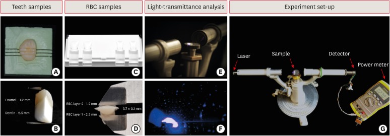
Materials and Methods B1-colored natural teeth slabs were compared to dental restoration build-ups with A2D and B1E-colored nanofilled, supra-nanofilled, microfilled, and microhybrid composites. The enamel layer varied from 0.3, 0.5, or 1.2 mm thick, and the dentin layer was varied to provide a standardized 3.7 mm overall sample thickness (
n = 10). All increments were light-cured to 16 J/cm2 with a multi-wave LED (Valo, Ultradent). Using a spectrophotometer, the samples were irradiated by an RGB laser beam. A voltmeter recorded the light output signal to calculate the light-transmittance through the specimens. The data were analyzed using 1-way analysis of variance followed by thepost hoc Tukey's test (p = 0.05).Results Mean light-transmittance observed at thicker final layers of enamel were significantly lower than those observed at thinner final layers. Within 1.2 mm final enamel resin layer (FERL) thickness, all composites were similar to the dental tissues, with exception of the nanofilled composite. However, within 0.5 mm FERL thickness, only the supra-nanofilled composite showed no difference from the dental tissues. Within 0.3 mm FERL thickness, none of the composites were similar to the dental tissues.
Conclusions The supra-nanofilled composite had the most similar light-transmittance pattern when compared to the natural teeth. However, for other composites, thicker FERL have a greater chance to match the light-transmittance of natural dental tissues.
-
Citations
Citations to this article as recorded by
- Impact of water absorption on the translucency of single-shade and conventional resin composites: an in vitro comparative study
Ceyda Sari, Elifnur Aydemir Aydın
Odontology.2026;[Epub] CrossRef - 3-year randomized clinical trial to evaluate the performance of posterior composite restorations lined with ion-releasing materials
Basma Ahmed, Ramy Ahmed Wafaie, Hamdi H. Hamama, Salah Hasab Mahmoud
Scientific Reports.2024;[Epub] CrossRef - Investigation on the Biaxial Flexural Strength of Universal Shade Resin-Based Composites
Keiko Sakuma, Taku Horie, Takafumi Kishimoto, Mayumi Maesako, Shigetaka Tomoda, Morioki Fujitani, Akimasa Tsujimoto
Polymers.2024; 16(13): 1853. CrossRef - Fabrication of color-graded feldspathic dental prosthetics for aesthetic and restorative dentistry
Imam Akbar Sutejo, Jeehwan Kim, Sinuo Zhang, Chang Woo Gal, Yeong-Jin Choi, Honghyun Park, Hui-suk Yun
Dental Materials.2023; 39(6): 568. CrossRef - Spectrophotometric evaluation of restorative composite shades and their match with a classical shade guide
Rafael Melara, Luciana Mendonça, Fábio Herrmann Coelho-de-Souza, Juliana Nunes Rolla, Luciano de Souza Gonçalves
Restorative Dentistry & Endodontics.2021;[Epub] CrossRef - In vitro wear of dual‐cured bulkfill composites and flowable bulkfill composites
Jean‐François Roulet, Snigdha Gummadi, Hind S. Hussein, Nader Abdulhameed, Chiayi Shen
Journal of Esthetic and Restorative Dentistry.2020; 32(5): 512. CrossRef
- Impact of water absorption on the translucency of single-shade and conventional resin composites: an in vitro comparative study
- 2,244 View
- 17 Download
- 6 Crossref

Original Article
- Comparison of the residual stress of the nanofilled composites
- Jeong-won Park
- J Korean Acad Conserv Dent 2008;33(5):457-462. Published online September 30, 2008
- DOI: https://doi.org/10.5395/JKACD.2008.33.5.457
-
Abstract
PDFPubReaderePub
"Residual stress" can be developed during polymerization of the dental composite and it can be remained after this process was completed. The total amount of the force which applied to the composite restoration can be calculated by the sum of external and internal force. For the complete understanding of the restoration failure behavior, these two factors should be considered. In this experiment, I compared the residual stress of the recently developed nanofilled dental composite by ring slitting methods.
The composites used in this study can be categorized in two groups, one is microhybrid type-Z250, as control group, and nanofilled type-Grandio, Filtek Supreme, Ceram-X, as experimental ones. Composite ring was made and marked two reference points on the surface. Then measure the change of the distance between these two points before and after ring slitting. From the distance change, average circumferential residual stress (σθ) was calculated. In 10 minutes and 1 hour measurement groups, Filtek Supreme showed higher residual stress than Z250 and Ceram-X. In 24 hour group, Filtek showed higher stress than the other groups.
Following the result of this experiment, nanofilled composite showed similar or higher residual stress than Z250, and when comparing the Z250 and Filtek Supreme, which have quite similar matrix components, Filtek Supreme groups showed higher residual stress.
-
Citations
Citations to this article as recorded by- Microleakage of the experimental composite resin with three component photoinitiator systems
Ji-Hoon Kim, Dong-Hoon Shin
Journal of Korean Academy of Conservative Dentistry.2009; 34(4): 333. CrossRef
- Microleakage of the experimental composite resin with three component photoinitiator systems
- 1,651 View
- 4 Download
- 1 Crossref
 First
First Prev
Prev

